Zimmerman Z et al. (AUG 2005)
Biology of Blood and Marrow Transplantation 11 8 576--86
Effector cells derived from host CD8 memory T cells mediate rapid resistance against minor histocompatibility antigen-mismatched allogeneic marrow grafts without participation of perforin, Fas ligand, and the simultaneous inhibition of 3 tumor necrosis Fa
Reduced-intensity conditioning regimens for transplant recipients have heightened awareness of immunologic resistance to allogeneic bone marrow transplants (BMT). Although T cell-mediated cytotoxicity has been assumed to play a role in the resistance against donor allogeneic hematopoietic stem and progenitor cell grafts,several studies have reported relatively unimpaired resistance by recipients who lack perforin,Fas ligand (FasL),and other cytotoxic mediators. This study compared the early kinetics of T cell-mediated resistance in B6 (H2b) cytotoxically normal versus deficient recipients after transplantation with major histocompatibility complex-matched,minor histocompatibility antigen (MiHA)-mismatched allogeneic marrow grafts. Wild-type B6 or cytotoxic double-deficient perforin-/-/ gld+/+ (B6-cdd) mice were sensitized against major histocompatibility complex-matched BALB.B or C3H.SW (H2b) MiHA and transplanted with a high dose (1 ?? 107) of T cell-depleted bone marrow. CD8 T memory cells were shown to be present in recipients before BMT,and anti-CD8 monoclonal antibody infusion abolished resistance,thus demonstrating that CD8 T cells are the host effector population. Donor-committed and high proliferative potential progenitor numbers were markedly diminished by 48 hours after transplantation in both wild-type B6 and B6-cdd anti-donor MiHA-sensitized recipients. These observations indicate that the resistance pathway used in the cytotoxic deficient mice was both potent and rapidly induced - consistent with a CD8 memory T-cell response. To examine the role of Tumor necrosis factor-like weak inducer of apoptosis (TWEAK)- and TL1A-mediated cytotoxicity in this strong resistance,newly generated monoclonal antibodies specific for these ligands were administered to B6-cdd recipients sensitized to donor antigens. Recipients of syngeneic B6-gfp bone marrow exhibited significant donor colony-forming unit numbers after BMT. In contrast,low or absent colony-forming unit levels were detected in allogeneic recipients,including those that lacked perforin and FasL and that received anti-TWEAK,anti-tumor necrosis factor-related apoptosis-inducing ligand,and anti-TL1A monoclonal antibodies. These findings extend previous observations by demonstrating the existence of a rapidly effected resistance pathway mediated by memory CD8 effector T cells independent of the 2 major pathways of cytotoxicity. Together with previous findings,these results support the notion that effector cells derived from memory CD8 T-cell populations can mediate strong resistance against donor allogeneic MiHA-disparate hematopoietic engraftment by using a mechanism that is independent of the contribution of perforin,FasL,and the known death ligand receptor pathways. ?? 2005 American Society for Blood and Marrow Transplantation.
View Publication
Verhoeyen E et al. (MAR 2003)
Blood 101 6 2167--74
IL-7 surface-engineered lentiviral vectors promote survival and efficient gene transfer in resting primary T lymphocytes.
Important gene therapy target cells such as resting human T cells are refractory to transduction with lentiviral vectors. Completion of reverse transcription,nuclear import,and subsequent integration of the lentiviral genome occur in these cells only if they have been activated. In T-cell-based gene therapy trials performed to date,cells have been activated via their cognate antigen receptor. To couple activation with gene transfer,we previously generated lentiviral vectors displaying an anti-CD3 scFv fragment that allowed up to 48% transduction of freshly isolated T cells. However,transduction of highly purified resting T cells with these anti-CD3-displaying lentiviral vectors was inefficient and shifted the T cells from the naive to the memory phenotype. Here,we describe interleukin-7 (IL-7)-displaying HIV-1-derived vectors. Like recombinant IL-7,these modified particles could promote the survival of primary T cells placed in culture without inducing a naive-to-memory phenotypic switch. Furthermore,a single exposure to the IL-7-displaying vectors resulted in efficient gene transfer in both resting memory adult T cells and naive cord blood T cells. With adult naive T cells,preactivation with recombinant IL-7 was necessary for efficient gene transfer. Altogether,these results suggest that IL-7-displaying vectors could constitute interesting tools for T-cell-targeted gene therapy.
View Publication
Veler H et al. (MAR 2007)
Journal of immunology (Baltimore,Md. : 1950) 178 6 3627--36
Superantigen presentation by airway smooth muscle to CD4+ T lymphocytes elicits reciprocal proasthmatic changes in airway function.
Microbial products serving as superantigens (SAgs) have been implicated in triggering various T cell-mediated chronic inflammatory disorders,including severe asthma. Given earlier evidence demonstrating that airway smooth muscle (ASM) cells express MHC class II molecules,we investigated whether ASM can present SAg to resting CD4(+) T cells,and further examined whether this action reciprocally elicits proasthmatic changes in ASM responsiveness. Coincubation of CD4(+) T cells with human ASM cells pulsed with the SAg,staphylococcal enterotoxin A (SEA),elicited adherence and clustering of class II and CD3 molecules at the ASM/T cell interface,indicative of immunological synapse formation,in association with T cell activation. This ASM/T cell interaction evoked up-regulated mRNA expression and pronounced release of the Th2-type cytokine,IL-13,into the coculture medium,which was MHC class II dependent. Moreover,when administering the conditioned medium from the SEA-stimulated ASM/T cell cocultures to isolated naive rabbit ASM tissues,the latter exhibited proasthmatic-like changes in their constrictor and relaxation responsiveness that were prevented by pretreating the tissues with an anti-IL-13 neutralizing Ab. Collectively,these observations are the first to demonstrate that ASM can present SAg to CD4(+) T cells,and that this MHC class II-mediated cooperative ASM/T cell interaction elicits release of IL-13 that,in turn,evokes proasthmatic changes in ASM constrictor and relaxant responsiveness. Thus,a new immuno-regulatory role for ASM is identified that potentially contributes to the pathogenesis of nonallergic (intrinsic) asthma and,accordingly,may underlie the reported association between microbial SAg exposure,T cell activation,and severe asthma.
View Publication
Beeton C et al. (MAR 2003)
The Journal of biological chemistry 278 11 9928--37
A novel fluorescent toxin to detect and investigate Kv1.3 channel up-regulation in chronically activated T lymphocytes.
T lymphocytes with unusually high expression of the voltage-gated Kv1.3 channel (Kv1.3(high) cells) have been implicated in the pathogenesis of experimental autoimmune encephalomyelitis,an animal model for multiple sclerosis. We have developed a fluoresceinated analog of ShK (ShK-F6CA),the most potent known inhibitor of Kv1.3,for detection of Kv1.3(high) cells by flow cytometry. ShK-F6CA blocked Kv1.3 at picomolar concentrations with a Hill coefficient of 1 and exhibited textgreater80-fold specificity for Kv1.3 over Kv1.1 and other K(V) channels. In flow cytometry experiments,ShK-F6CA specifically stained Kv1.3-expressing cells with a detection limit of approximately 600 channels per cell. Rat and human T cells that had been repeatedly stimulated 7-10 times with antigen were readily distinguished on the basis of their high levels of Kv1.3 channels (textgreater600 channels/cell) and ShK-F6CA staining from resting T cells or cells that had undergone 1-3 rounds of activation. Functional Kv1.3 expression levels increased substantially in a myelin-specific rat T cell line following myelin antigen stimulation,peaking at 15-20 h and then declining to baseline over the next 7 days,in parallel with the acquisition and loss of encephalitogenicity. Both calcium- and protein kinase C-dependent pathways were required for the antigen-induced Kv1.3 up-regulation. ShK-F6CA might be useful for rapid and quantitative detection of Kv1.3(high) expressing cells in normal and diseased tissues,and to visualize the distribution of functional channels in intact cells.
View Publication


 EasySep™小鼠TIL(CD45)正选试剂盒




 人IL-13 ELISA试剂盒 用于人白细胞介素13的检测和测定
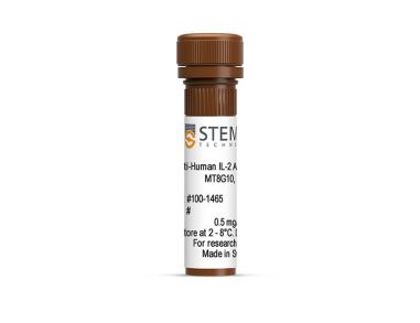 抗人IL-2抗体,clone MT8G10 小鼠Monoclonal IgG1抗体,抗人、食蟹猴IL-2
 小鼠IL-5 ELISA试剂盒 用于小鼠白细胞介素5的检测和测定
 科学海报Cell Enrichment to Obtain Mouse NK Cell Populations By Negative Selection
 抗小鼠CD69抗体,clone H1.2F3 仓鼠(亚美尼亚)抗小鼠CD69 Monoclonal 抗体
 III型胶原酶 用于消化天然胶原纤维
 小鼠IL-2 ELISA试剂盒 用于小鼠白细胞介素2的检测和测定

 沪公网安备31010102008431号