Garg TK et al. (SEP 2012)
Haematologica 97 9 1348--56
Highly activated and expanded natural killer cells for multiple myeloma immunotherapy.
BACKGROUND Patients with gene expression profiling-defined high-risk myeloma in relapse have poor outcomes with current therapies. We tested whether natural killer cells expanded by co-culture with K562 cells transfected with 41BBL and membrane-bound interleukin-15 could kill myeloma cells with a high-risk gene expression profile in vitro and in a unique model which recapitulates human myeloma. DESIGN AND METHODS OPM2 and high-risk primary myeloma tumors were grown in human fetal bone implanted into non-obese diabetic severe combined immunodeficiency mice with a deficient interleukin-2 receptor gamma chain. These mice are devoid of endogenous natural killer and T-cell activity and were used to determine whether adoptively transferred expanded natural killer cells could inhibit myeloma growth and myeloma-associated bone destruction. RESULTS Natural killer cells from healthy donors and myeloma patients expanded a median of 804- and 351-fold,respectively,without significant T-cell expansion. Expanded natural killer cells killed both allogeneic and autologous primary myeloma cells avidly via a perforin-mediated mechanism in which the activating receptor NKG2D,natural cytotoxicity receptors,and DNAX-accessory molecule-1 played a central role. Adoptive transfer of expanded natural killer cells inhibited the growth of established OPM2 and high-risk primary myeloma tumors grown in the murine model. The transferred,expanded natural killer cells proliferated in vivo in an interleukin-2 dose-dependent fashion,persisted up to 4 weeks,were readily detectable in the human bone,inhibited myeloma growth and protected bone from myeloma-induced osteolysis. CONCLUSIONS These studies provide the rationale for testing expanded natural killer cells in humans.
View Publication
产品类型:
产品号#:
19055
19055RF
产品名:
EasySep™人NK细胞富集试剂盒
RoboSep™ 人NK细胞富集试剂盒含滤芯吸头
Zieliʼn et al. ( 2013)
Transplantation proceedings 45 1 88--94
Modified flow cytometry crossmatch detecting alloantibody-related cytotoxicity as a way to distinguish lytic antibodies from harmless in allosensitised kidney recipients.
The serological complement-dependent cytotoxicity crossmatch (CDC-XM) permits routine identification of anti-donor alloantibodies in the sera of allotransplant recipients. However,in a small group of recipients,antibodies below the threshold of detection may still be responsible for hyperacute rejection. For the same reason,approximately 20% of recipients develop acute rejection episodes. The flow cytometry crossmatch (FCXM) was designed to address these problems,but because of the presence of clinically insignificant antibodies (linked,non-lytic),the FCXM appears to be too sensitive yielding false-positive results. We compared FCXM with its modified version assessing cell viability (cytolytic flow cytometry crossmatch; cFCXM) using sera from previously sensitised kidney recipients. The presence of alloantibodies was detected using the Luminex platform. The cFCXM proved to be of greater sensitivity than CDC-XM,which was additionally confirmed with bead-based Luminex techniques. The cFCXM was also superior to FCXM because it distinguished lytic from non-lytic antibodies. The cFCXM was superior to assess donor specificity,sensitivity,and detection of clinically relevant lytic antibodies.
View Publication
产品类型:
产品号#:
19054HLA
19054HLARF
19051HLA
19051HLARF
产品名:
EasySep™ HLA B细胞富集试剂盒
RoboSep™ HLA B细胞富集试剂盒含滤芯吸头
EasySep™ HLA T细胞富集试剂盒
RoboSep™ HLA T细胞富集试剂盒含滤芯吸头
Kang L et al. ( 2013)
Frontiers in immunology 4 MAY 101
Characterization and ex vivo Expansion of Human Placenta-Derived Natural Killer Cells for Cancer Immunotherapy.
Recent clinical studies suggest that adoptive transfer of donor-derived natural killer (NK) cells may improve clinical outcome in hematological malignancies and some solid tumors by direct anti-tumor effects as well as by reduction of graft versus host disease (GVHD). NK cells have also been shown to enhance transplant engraftment during allogeneic hematopoietic stem cell transplantation (HSCT) for hematological malignancies. The limited ex vivo expansion potential of NK cells from peripheral blood (PB) or umbilical cord blood (UCB) has however restricted their therapeutic potential. Here we define methods to efficiently generate NK cells from donor-matched,full-term human placenta perfusate (termed Human Placenta-Derived Stem Cell,HPDSC) and UCB. Following isolation from cryopreserved donor-matched HPDSC and UCB units,CD56+CD3- placenta-derived NK cells,termed pNK cells,were expanded in culture for up to 3 weeks to yield an average of 1.2 billion cells per donor that were textgreater80% CD56+CD3-,comparable to doses previously utilized in clinical applications. Ex vivo-expanded pNK cells exhibited a marked increase in anti-tumor cytolytic activity coinciding with the significantly increased expression of NKG2D,NKp46,and NKp44 (p textless 0.001,p textless 0.001,and p textless 0.05,respectively). Strong cytolytic activity was observed against a wide range of tumor cell lines in vitro. pNK cells display a distinct microRNA (miRNA) expression profile,immunophenotype,and greater anti-tumor capacity in vitro compared to PB NK cells used in recent clinical trials. With further development,pNK may represent a novel and effective cellular immunotherapy for patients with high clinical needs and few other therapeutic options.
View Publication
产品类型:
产品号#:
07900
19055
19055RF
100-0762
产品名:
DNase I 溶液(1 mg/mL)
EasySep™人NK细胞富集试剂盒
RoboSep™ 人NK细胞富集试剂盒含滤芯吸头
DNase I溶液(1mg /mL)
Jiang S et al. (JAN 2018)
Cell metabolism
Let-7 Suppresses B Cell Activation through Restricting the Availability of Necessary Nutrients.
The control of uptake and utilization of necessary extracellular nutrients-glucose and glutamine-is an important aspect of B cell activation. Let-7 is a family of microRNAs known to be involved in metabolic control. Here,we employed several engineered mouse models,including B cell-specific overexpression of Lin28a or the let-7a-1/let-7d/let-7f-1 cluster (let-7adf) and knockout of individual let-7 clusters to show that let-7adf specifically inhibits T cell-independent (TI) antigen-induced immunoglobulin (Ig)M antibody production. Both overexpression and deletion of let-7 in this cluster leads to altered TI-IgM production. Mechanistically,let-7adf suppresses the acquisition and utilization of key nutrients,including glucose and glutamine,through directly targeting hexokinase 2 (Hk2) and by repressing a glutamine transporter Slc1a5 and a key degradation enzyme,glutaminase (Gls),a mechanism mediated by regulation of c-Myc. Our results suggest a novel role of let-7adf as a metabolic brake" on B cell antibody production."
View Publication
BiHC, a T-Cell-Engaging Bispecific Recombinant Antibody, Has Potent Cytotoxic Activity Against Her2 Tumor Cells.
Among different cancer immunotherapy approaches,bispecific antibodies (BsAbs) are of great interest due to their ability to recruit immune cells to kill tumor cells directly. Various BsAbs against Her2 tumor cells have been proposed with potent cytotoxic activities. However,most of these formats require extensive processing to obtain heterodimeric bispecific antibodies. In this study,we describe a bispecific antibody,BiHC (bispecific Her2-CD3 antibody),constructed with a single-domain anti-Her2 and a single-chain Fv (variable fragment) of anti-CD3 in an IgG-like format. In contrast to most IgG-like BsAbs,the two arms in BiHC have different molecular weights,making it easier to separate hetero- or homodimers. BiHC can be expressed in Escherichia coli and purified via Protein A affinity chromatography. The purified BiHC can recruit T cells and induce specific cytotoxicity of Her2-expressing tumor cells in vitro. The BiHC can also efficiently inhibit the tumor growth in vivo. Thus,BiHC is a promising candidate for the treatment of Her2-positive cancers.
View Publication
产品类型:
产品号#:
17851
17851RF
100-0692
产品名:
EasySep™人CD3正选试剂盒II
RoboSep™ 人CD3正选试剂盒II
EasySep™人CD3正选试剂盒II
Dewhurst JA et al. (AUG 2017)
Scientific reports 7 1 7143
Characterisation of lung macrophage subpopulations in COPD patients and controls.
Lung macrophage subpopulations have been identified based on size. We investigated characteristics of small and large macrophages in the alveolar spaces and lung interstitium of COPD patients and controls. Alveolar and interstitial cells were isolated from lung resection tissue from 88 patients. Macrophage subpopulation cell-surface expression of immunological markers and phagocytic ability were assessed by flow cytometry. Inflammatory related gene expression was measured. Alveolar and interstitial macrophages had subpopulations of small and large macrophages based on size and granularity. Alveolar macrophages had similar numbers of small and large cells; interstitial macrophages were mainly small. Small macrophages expressed significantly higher cell surface HLA-DR,CD14,CD38 and CD36 and lower CD206 compared to large macrophages. Large alveolar macrophages showed lower marker expression in COPD current compared to ex-smokers. Small interstitial macrophages had the highest pro-inflammatory gene expression levels,while large alveolar macrophages had the lowest. Small alveolar macrophages had the highest phagocytic ability. Small alveolar macrophage CD206 expression was lower in COPD patients compared to smokers. COPD lung macrophages include distinct subpopulations; Small interstitial and small alveolar macrophages with more pro-inflammatory and phagocytic function respectively,and large alveolar macrophages with low pro-inflammatory and phagocytic ability.
View Publication
Direct interaction of whole-inactivated influenza A and pneumococcal vaccines enhances influenza-specific immunity.
The upper respiratory tract is continuously exposed to a vast array of potentially pathogenic viruses and bacteria. Influenza A virus (IAV) has particular synergism with the commensal bacterium Streptococcus pneumoniae in this niche,and co-infection exacerbates pathogenicity and causes significant mortality. However,it is not known whether this synergism is associated with a direct interaction between the two pathogens. We have previously reported that co-administration of a whole-inactivated IAV vaccine (gamma-Flu) with a whole-inactivated pneumococcal vaccine (gamma-PN) enhances pneumococcal-specific responses. In this study,we show that mucosal co-administration of gamma-Flu and gamma-PN similarly augments IAV-specific immunity,particularly tissue-resident memory cell responses in the lung. In addition,our in vitro analysis revealed that S. pneumoniae directly interacts with both gamma-Flu and with live IAV,facilitating increased uptake by macrophages as well as increased infection of epithelial cells by IAV. These observations provide an additional explanation for the synergistic pathogenicity of IAV and S. pneumoniae,as well as heralding the prospect of exploiting the phenomenon to develop better vaccine strategies for both pathogens.
View Publication
产品类型:
产品号#:
19858
19858RF
产品名:
EasySep™小鼠Naïve CD8+ T细胞分选试剂盒
RoboSep™ 小鼠Naïve CD8+ T细胞分选试剂盒
E. Giuliani et al. (mar 2019)
Scientific reports 9 1 4373
Hexamethylene bisacetamide impairs NK cell-mediated clearance of acute T lymphoblastic leukemia cells and HIV-1-infected T cells that exit viral latency.
The hexamethylene bisacetamide (HMBA) anticancer drug was dismissed due to limited efficacy in leukemic patients but it may re-enter into the clinics in HIV-1 eradication strategies because of its recently disclosed capacity to reactivate latent virus. Here,we investigated the impact of HMBA on the cytotoxicity of natural killer (NK) cells against acute T lymphoblastic leukemia (T-ALL) cells or HIV-1-infected T cells that exit from latency. We show that in T-ALL cells HMBA upmodulated MICB and ULBP2 ligands for the NKG2D activating receptor. In a primary CD4+ T cell-based latency model,HMBA did not reactivate HIV-1,yet enhanced ULBP2 expression on cells harboring virus reactivated by prostratin (PRO). However,HMBA reduced the expression of NKG2D and its DAP10 adaptor in NK cells,hence impairing NKG2D-mediated cytotoxicity and DAP10-dependent response to IL-15 stimulation. Alongside,HMBA dampened killing of T-ALL targets by IL-15-activated NK cells and impaired NK cell-mediated clearance of PRO-reactivated HIV-1+ cells. Overall,our results demonstrate a dominant detrimental effect of HMBA on the NKG2D pathway that crucially controls NK cell-mediated killing of tumors and virus-infected cells,providing one possible explanation for poor clinical outcome in HMBA-treated cancer patients and raising concerns for future therapeutic application of this drug.
View Publication
产品类型:
产品号#:
19052
19052RF
产品名:
EasySep™人CD4+ T细胞富集试剂盒
RoboSep™ 人CD4+ T细胞富集试剂盒含滤芯吸头
J. Jung et al. (apr 2019)
Cell host microbe 25 4 513--525.e6
Cleaved Cochlin Sequesters Pseudomonas aeruginosa and Activates Innate Immunity in the Inner Ear.
In the inner ear,endolymph fluid surrounds the organ of Corti,which is important for auditory function; notably,even slight environmental changes mediated by trauma or infection can have significant consequences. However,it is unclear how the immune response is modulated in these tissues. Here,we report the local immune surveillance role of cleaved cochlin LCCL (Limulus factor C,Cochlin,and Lgl1) during Pseudomonas aeruginosa infection in the cochlea. Upon infection,the LCCL domain is cleaved from cochlin and secreted into the perilymph. This cleaved fragment sequesters infiltrating bacteria in the scala tympani and subsequently recruits resident immune cells to eliminate the bacteria. Importantly,hearing loss in a cochlin knockout mouse model is remedied by treatment with a cochlin LCCL peptide. These findings suggest cleaved cochlin LCCL constitutes a critical factor in innate immunity and auditory function and may be a potential therapeutic target to treat chronic otitis media-induced hearing loss.
View Publication
产品类型:
产品号#:
19762
19762RF
产品名:
EasySep™小鼠中性粒细胞富集试剂盒
RoboSep™ 小鼠中性粒细胞富集试剂盒含滤芯吸头
N. Kuroda et al. (jun 2019)
Scientific reports 9 1 8568
Infiltrating CCR2+ monocytes and their progenies, fibrocytes, contribute to colon fibrosis by inhibiting collagen degradation through the production of TIMP-1.
Intestinal fibrosis is a serious complication in inflammatory bowel disease (IBD). Despite the remarkable success of recent anti-inflammatory therapies for IBD,incidence of intestinal fibrosis and need for bowel resection have not significantly changed. To clarify the contribution of haematopoietic-derived cells in intestinal fibrosis,we prepared bone marrow (BM) chimeric mice (chimeras),which were reconstituted with BM cells derived from enhanced green fluorescent protein (EGFP)-transgenic mice or CC chemokine receptor 2 (CCR2)-deficient mice. After 2 months of transplantation,BM chimeras were treated with azoxymethane/dextran sodium sulphate. During chronic inflammation,CCR2+ BM-derived monocyte and fibrocyte infiltration into the colon and CC chemokine ligand 2 production increased,leading to colon fibrosis in EGFP BM chimeras. In CCR2-deficient BM chimeras,monocyte and fibrocyte numbers in the colonic lamina propria significantly decreased,and colon fibrosis was attenuated. In colon tissue,mRNA expression of tissue inhibitor of metalloproteinase (TIMP)-1 but not of collagen I,transforming growth factor-beta1 or matrix metalloproteinases was significantly different between the two chimeras. CCR2+ monocytes and fibrocytes showed high Timp1 mRNA expression. Our results suggest that infiltrating CCR2+ monocytes and their progenies,fibrocytes,promote colon fibrosis by inhibiting collagen degradation through TIMP-1 production.
View Publication

 EasySep™小鼠TIL(CD45)正选试剂盒


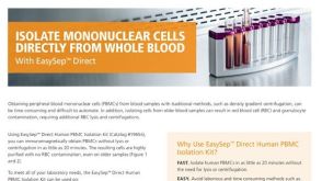 产品手册从全血中直接分离单个核细胞
 挂图Frequencies of Immune Cells in Rat Tissue Lists the estimated frequencies of more than 15 immune cell types in Sprague Dawley rats
 沪公网安备31010102008431号