
 EasySep™小鼠TIL(CD45)正选试剂盒


搜索结果: 'methocult media formulations for mouse hematopoietic cells serum containing'
-

产品类型:
产品号#:
15021
15061
15028
15068
产品名:
RosetteSep™人T细胞富集抗体混合物
RosetteSep™人T细胞富集抗体混合物
RosetteSep™人单核细胞富集抗体混合物
RosetteSep™人单核细胞富集抗体混合物
-
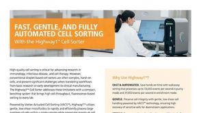 产品手册Highway1™: Fast, Gentle, and Automated Cell Sorting for Every Lab
产品类型:
品牌:
Highway1
产品号#:
产品名:
发布日期: 04/14/2026 -
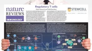 挂图Regulatory T Cells Overview of the development, phenotype and functions of regulatory T cells
 -
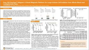 科学海报Easy 250 EasySep™ Magnet: A Novel Magnetic Platform for Large-Volume Cell Isolation
产品类型:
产品号#:
产品名:
-
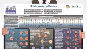 挂图Natural Killer Cells Overview of NK cell receptors, subsets, activation and function
 -
产品类型:
产品号#:
18553
18553RF
15024
15064
产品名:
RosetteSep™人B细胞富集抗体混合物
RosetteSep™人B细胞富集抗体混合物
-
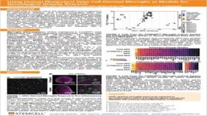 科学海报Using Human Pluripotent Stem Cell-Derived Microglia As Models For Neurological Disease Research
产品类型:
Conference:
FENS 2020
产品号#:
产品名:
发布日期: 07/24/2020 -
 实验方案How to Process Leukocyte Reduction System (LRS) Cones/Chambers for Downstream Cell Isolation
产品类型:
研究方向:
免疫学,药物发现和毒性检测,传染病
产品号#:
产品名:
-
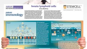 挂图Innate Lymphoid Cells Overview of innate lymphoid cells (ILCs) development, classification, plasticity and functional diversity


 沪公网安备31010102008431号