Ayasoufi K et al. (APR 2016)
Journal of Immunology 196 7 3180--90
CD4 T Cell Help via B Cells Is Required for Lymphopenia-Induced CD8 T Cell Proliferation.
Ab-mediated lymphoablation is commonly used in solid organ and hematopoietic cell transplantation. However,these strategies fail to control pathogenic memory T cells efficiently and to improve long-term transplant outcomes significantly. Understanding the mechanisms of T cell reconstitution is critical for enhancing the efficacy of Ab-mediated depletion in sensitized recipients. Using a murine analog of anti-thymocyte globulin (mATG) in a mouse model of cardiac transplantation,we previously showed that peritransplant lymphocyte depletion induces rapid memory T cell proliferation and only modestly prolongs allograft survival. We now report that T cell repertoire following depletion is dominated by memory CD4 T cells. Additional depletion of these residual CD4 T cells severely impairs the recovery of memory CD8 T cells after mATG treatment. The CD4 T cell help during CD8 T cell recovery depends on the presence of B cells expressing CD40 and intact CD40/CD154 interactions. The requirement for CD4 T cell help is not limited to the use of mATG in heart allograft recipients,and it is observed in nontransplanted mice and after CD8 T cell depletion with mAb instead of mATG. Most importantly,limiting helper signals increases the efficacy of mATG in controlling memory T cell expansion and significantly extends heart allograft survival in sensitized recipients. Our findings uncover the novel role for helper memory CD4 T cells during homeostatic CD8 T cell proliferation and open new avenues for optimizing lymphoablative therapies in allosensitized patients.
View Publication
产品类型:
产品号#:
19851
19851RF
产品名:
EasySep™小鼠T细胞分选试剂盒
RoboSep™ 小鼠T细胞分选试剂盒
Pu Y et al. (APR 2016)
Science Translational Medicine 8 333 333ra47
Androgen receptor antagonists compromise T cell response against prostate cancer leading to early tumor relapse.
Surgical and medical androgen deprivation therapy (ADT) is a cornerstone for prostate cancer treatment,but relapse usually occurs. We herein show that orchiectomy synergizes with immunotherapy,whereas the more widely used treatment of medical ADT involving androgen receptor (AR) antagonists suppresses immunotherapy. Furthermore,we observed that the use of medical ADT could unexpectedly impair the adaptive immune responses through interference with initial T cell priming rather than in the reactivation or expansion phases. Mechanistically,we have revealed that inadvertent immunosuppression might be potentially mediated by a receptor shared with γ-aminobutyric acid. Our data demonstrate that the timing and dosing of antiandrogens are critical to maximizing the antitumor effects of combination therapy. This study highlights an underappreciated mechanism of AR antagonist-mediated immunosuppression and provides a new strategy to enhance immune response and prevent the relapse of advanced prostate cancer.
View Publication
产品类型:
产品号#:
19853
19853RF
产品名:
EasySep™小鼠CD8+ T细胞分选试剂盒
RoboSep™ 小鼠CD8+ T细胞分选试剂盒
Rega A et al. (MAR 2013)
Journal of immunology (Baltimore,Md. : 1950) 190 5 2391--402
Plasmacytoid dendritic cells play a key role in tumor progression in lipopolysaccharide-stimulated lung tumor-bearing mice.
The antitumor activity of LPS was first described by Dr. William Coley. However,its role in lung cancer remains unclear. The aim of our study was to elucidate the dose-dependent effects of LPS (0.1-10 μg/mouse) in a mouse model of B16-F10-induced metastatic lung cancer. Lung tumor growth increased at 3 and 7 d after the administration of low-dose LPS (0.1 μg/mouse) compared with control mice. This was associated with an influx of plasmacytoid dendritic cells (pDCs),regulatory T cells,myeloid-derived suppressor cells,and CD8(+) regulatory T cells. In contrast,high-dose LPS (10 μg/mouse) reduced lung tumor burden and was associated with a greater influx of pDCs,as well as a stronger Th1 and Th17 polarization. Depletion of pDCs during low-dose LPS administration resulted in a decreased lung tumor burden. Depletion of pDCs during high-dose LPS treatment resulted in an increased tumor burden. The dichotomy in LPS effects was due to the phenotype of pDCs,which were immunosuppressive after the low-dose LPS,and Th1- and T cytotoxic-polarizing cells after the high-dose LPS. Adoptive transfer of T cells into nude mice demonstrated that CD8(+) T cells were responsible for pDC recruitment following low-dose LPS administration,whereas CD4(+) T cells were required for pDC influx after the high-dose LPS. In conclusion,our data suggest differential effects of low-dose versus high-dose LPS on pDC phenotype and tumor progression or regression in the lungs of mice.
View Publication
S. Balu et al. ( 2011)
The Journal of Immunology 186 3113-3119
A novel human IgA monoclonal antibody protects against tuberculosis
Abs have been shown to be protective in passive immunotherapy of tuberculous infection using mouse experimental models. In this study,we report on the properties of a novel human IgA1,constructed using a single-chain variable fragment clone (2E9),selected from an Ab phage library. The purified Ab monomer revealed high binding affinities for the mycobacterial ?-crystallin Ag and for the human Fc?RI (CD89) IgA receptor. Intranasal inoculations with 2E9IgA1 and recombinant mouse IFN-? significantly inhibited pulmonary H37Rv infection in mice transgenic for human CD89 but not in CD89-negative littermate controls,suggesting that binding to CD89 was necessary for the IgA-imparted passive protection. 2E9IgA1 added to human whole-blood or monocyte cultures inhibited luciferase-tagged H37Rv infection although not for all tested blood donors. Inhibition by 2E9IgA1 was synergistic with human rIFN-? in cultures of purified human monocytes but not in whole-blood cultures. The demonstration of the mandatory role of Fc?RI (CD89) for human IgA-mediated protection is important for understanding of the mechanisms involved and also for translation of this approach toward development of passive immunotherapy of tuberculosis.
View Publication


 EasySep™小鼠TIL(CD45)正选试剂盒





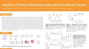 科学海报Isolation of Tumor-Infiltrating Leukocytes from Mouse Tumors
 技术窍门从小鼠健康组织和肿瘤中分离CD45+细胞
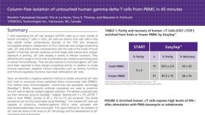 科学海报Immunomagnetic Cell Isolation of Human Gamma-Delta T Cells from PBMC
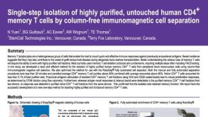 科学海报Cell Isolation of Human CD4+ Memory T Cells By Column-Free Immunomagnetic Cell Separation
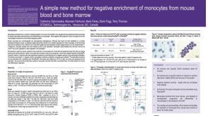 科学海报Method for Negative Enrichment of Monocytes from Mouse Blood and Bone Marrow


 沪公网安备31010102008431号