Xue D et al. (NOV 2016)
Journal of immunology (Baltimore,Md. : 1950)
Semaphorin 4C Protects against Allergic Inflammation: Requirement of Regulatory CD138+ Plasma Cells.
The regulatory properties of B cells have been studied in autoimmune diseases; however,their role in allergic diseases is poorly understood. We demonstrate that Semaphorin 4C (Sema4C),an axonal guidance molecule,plays a crucial role in B cell regulatory function. Mice deficient in Sema4C exhibited increased airway inflammation after allergen exposure,with massive eosinophilic lung infiltrates and increased Th2 cytokines. This phenotype was reproduced by mixed bone marrow chimeric mice with Sema4C deficient only in B cells,indicating that B lymphocytes were the key cells affected by the absence of Sema4C expression in allergic inflammation. We determined that Sema4C-deficient CD19(+)CD138(+) cells exhibited decreased IL-10 and increased IL-4 expression in vivo and in vitro. Adoptive transfer of Sema4c(-/-) CD19(+)CD138(+) cells induced marked pulmonary inflammation,eosinophilia,and increased bronchoalveolar lavage fluid IL-4 and IL-5,whereas adoptive transfer of wild-type CD19(+)CD138(+)IL-10(+) cells dramatically decreased allergic airway inflammation in wild-type and Sema4c(-/-) mice. This study identifies a novel pathway by which Th2-mediated immune responses are regulated. It highlights the importance of plasma cells as regulatory cells in allergic inflammation and suggests that CD138(+) B cells contribute to cytokine balance and are important for maintenance of immune homeostasis in allergic airways disease. Furthermore,we demonstrate that Sema4C is critical for optimal regulatory cytokine production in CD138(+) B cells.
View Publication
产品类型:
产品号#:
19854
19854RF
产品名:
EasySep™小鼠B细胞分选试剂盒
RoboSep™ 小鼠B细胞分选试剂盒
Jitprasertwong P et al. (FEB 2014)
Cytokine 65 2 222--30
Leptin enhances the secretion of interleukin (IL)-18, but not IL-1β, from human monocytes via activation of caspase-1.
Circulating levels of leptin are elevated in type-2 diabetes mellitus (T2DM) and leptin plays a role in immune responses. Elevated circulating IL-18 levels are associated with clinical complications of T2DM. IL-18 regulates cytokine secretion and the function of a number of immune cells including T-cells,neutrophils and macrophages and as such has a key role in immunity and inflammation. Pro-inflammatory monocytes exhibiting elevated cytokine secretion are closely associated with inflammation in T2DM,however,little is known about the role of leptin in modifying monocyte IL-18 secretion. We therefore aimed to investigate the effect of leptin on IL-18 secretion by monocytes. We report herein that leptin increases IL-18 secretion in THP-1 and primary human monocytes but has no effect on IL-18mRNA. Leptin and LPS signalling in monocytes occurs by overlapping but distinct pathways. Thus,in contrast to a strong stimulation by LPS,leptin has no effect on IL-1βmRNA levels or IL-1β secretion. In addition,LPS stimulates the secretion of IL-6 but leptin did not whereas both treatments up regulate IL-8 secretion from the same cells. Although leptin (and LPS) has a synergistic effect with exogenous ATP on IL-18 secretion in both THP-1 and primary monocytes,experiments involving ATP assays and pharmacological inhibition of ATP signalling failed to provide any evidence that endogenous ATP secreted by leptin-stimulated monocytes was responsible for enhancement of monocyte IL-18 secretion by leptin. Analysis of the action of caspase-1 revealed that leptin up regulates caspase-1 activity and the effect of leptin on IL-18 release is prevented by caspase-1 inhibitor (Ac-YVAD-cmk). These data suggest that leptin activates IL-18 processing rather than IL-18 transcription. In conclusion,leptin enhances IL-18 secretion via modulation of the caspase-1 inflammasome function and acts synergistically with ATP in this regard. This process may contribute to aberrant immune responses in T2DM and other conditions of hyperleptinemia.
View Publication
产品类型:
产品号#:
18058
18058RF
产品名:
Kubala SA et al. ( 2014)
Prostaglandins and Other Lipid Mediators 108 1--8
Pathogen induced chemo-attractant hepoxilin A3 drives neutrophils, but not eosinophils across epithelial barriers
Pathogen induced migration of neutrophils across mucosal epithelial barriers requires epithelial production of the chemotactic lipid mediator,hepoxilin A3 (HXA3). HXA3 is an eicosanoid derived from arachidonic acid. Although eosinophils are also capable of penetrating mucosal surfaces,eosinophilic infiltration occurs mainly during allergic processes whereas neutrophils dominate mucosal infection. Both neutrophils and eosinophils can respond to chemotactic gradients of certain eicosanoids,however,it is not known whether eosinophils respond to pathogen induced lipid mediators such as HXA3. In this study,neutrophils and eosinophils were isolated from human blood and placed on the basolateral side of polarized epithelial monolayers grown on permeable Transwell filters and challenged by various chemotactic gradients of distinct lipid mediators. We observed that both cell populations migrated across epithelial monolayers in response to a leukotriene B4 (LTB4) gradient,whereas only eosinophils migrated toward a prostaglandin D2 (PGD2) gradient. Interestingly,while pathogen induced neutrophil trans-epithelial migration was substantial,pathogen induced eosinophil trans-epithelial migration was not observed. Further,gradients of chemotactic lipids derived from pathogen infected epithelial cells known to be enriched for HXA3 as well as purified HXA3 drove significant numbers of neutrophils across epithelial barriers,whereas eosinophils failed to respond to these gradients. These data suggest that although the eicosanoid HXA3 serves as an important neutrophil chemo-attractant at mucosal surfaces during pathogenic infection,HXA3 does not appear to exhibit chemotactic activity toward eosinophils. ?? 2013 Elsevier Ltd. All rights reserved.
View Publication
产品类型:
产品号#:
19256
19256RF
产品名:
Long T et al. (MAR 2014)
Biomaterials 35 9 2752--9
The effect of mesenchymal stem cell sheets on structural allograft healing of critical sized femoral defects in mice.
Structural bone allografts are widely used in the clinic to treat critical sized bone defects,despite lacking the osteoinductive characteristics of live autografts. To address this,we generated revitalized structural allografts wrapped with mesenchymal stem/progenitor cell (MSC) sheets,which were produced by expanding primary syngenic bone marrow derived cells on temperature-responsive plates,as a tissue-engineered periosteum. In vitro assays demonstrated maintenance of the MSC phenotype in the sheets,suggesting that short-term culturing of MSC sheets is not detrimental. To test their efficacy in vivo,allografts wrapped with MSC sheets were transplanted into 4-mm murine femoral defects and compared to allografts with direct seeding of MSCs and allografts without cells. Evaluations consisted of X-ray plain radiography,3D microCT,histology,and biomechanical testing at 4- and 6-weeks post-surgery. Our findings demonstrate that MSC sheets induce prolonged cartilage formation at the graft-host junction and enhanced bone callus formation,as well as graft-host osteointegration. Moreover,a large periosteal callus was observed spanning the allografts with MSC sheets,which partially mimics live autograft healing. Finally,biomechanical testing showed a significant increase in the structural and functional properties of MSC sheet grafted femurs. Taken together,MSC sheets exhibit enhanced osteogenicity during critical sized bone defect repair,demonstrating the feasibility of this tissue engineering solution for massive allograft healing.
View Publication
产品类型:
产品号#:
19771
产品名:
EasySep™ 小鼠间充质干/祖细胞富集试剂盒
Malhotra D et al. (FEB 2016)
Nature Immunology 17 2 187--95
Tolerance is established in polyclonal CD4(+) T cells by distinct mechanisms, according to self-peptide expression patterns.
Studies of repertoires of mouse monoclonal CD4(+) T cells have revealed several mechanisms of self-tolerance; however,which mechanisms operate in normal repertoires is unclear. Here we studied polyclonal CD4(+) T cells specific for green fluorescent protein expressed in various organs,which allowed us to determine the effects of specific expression patterns on the same epitope-specific T cells. Peptides presented uniformly by thymic antigen-presenting cells were tolerated by clonal deletion,whereas peptides excluded from the thymus were ignored. Peptides with limited thymic expression induced partial clonal deletion and impaired effector T cell potential but enhanced regulatory T cell potential. These mechanisms were also active for T cell populations specific for endogenously expressed self antigens. Thus,the immunotolerance of polyclonal CD4(+) T cells was maintained by distinct mechanisms,according to self-peptide expression patterns.
View Publication


 EasySep™小鼠TIL(CD45)正选试剂盒




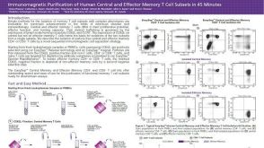 科学海报Immunomagnetic Purification of Human Central and Effector Memory T Cell Subsets in 45 Minutes
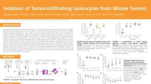 科学海报Isolation of Tumor-Infiltrating Leukocytes from Mouse Tumors
 技术窍门从小鼠健康组织和肿瘤中分离CD45+细胞
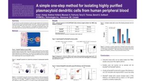 科学海报Isolation of Plasmacytoid Dendritic Cells from Human Peripheral Blood
 科学海报Pre-Enrichment of Dendritic Cells from Human Peripheral Blood Samples

 沪公网安备31010102008431号