D. G. W. Alanine et al. (jun 2019)
Cell 178 1 216--228
Human Antibodies that Slow Erythrocyte Invasion Potentiate Malaria-Neutralizing Antibodies.
The Plasmodium falciparum reticulocyte-binding protein homolog 5 (PfRH5) is the leading target for next-generation vaccines against the disease-causing blood-stage of malaria. However,little is known about how human antibodies confer functional immunity against this antigen. We isolated a panel of human monoclonal antibodies (mAbs) against PfRH5 from peripheral blood B cells from vaccinees in the first clinical trial of a PfRH5-based vaccine. We identified a subset of mAbs with neutralizing activity that bind to three distinct sites and another subset of mAbs that are non-functional,or even antagonistic to neutralizing antibodies. We also identify the epitope of a novel group of non-neutralizing antibodies that significantly reduce the speed of red blood cell invasion by the merozoite,thereby potentiating the effect of all neutralizing PfRH5 antibodies as well as synergizing with antibodies targeting other malaria invasion proteins. Our results provide a roadmap for structure-guided vaccine development to maximize antibody efficacy against blood-stage malaria.
View Publication
产品类型:
产品号#:
19054
19054RF
17963
17963RF
产品名:
EasySep™人B细胞富集试剂盒
RoboSep™ 人B细胞富集试剂盒含滤芯吸头
EasySep™人B细胞富集试剂盒II(不去除CD43)
RoboSep™ 人B细胞富集试剂盒II(不去除CD43)
R. Bertolio et al. ( 2019)
Nature communications 10 1 1326
Sterol regulatory element binding protein 1 couples mechanical cues and lipid metabolism.
Sterol regulatory element binding proteins (SREBPs) are a family of transcription factors that regulate lipid biosynthesis and adipogenesis by controlling the expression of several enzymes required for cholesterol,fatty acid,triacylglycerol and phospholipid synthesis. In vertebrates,SREBP activation is mainly controlled by a complex and well-characterized feedback mechanism mediated by cholesterol,a crucial bio-product of the SREBP-activated mevalonate pathway. In this work,we identified acto-myosin contractility and mechanical forces imposed by the extracellular matrix (ECM) as SREBP1 regulators. SREBP1 control by mechanical cues depends on geranylgeranyl pyrophosphate,another key bio-product of the mevalonate pathway,and impacts on stem cell fate in mouse and on fat storage in Drosophila. Mechanistically,we show that activation of AMP-activated protein kinase (AMPK) by ECM stiffening and geranylgeranylated RhoA-dependent acto-myosin contraction inhibits SREBP1 activation. Our results unveil an unpredicted and evolutionary conserved role of SREBP1 in rewiring cell metabolism in response to mechanical cues.
View Publication
产品类型:
产品号#:
05610
19868
产品名:
EpiCult™-B 小鼠培养基
EasySep™小鼠上皮细胞富集试剂盒II
S. Bhatia et al. (may 2019)
Cancer research 79 10 2722--2735
Inhibition of EphB4-Ephrin-B2 Signaling Reprograms the Tumor Immune Microenvironment in Head and Neck Cancers.
Identifying targets present in the tumor microenvironment that contribute to immune evasion has become an important area of research. In this study,we identified EphB4-ephrin-B2 signaling as a regulator of both innate and adaptive components of the immune system. EphB4 belongs to receptor tyrosine kinase family that interacts with ephrin-B2 ligand at sites of cell-cell contact,resulting in bidirectional signaling. We found that EphB4-ephrin-B2 inhibition alone or in combination with radiation (RT) reduced intratumoral regulatory T cells (Tregs) and increased activation of both CD8+ and CD4+Foxp3- T cells compared with the control group in an orthotopic head and neck squamous cell carcinoma (HNSCC) model. We also compared the effect of EphB4-ephrin-B2 inhibition combined with RT with combined anti-PDL1 and RT and observed similar tumor growth suppression,particularly at early time-points. A patient-derived xenograft model showed reduction of tumor-associated M2 macrophages and favored polarization towards an antitumoral M1 phenotype following EphB4-ephrin-B2 inhibition with RT. In vitro,EphB4 signaling inhibition decreased Ki67-expressing Tregs and Treg activation compared with the control group. Overall,our study is the first to implicate the role of EphB4-ephrin-B2 in tumor immune response. Moreover,our findings suggest that EphB4-ephrin-B2 inhibition combined with RT represents a potential alternative for patients with HNSCC and could be particularly beneficial for patients who are ineligible to receive or cannot tolerate anti-PDL1 therapy. SIGNIFICANCE: These findings present EphB4-ephrin-B2 inhibition as an alternative to anti-PDL1 therapeutics that can be used in combination with radiation to induce an effective antitumor immune response in patients with HNSCC.
View Publication
产品类型:
产品号#:
17952
17952RF
100-0696
产品名:
EasySep™人CD4+ T细胞分选试剂盒
RoboSep™ 人CD4+ T细胞分选试剂盒
EasySep™人CD4+ T细胞分离试剂盒
L. Hang et al. (apr 2019)
Journal of immunology (Baltimore,Md. : 1950) 202 8 2473--2481
Heligmosomoides polygyrus bakeri Infection Decreases Smad7 Expression in Intestinal CD4+ T Cells, Which Allows TGF-beta to Induce IL-10-Producing Regulatory T Cells That Block Colitis.
Helminthic infections modulate host immunity and may protect their hosts from developing immunological diseases like inflammatory bowel disease. Induction of regulatory T cells (Tregs) may be an important part of this protective process. Heligmosomoides polygyrus bakeri infection also promotes the production of the regulatory cytokines TGF-beta and IL-10 in the gut. In the intestines,TGF-beta helps induce regulatory T cells. This study used Foxp3/IL-10 double reporter mice to investigate the effect of TGF-beta on the differentiation of colon and mesenteric lymph node-derived murine Foxp3- IL-10- CD4+ T cells into their regulatory phenotypes. Foxp3- IL-10- CD4+ T cells from H. polygyrus bakeri-infected mice,as opposed to T cells from uninfected animals,cultured in vitro with TGF-beta and anti-CD3/CD28 mAb differentiated into Foxp3+ and/or IL-10+ T cells. The IL-10-producing T cells nearly all displayed CD25. Smad7 is a natural inhibitor of TGF-beta signaling. In contrast to gut T cells from uninfected mice,Foxp3- IL10- CD4+ T cells from H. polygyrus bakeri-infected mice displayed reduced Smad7 expression and responded to TGF-beta with Smad2/3 phosphorylation. The TGF-beta-induced Tregs that express IL-10 blocked colitis when transferred into the Rag/CD25- CD4+ T cell transfer model of inflammatory bowel disease. TGF-beta had a greatly diminished capacity to induce Tregs in H. polygyrus bakeri-infected transgenic mice with constitutively high T cell-specific Smad7 expression. Thus,infection with H. polygyrus bakeri causes down-modulation in Smad7 expression in intestinal CD4+ T cells,which allows the TGF-beta produced in response to the infection to induce the Tregs that prevent colitis.
View Publication
产品类型:
产品号#:
19751
19751RF
产品名:
B. L. Jamison et al. (jul 2019)
Journal of immunology (Baltimore,Md. : 1950) 203 1 48--57
Nanoparticles Containing an Insulin-ChgA Hybrid Peptide Protect from Transfer of Autoimmune Diabetes by Shifting the Balance between Effector T Cells and Regulatory T Cells.
CD4 T cells play a critical role in promoting the development of autoimmunity in type 1 diabetes. The diabetogenic CD4 T cell clone BDC-2.5,originally isolated from a NOD mouse,has been widely used to study the contribution of autoreactive CD4 T cells and relevant Ags to autoimmune diabetes. Recent work from our laboratory has shown that the Ag for BDC-2.5 T cells is a hybrid insulin peptide (2.5HIP) consisting of an insulin C-peptide fragment fused to a peptide from chromogranin A (ChgA) and that endogenous 2.5HIP-reactive T cells are major contributors to autoimmune pathology in NOD mice. The objective of this study was to determine if poly(lactide-co-glycolide) (PLG) nanoparticles (NPs) loaded with the 2.5HIP Ag (2.5HIP-coupled PLG NPs) can tolerize BDC-2.5 T cells. Infusion of 2.5HIP-coupled PLG NPs was found to prevent diabetes in an adoptive transfer model by impairing the ability of BDC-2.5 T cells to produce proinflammatory cytokines through induction of anergy,leading to an increase in the ratio of Foxp3+ regulatory T cells to IFN-gamma+ effector T cells. To our knowledge,this work is the first to use a hybrid insulin peptide,or any neoepitope,to re-educate diabetogenic T cells and may have significant implications for the development of an Ag-specific therapy for type 1 diabetes patients.
View Publication
产品类型:
产品号#:
19852
19852RF
18783
18783RF
18765
18765RF
产品名:
EasySep™小鼠CD4+ T细胞分选试剂盒
RoboSep™ 小鼠CD4+ T细胞分选试剂盒
EasySep™小鼠CD4+CD25+调节性T细胞分选试剂盒II
RoboSep™ 小鼠CD4+CD25+调节性T细胞分选试剂盒II
EasySep™小鼠CD4+ CD62L+ T细胞分选试剂盒
RoboSep™ 小鼠CD4+ CD62L+ T细胞分选试剂盒
S. Omenetti et al. (jun 2019)
Immunity
The Intestine Harbors Functionally Distinct Homeostatic Tissue-Resident and Inflammatory Th17 Cells.
T helper 17 (Th17) cells are pathogenic in many inflammatory diseases,but also support the integrity of the intestinal barrier in a non-inflammatory manner. It is unclear what distinguishes inflammatory Th17 cells elicited by pathogens and tissue-resident homeostatic Th17 cells elicited by commensals. Here,we compared the characteristics of Th17 cells differentiating in response to commensal bacteria (SFB) to those differentiating in response to a pathogen (Citrobacter rodentium). Homeostatic Th17 cells exhibited little plasticity towards expression of inflammatory cytokines,were characterized by a metabolism typical of quiescent or memory T cells,and did not participate in inflammatory processes. In contrast,infection-induced Th17 cells showed extensive plasticity towards pro-inflammatory cytokines,disseminated widely into the periphery,and engaged aerobic glycolysis in addition to oxidative phosphorylation typical for inflammatory effector cells. These findings will help ensure that future therapies directed against inflammatory Th17 cells do not inadvertently damage the resident gut population.
View Publication
产品类型:
产品号#:
19752
19752RF
19052
19052RF
产品名:
EasySep™人CD4+ T细胞富集试剂盒
RoboSep™ 人CD4+ T细胞富集试剂盒含滤芯吸头
C. Onyilagha et al. (jun 2019)
Journal of immunology (Baltimore,Md. : 1950)
NK Cells Are Critical for Optimal Immunity to Experimental Trypanosoma congolense Infection.
NK cells are key innate immune cells that play critical roles in host defense. Although NK cells have been shown to regulate immunity to some infectious diseases,their role in immunity to Trypanosoma congolense has not been investigated. NK cells are vital sources of IFN-gamma and TNF-alpha; two key cytokines that are known to play important roles in resistance to African trypanosomes. In this article,we show that infection with T. congolense leads to increased levels of activated and functional NK cells in multiple tissue compartments. Systemic depletion of NK cells with anti-NK1.1 mAb led to increased parasitemia,which was accompanied by significant reduction in IFN-gamma production by immune cells in the spleens and liver of infected mice. Strikingly,infected NFIL3-/- mice (which genetically lack NK cell development and function) on the normally resistant background were highly susceptible to T. congolense infection. These mice developed fulminating and uncontrolled parasitemia and died significantly earlier (13 ± 1 d) than their wild-type control mice (106 ± 26 d). The enhanced susceptibility of NFIL3-/- mice to infection was accompanied by significantly impaired cytokine (IFN-gamma and TNF-alpha) response by CD3+ T cells in the spleens and liver. Adoptive transfer of NK cells into NFIL3-/- mice before infection rescued them from acute death in a perforin-dependent manner. Collectively,these studies show that NK cells are critical for optimal resistance to T. congolense,and its deficiency leads to enhanced susceptibility in infected mice.
View Publication
产品类型:
产品号#:
19855
19855RF
产品名:
EasySep™小鼠NK细胞分选试剂盒
RoboSep™ 小鼠NK细胞分选试剂盒
Z. Yan et al. (apr 2019)
JCI insight 5
Deficiency of Socs3 leads to brain-targeted EAE via enhanced neutrophil activation and ROS production.
Dysregulation of the JAK/STAT signaling pathway is associated with Multiple Sclerosis (MS) and its mouse model,Experimental Autoimmune Encephalomyelitis (EAE). Suppressors Of Cytokine Signaling (SOCS) negatively regulate the JAK/STAT pathway. We previously reported a severe,brain-targeted,atypical form of EAE in mice lacking Socs3 in myeloid cells (Socs3DeltaLysM),which is associated with cerebellar neutrophil infiltration. There is emerging evidence that neutrophils are detrimental in the pathology of MS/EAE,however,their exact function is unclear. Here we demonstrate that neutrophils from the cerebellum of Socs3DeltaLysM mice show a hyper-activated phenotype with excessive production of reactive oxygen species (ROS) at the peak of EAE. Neutralization of ROS in vivo delayed the onset and reduced severity of atypical EAE. Mechanistically,Socs3-deficient neutrophils exhibit enhanced STAT3 activation,a hyper-activated phenotype in response to G-CSF,and upon G-CSF priming,increased ROS production. Neutralization of G-CSF in vivo significantly reduced the incidence and severity of the atypical EAE phenotype. Overall,our work elucidates that hypersensitivity of G-CSF/STAT3 signaling in Socs3DeltaLysM mice leads to atypical EAE by enhanced neutrophil activation and increased oxidative stress,which may explain the detrimental role of G-CSF in MS patients.
View Publication
产品类型:
产品号#:
19762
19762RF
产品名:
EasySep™小鼠中性粒细胞富集试剂盒
RoboSep™ 小鼠中性粒细胞富集试剂盒含滤芯吸头
C. Yang et al. (may 2019)
The Journal of experimental medicine 216 5 1182--1198
Thyrotropin aggravates atherosclerosis by promoting macrophage inflammation in plaques.
Subclinical hypothyroidism is associated with cardiovascular diseases,yet the underlying mechanism remains largely unknown. Herein,in a common population (n = 1,103),TSH level was found to be independently correlated with both carotid plaque prevalence and intima-media thickness. Consistently,TSH receptor ablation in ApoE-/- mice attenuated atherogenesis,accompanied by decreased vascular inflammation and macrophage burden in atherosclerotic plaques. These results were also observed in myeloid-specific Tshr-deficient ApoE-/- mice,which indicated macrophages to be a critical target of the proinflammatory and atherogenic effects of TSH. In vitro experiments further revealed that TSH activated MAPKs (ERK1/2,p38alpha,and JNK) and IkappaB/p65 pathways in macrophages and increased inflammatory cytokine production and their recruitment of monocytes. Thus,the present study has elucidated the new mechanisms by which TSH,as an independent risk factor of atherosclerosis,aggravates vascular inflammation and contributes to atherogenesis.
View Publication

 EasySep™小鼠TIL(CD45)正选试剂盒


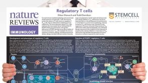 挂图Regulatory T Cells Overview of the development, phenotype and functions of regulatory T cells
 科学海报Depletion of Dead Cells from Primary Tissue in 6 Minutes

 沪公网安备31010102008431号