Lee DJ et al. (NOV 2016)
Scientific reports 6 37790
MC5r and A2Ar Deficiencies During Experimental Autoimmune Uveitis Identifies Distinct T cell Polarization Programs and a Biphasic Regulatory Response.
Autoantigen-specific regulatory immunity emerges in the spleen of mice recovering from experimental autoimmune uveitis (EAU),a murine model for human autoimmune uveoretinitis. This regulatory immunity provides induced tolerance to ocular autoantigen,and requires melanocortin 5 receptor (MC5r) expression on antigen presenting cells with adenosine 2 A receptor (A2Ar) expression on T cells. During EAU it is not well understood what roles MC5r and A2Ar have on promoting regulatory immunity. Cytokine profile analysis during EAU revealed MC5r and A2Ar each mediate distinct T cell responses,and are responsible for a functional regulatory immune response in the spleen. A2Ar stimulation at EAU onset did not augment this regulatory response,nor bypass the MC5r requirement to induce regulatory immunity. The importance of this pathway in human autoimmune uveitis was assayed. PBMC from uveitis patients were assayed for MC5r expression on monocytes and A2Ar on T cells,and comparison between uveitis patients and healthy controls had no significant difference. The importance for MC5r and A2Ar expression in EAU to promote the induction of protective regulatory immunity,and the expression of MC5r and A2Ar on human immune cells,suggests that it may be possible to utilize the melanocortin-adenosinergic pathways to induce protective immunity in uveitic patients.
View Publication
产品类型:
产品号#:
85450
85460
86450
86460
产品名:
SepMate™-50 (IVD)
SepMate™-50 (IVD)
SepMate™-50 (RUO)
SepMate™-50 (RUO)
Abdelwahab SF et al. (DEC 2003)
Proceedings of the National Academy of Sciences of the United States of America 100 25 15006--10
HIV-1-suppressive factors are secreted by CD4+ T cells during primary immune responses.
CD4+ T cells are required for immunity against many viral infections,including HIV-1 where a positive correlation has been observed between strong recall responses and low HIV-1 viral loads. Some HIV-1-specific CD4+ T cells are preferentially infected with HIV-1,whereas others escape infection by unknown mechanisms. One possibility is that some CD4+ T cells are protected from infection by the secretion of soluble HIV-suppressive factors,although it is not known whether these factors are produced during primary antigen-specific responses. Here,we show that soluble suppressive factors are produced against CXCR4 and CCR5 isolates of HIV-1 during the primary immune response of human CD4+ T cells. This activity requires antigenic stimulation of naïve CD4+ T cells. One anti-CXCR4 factor is macrophage-derived chemokine (chemokine ligand 22,CCL22),and anti-CCR5 factors include macrophage inflammatory protein-1 alpha (CCL3),macrophage inflammatory protein-1 beta (CCL4),and RANTES (regulated upon activation of normal T cells expressed and secreted) (CCL5). Intracellular staining confirms that CD3+CD4+ T cells are the source of the prototype HIV-1-inhibiting chemokines CCL22 and CCL4. These results show that CD4+ T cells secrete an evolving HIV-1-suppressive activity during the primary immune response and that this activity is comprised primarily of CC chemokines. The data also suggest that production of such factors should be considered in the design of vaccines against HIV-1 and as a mechanism whereby the host can control infections with this virus.
View Publication
产品类型:
产品号#:
09500
09600
09650
19155
19155RF
产品名:
BIT 9500血清替代物
StemSpan™ SFEM
StemSpan™ SFEM
Fang H et al. (APR 2005)
Journal of immunology (Baltimore,Md. : 1950) 174 8 4966--71
Anthrax lethal toxin blocks MAPK kinase-dependent IL-2 production in CD4+ T cells.
Anthrax lethal toxin (LT) is a critical virulence factor that cleaves and inactivates MAPK kinases (MAPKKs) in host cells and has been proposed as a therapeutic target in the treatment of human anthrax infections. Despite the potential use of anti-toxin agents in humans,the standard activity assays for anthrax LT are currently based on cytotoxic actions of anthrax LT that are cell-,strain-,and species-specific,which have not been demonstrated to occur in human cells. We now report that T cell proliferation and IL-2 production inversely correlate with anthrax LT levels in human cell assays. The model CD4+ T cell tumor line,Jurkat,is a susceptible target for the specific protease action of anthrax LT. Anthrax LT cleaves and inactivates MAPKKs in Jurkat cells,whereas not affecting proximal or parallel TCR signal transduction pathways. Moreover,anthrax LT specifically inhibits PMA/ionomycin- and anti-CD3-induced IL-2 production in Jurkat cells. An inhibitor of the protease activity of anthrax LT completely restores IL-2 production by anthrax LT-treated Jurkat cells. Anthrax LT acts on primary CD4+ T cells as well,cleaving MAPKKs and leading to a 95% reduction in anti-CD3-induced proliferation and IL-2 production. These findings not only will be useful in the development of new human cell-based bioassays for the activity of anthrax LT,but they also suggest new mechanisms that facilitate immune evasion by Bacillus anthracis. Specifically,anthrax LT inhibits IL-2 production and proliferative responses in CD4+ T cells,thereby blocking functions that are pivotal in the regulation of immune responses.
View Publication
产品类型:
产品号#:
15022
15062
产品名:
RosetteSep™人CD4+ T细胞富集抗体混合物
RosetteSep™人CD4+ T细胞富集抗体混合物
E. Drent et al. (jul 2019)
Clinical cancer research : an official journal of the American Association for Cancer Research 25 13 4014--4025
Combined CD28 and 4-1BB Costimulation Potentiates Affinity-tuned Chimeric Antigen Receptor-engineered T Cells.
PURPOSE Targeting nonspecific,tumor-associated antigens (TAA) with chimeric antigen receptors (CAR) requires specific attention to restrict possible detrimental on-target/off-tumor effects. A reduced affinity may direct CAR-engineered T (CAR-T) cells to tumor cells expressing high TAA levels while sparing low expressing normal tissues. However,decreasing the affinity of the CAR-target binding may compromise the overall antitumor effects. Here,we demonstrate the prime importance of the type of intracellular signaling on the function of low-affinity CAR-T cells. EXPERIMENTAL DESIGN We used a series of single-chain variable fragments (scFv) with five different affinities targeting the same epitope of the multiple myeloma-associated CD38 antigen. The scFvs were incorporated in three different CAR costimulation designs and we evaluated the antitumor functionality and off-tumor toxicity of the generated CAR-T cells in vitro and in vivo. RESULTS We show that the inferior cytotoxicity and cytokine secretion mediated by CD38 CARs of very low-affinity (Kd {\textless} 1.9 × 10⁻⁶ mol/L) bearing a 4-1BB intracellular domain can be significantly improved when a CD28 costimulatory domain is used. Additional 4-1BB signaling mediated by the coexpression of 4-1BBL provided the CD28-based CD38 CAR-T cells with superior proliferative capacity,preservation of a central memory phenotype,and significantly improved in vivo antitumor function,while preserving their ability to discriminate target antigen density. CONCLUSIONS A combinatorial costimulatory design allows the use of very low-affinity binding domains (Kd {\textless} 1 mumol/L) for the construction of safe but also optimally effective CAR-T cells. Thus,very-low-affinity scFvs empowered by selected costimulatory elements can enhance the clinical potential of TAA-targeting CARs.
View Publication
产品类型:
产品号#:
04534
04544
17856
17856RF
100-1569
产品名:
MethoCult™ H4534 Classic(不含 EPO)
MethoCult™ H4534 Classic(不含 EPO)
EasySep™人CD34正选试剂盒 II
EasySep™人CD34正选试剂盒 II
EasySep™人CD34正选试剂盒 II
L. Hang et al. (apr 2019)
Journal of immunology (Baltimore,Md. : 1950) 202 8 2473--2481
Heligmosomoides polygyrus bakeri Infection Decreases Smad7 Expression in Intestinal CD4+ T Cells, Which Allows TGF-beta to Induce IL-10-Producing Regulatory T Cells That Block Colitis.
Helminthic infections modulate host immunity and may protect their hosts from developing immunological diseases like inflammatory bowel disease. Induction of regulatory T cells (Tregs) may be an important part of this protective process. Heligmosomoides polygyrus bakeri infection also promotes the production of the regulatory cytokines TGF-beta and IL-10 in the gut. In the intestines,TGF-beta helps induce regulatory T cells. This study used Foxp3/IL-10 double reporter mice to investigate the effect of TGF-beta on the differentiation of colon and mesenteric lymph node-derived murine Foxp3- IL-10- CD4+ T cells into their regulatory phenotypes. Foxp3- IL-10- CD4+ T cells from H. polygyrus bakeri-infected mice,as opposed to T cells from uninfected animals,cultured in vitro with TGF-beta and anti-CD3/CD28 mAb differentiated into Foxp3+ and/or IL-10+ T cells. The IL-10-producing T cells nearly all displayed CD25. Smad7 is a natural inhibitor of TGF-beta signaling. In contrast to gut T cells from uninfected mice,Foxp3- IL10- CD4+ T cells from H. polygyrus bakeri-infected mice displayed reduced Smad7 expression and responded to TGF-beta with Smad2/3 phosphorylation. The TGF-beta-induced Tregs that express IL-10 blocked colitis when transferred into the Rag/CD25- CD4+ T cell transfer model of inflammatory bowel disease. TGF-beta had a greatly diminished capacity to induce Tregs in H. polygyrus bakeri-infected transgenic mice with constitutively high T cell-specific Smad7 expression. Thus,infection with H. polygyrus bakeri causes down-modulation in Smad7 expression in intestinal CD4+ T cells,which allows the TGF-beta produced in response to the infection to induce the Tregs that prevent colitis.
View Publication
产品类型:
产品号#:
19751
19751RF
产品名:
B. L. Jamison et al. (jul 2019)
Journal of immunology (Baltimore,Md. : 1950) 203 1 48--57
Nanoparticles Containing an Insulin-ChgA Hybrid Peptide Protect from Transfer of Autoimmune Diabetes by Shifting the Balance between Effector T Cells and Regulatory T Cells.
CD4 T cells play a critical role in promoting the development of autoimmunity in type 1 diabetes. The diabetogenic CD4 T cell clone BDC-2.5,originally isolated from a NOD mouse,has been widely used to study the contribution of autoreactive CD4 T cells and relevant Ags to autoimmune diabetes. Recent work from our laboratory has shown that the Ag for BDC-2.5 T cells is a hybrid insulin peptide (2.5HIP) consisting of an insulin C-peptide fragment fused to a peptide from chromogranin A (ChgA) and that endogenous 2.5HIP-reactive T cells are major contributors to autoimmune pathology in NOD mice. The objective of this study was to determine if poly(lactide-co-glycolide) (PLG) nanoparticles (NPs) loaded with the 2.5HIP Ag (2.5HIP-coupled PLG NPs) can tolerize BDC-2.5 T cells. Infusion of 2.5HIP-coupled PLG NPs was found to prevent diabetes in an adoptive transfer model by impairing the ability of BDC-2.5 T cells to produce proinflammatory cytokines through induction of anergy,leading to an increase in the ratio of Foxp3+ regulatory T cells to IFN-gamma+ effector T cells. To our knowledge,this work is the first to use a hybrid insulin peptide,or any neoepitope,to re-educate diabetogenic T cells and may have significant implications for the development of an Ag-specific therapy for type 1 diabetes patients.
View Publication
Beeton C et al. (NOV 2006)
Proceedings of the National Academy of Sciences of the United States of America 103 46 17414--9
Kv1.3 channels are a therapeutic target for T cell-mediated autoimmune diseases.
Autoreactive memory T lymphocytes are implicated in the pathogenesis of autoimmune diseases. Here we demonstrate that disease-associated autoreactive T cells from patients with type-1 diabetes mellitus or rheumatoid arthritis (RA) are mainly CD4+ CCR7- CD45RA- effector memory T cells (T(EM) cells) with elevated Kv1.3 potassium channel expression. In contrast,T cells with other antigen specificities from these patients,or autoreactive T cells from healthy individuals and disease controls,express low levels of Kv1.3 and are predominantly naïve or central-memory (T(CM)) cells. In T(EM) cells,Kv1.3 traffics to the immunological synapse during antigen presentation where it colocalizes with Kvbeta2,SAP97,ZIP,p56(lck),and CD4. Although Kv1.3 inhibitors [ShK(L5)-amide (SL5) and PAP1] do not prevent immunological synapse formation,they suppress Ca2+-signaling,cytokine production,and proliferation of autoantigen-specific T(EM) cells at pharmacologically relevant concentrations while sparing other classes of T cells. Kv1.3 inhibitors ameliorate pristane-induced arthritis in rats and reduce the incidence of experimental autoimmune diabetes in diabetes-prone (DP-BB/W) rats. Repeated dosing with Kv1.3 inhibitors in rats has not revealed systemic toxicity. Further development of Kv1.3 blockers for autoimmune disease therapy is warranted.
View Publication
产品类型:
产品号#:
19051
19051RF
产品名:
EasySep™人T细胞富集试剂盒
RoboSep™ 人T细胞富集试剂盒含滤芯吸头
Hale JS et al. (JUN 2010)
Journal of immunology (Baltimore,Md. : 1950) 184 11 5964--8
Cutting Edge: Rag deletion in peripheral T cells blocks TCR revision.
Mature CD4(+)Vbeta5(+) T cells that recognize a peripherally expressed endogenous superantigen are tolerized either by deletion or TCR revision. In Vbeta5 transgenic mice,this latter tolerance pathway results in the appearance of CD4(+)Vbeta5(-)TCRbeta(+) T cells,coinciding with Rag1,Rag2,and TdT expression and the accumulation of V(beta)-DJ(beta) recombination intermediates in peripheral CD4(+) T cells. Because postthymic RAG-dependent TCR rearrangement has remained controversial,we sought to definitively determine whether TCR revision is an extrathymic process that occurs in mature peripheral T cells. We show in this study that Rag deletion in post-positive selection T cells in Vbeta5 transgenic mice blocks TCR revision in vivo and that mature peripheral T cells sorted to remove cells bearing endogenous TCRbeta-chains can express newly generated TCRbeta molecules in adoptive hosts. These findings unambiguously demonstrate postthymic,RAG-dependent TCR rearrangement and define TCR revision as a tolerance pathway that targets mature peripheral CD4(+) T cells.
View Publication


 EasySep™小鼠TIL(CD45)正选试剂盒





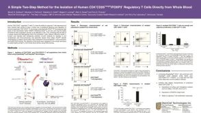 科学海报Isolation of Human CD4+CD25+Bright/Foxp3+ Regulatory T Cells Directly from Whole Blood


 沪公网安备31010102008431号