E. Drent et al. (jul 2019)
Clinical cancer research : an official journal of the American Association for Cancer Research 25 13 4014--4025
Combined CD28 and 4-1BB Costimulation Potentiates Affinity-tuned Chimeric Antigen Receptor-engineered T Cells.
PURPOSE Targeting nonspecific,tumor-associated antigens (TAA) with chimeric antigen receptors (CAR) requires specific attention to restrict possible detrimental on-target/off-tumor effects. A reduced affinity may direct CAR-engineered T (CAR-T) cells to tumor cells expressing high TAA levels while sparing low expressing normal tissues. However,decreasing the affinity of the CAR-target binding may compromise the overall antitumor effects. Here,we demonstrate the prime importance of the type of intracellular signaling on the function of low-affinity CAR-T cells. EXPERIMENTAL DESIGN We used a series of single-chain variable fragments (scFv) with five different affinities targeting the same epitope of the multiple myeloma-associated CD38 antigen. The scFvs were incorporated in three different CAR costimulation designs and we evaluated the antitumor functionality and off-tumor toxicity of the generated CAR-T cells in vitro and in vivo. RESULTS We show that the inferior cytotoxicity and cytokine secretion mediated by CD38 CARs of very low-affinity (Kd {\textless} 1.9 × 10⁻⁶ mol/L) bearing a 4-1BB intracellular domain can be significantly improved when a CD28 costimulatory domain is used. Additional 4-1BB signaling mediated by the coexpression of 4-1BBL provided the CD28-based CD38 CAR-T cells with superior proliferative capacity,preservation of a central memory phenotype,and significantly improved in vivo antitumor function,while preserving their ability to discriminate target antigen density. CONCLUSIONS A combinatorial costimulatory design allows the use of very low-affinity binding domains (Kd {\textless} 1 mumol/L) for the construction of safe but also optimally effective CAR-T cells. Thus,very-low-affinity scFvs empowered by selected costimulatory elements can enhance the clinical potential of TAA-targeting CARs.
View Publication
产品类型:
产品号#:
04534
04544
17856
17856RF
100-1569
产品名:
MethoCult™ H4534 Classic(不含 EPO)
MethoCult™ H4534 Classic(不含 EPO)
EasySep™人CD34正选试剂盒 II
EasySep™人CD34正选试剂盒 II
EasySep™人CD34正选试剂盒 II
C. Gu et al. (jul 2019)
Journal of immunology (Baltimore,Md. : 1950) 203 2 389--399
Signaling Cascade through DC-ASGPR Induces Transcriptionally Active CREB for IL-10 Induction and Immune Regulation.
The types and magnitude of Ag-specific immune responses can be determined by the functional plasticity of dendritic cells (DCs). However,how DCs display functional plasticity and control host immune responses have not been fully understood. In this study,we report that ligation of DC-asialoglycoprotein receptor (DC-ASGPR),a C-type lectin receptor (CLR) expressed on human DCs,resulted in rapid activation of Syk,followed by PLCgamma2 and PKCdelta engagements. However,different from other Syk-coupled CLRs,including Dectin-1,signaling cascade through DC-ASGPR did not trigger NF-kappaB activation. Instead,it selectively activated MAPK ERK1/2 and JNK. Rapid and prolonged phosphorylation of ERK1/2 led to sequential activation of p90RSK and CREB,which consequently bound to IL10 promoter and initiated cytokine expression. In addition,DC-ASGPR ligation activated Akt,which differentially regulated the activities of GSK-3alpha/beta and beta-catenin and further contributed to IL-10 expression. Our observations demonstrate that DC-ASGPR induces IL-10 expression via an intrinsic signaling pathway,which provides a molecular explanation for DC-ASGPR-mediated programing of DCs to control host immune responses.
View Publication
产品类型:
产品号#:
19251
19251RF
19052
19052RF
产品名:
EasySep™人Pan-DC预富集试剂盒
RoboSep™ 人Pan-DC预富集试剂盒含滤芯吸头
EasySep™人CD4+ T细胞富集试剂盒
RoboSep™ 人CD4+ T细胞富集试剂盒含滤芯吸头
Q. Haas et al. ( 2019)
Cancer immunology research 7 5 707--718
Siglec-9 Regulates an Effector Memory CD8+ T-cell Subset That Congregates in the Melanoma Tumor Microenvironment.
Emerging evidence suggests an immunosuppressive role of altered tumor glycosylation due to downregulation of innate immune responses via immunoregulatory Siglecs. In contrast,human T cells,a major anticancer effector cell,only rarely express Siglecs. However,here,we report that the majority of intratumoral,but not peripheral blood,cytotoxic CD8+ T cells expressed Siglec-9 in melanoma. We identified Siglec-9+ CD8+ T cells as a subset of effector memory cells with high functional capacity and signatures of clonal expansion. This cytotoxic T-cell subset was functionally inhibited in the presence of Siglec-9 ligands or by Siglec-9 engagement by specific antibodies. TCR signaling pathways and key effector functions (cytotoxicity,cytokine production) of CD8+ T cells were suppressed by Siglec-9 engagement,which was associated with the phosphorylation of the inhibitory protein tyrosine phosphatase SHP-1,but not SHP-2. Expression of cognate Siglec-9 ligands was observed on the majority of tumor cells in primary and metastatic melanoma specimens. Targeting the tumor-restricted,glycosylation-dependent Siglec-9 axis may unleash this intratumoral T-cell subset,while confining T-cell activation to the tumor microenvironment.
View Publication
产品类型:
产品号#:
17953
17953RF
100-0710
产品名:
EasySep™人CD8+ T细胞分选试剂盒
RoboSep™ 人CD8+ T细胞分选试剂盒
EasySep™人CD8+ T细胞分选试剂盒
C. Imbratta et al. (apr 2019)
Scientific reports 9 1 6135
Maf deficiency in T cells dysregulates Treg - TH17 balance leading to spontaneous colitis.
The maintenance of homeostasis in the gut is a major challenge for the immune system. Here we demonstrate that the transcription factor MAF plays a central role in T cells for the prevention of gastro-intestinal inflammation. Conditional knock out mice lacking Maf in all T cells developed spontaneous late-onset colitis,correlating with a decrease of FOXP3+RORgammat+ T cells proportion,dampened IL-10 production in the colon and an increase of inflammatory TH17 cells. Strikingly,FOXP3+ specific conditional knock out mice for MAF did not develop colitis and demonstrated normal levels of IL-10 in their colon,despite the incapacity of regulatory T cells lacking MAF to suppress colon inflammation in Rag1-/- mice transferred with na{\{i}}ve CD4+ T cells. We showed that one of the cellular sources of IL-10 in the colon of these mice are TH17 cells. Thus MAF is critically involved in the maintenance of the gut homeostasis by regulating the balance between Treg and TH17 cells either at the level of their differentiation or through the modulation of their functions."
View Publication
产品类型:
产品号#:
19765
19765RF
19852
19852RF
产品名:
EasySep™小鼠Naïve CD4+ T细胞分选试剂盒
RoboSep™ 小鼠Naïve CD4+ T细胞分选试剂盒
EasySep™小鼠CD4+ T细胞分选试剂盒
RoboSep™ 小鼠CD4+ T细胞分选试剂盒
H.-W. Wu et al. (may 2019)
Clinical cancer research : an official journal of the American Association for Cancer Research
Anti-CD105 Antibody Eliminates Tumor Microenvironment Cells and Enhances Anti-GD2 Antibody Immunotherapy of Neuroblastoma with Activated Natural Killer Cells.
Purpose: We determined whether elimination of CD105+ cells in the tumor microenvironment (TME) with anti-CD105 antibodies enhanced anti-disialoganglioside (GD2) antibody dinutuximab therapy of neuroblastoma when combined with activated natural killer (aNK) cells.Experimental Design: The effect of MSCs and monocytes on antibody-dependent cellular cytotoxicity (ADCC) mediated by dinutuximab with aNK cells against neuroblastoma cells was determined in vitro. ADCC with anti-CD105 mAb TRC105 and aNK cells against MSCs,monocytes,and endothelial cells,which express CD105,was evaluated. Anti-neuroblastoma activity in immunodeficient NSG mice of dinutuximab with aNK cells without or with anti-CD105 mAbs was determined using neuroblastoma cell lines and a patient-derived xenograft.Results: ADCC mediated by dinutuximab with aNK cells against neuroblastoma cells in vitro was suppressed by addition of MSCs and monocytes,and dinutuximab with aNK cells was less effective against neuroblastomas formed with coinjected MSCs and monocytes in NSG mice than against those formed by tumor cells alone. Anti-CD105 antibody TRC105 with aNK cells mediated ADCC against MSCs,monocytes,and endothelial cells. Neuroblastomas formed in NSG mice by two neuroblastoma cell lines or a patient-derived xenograft coinjected with MSCs and monocytes were most effectively treated with dinutuximab and aNK cells when anti-human (TRC105) and anti-mouse (M1043) CD105 antibodies were added,which depleted human MSCs and murine endothelial cells and macrophages from the TME.Conclusions: Immunotherapy of neuroblastoma with anti-GD2 antibody dinutuximab and aNK cells is suppressed by CD105+ cells in the TME,but suppression is overcome by adding anti-CD105 antibodies to eliminate CD105+ cells.
View Publication
F. Stehle et al. ( 2013)
The Journal of Biological Chemistry 288 16334-16347
Reduced immunosuppressive properties of axitinib in comparison with other tyrosine kinase inhibitors
The multikinase inhibitors sunitinib,sorafenib,and axitinib have an impact not only on tumor growth and angiogenesis,but also on the activity and function of immune effector cells. In this study,a comparative analysis of the growth inhibitory properties and apoptosis induction potentials of tyrosine kinase inhibitors on T cells was performed. Tyrosine kinase inhibitor treatment resulted in a dramatic decrease in T cell proliferation along with distinct impacts on the cell cycle progression. This was at least partially associated with an enhanced induction of apoptosis although triggered by distinct apoptotic mechanisms. In contrast to sunitinib and sorafenib,axitinib did not affect the mitochondrial membrane potential but resulted in an induction or stabilization of the induced myeloid leukemia cell differentiation protein (Mcl-1),leading to an irreversible arrest in the G2/M cell cycle phase and delayed apoptosis. Furthermore,the sorafenib-mediated suppression of immune effector cells,in particular the reduction of the CD8(+) T cell subset along with the down-regulation of key immune cell markers such as chemokine CC motif receptor 7 (CCR7),CD26,CD69,CD25,and CXCR3,was not observed in axitinib-treated immune effector cells. Therefore,axitinib rather than sorafenib seems to be suitable for implementation in complex treatment regimens of cancer patients including immunotherapy.
View Publication

 EasySep™小鼠TIL(CD45)正选试剂盒


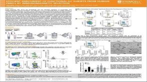 科学海报Efficient Enrichment of Functional ILC Subsets from Human PBMCs by Immunomagnetic Selection
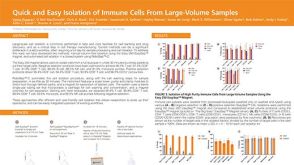 科学海报Quick and Easy Isolation of Immune Cells From Large-Volume Samples


 沪公网安备31010102008431号