Summers-DeLuca LE et al. (MAY 2007)
The Journal of experimental medicine 204 5 1071--81
Expression of lymphotoxin-alphabeta on antigen-specific T cells is required for DC function.
During an immune response,activated antigen (Ag)-specific T cells condition dendritic cells (DCs) to enhance DC function and survival within the inflamed draining lymph node (LN). It has been difficult to ascertain the role of the tumor necrosis factor (TNF) superfamily member lymphotoxin-alphabeta (LTalphabeta) in this process because signaling through the LTbeta-receptor (LTbetaR) controls multiple aspects of lymphoid tissue organization. To resolve this,we have used an in vivo system where the expression of TNF family ligands is manipulated only on the Ag-specific T cells that interact with and condition Ag-bearing DCs. We report that LTalphabeta is a critical participant required for optimal DC function,independent of its described role in maintaining lymphoid tissue organization. In the absence of LTalphabeta or CD40L on Ag-specific T cells,DC dysfunction could be rescued in vivo via CD40 or LTbetaR stimulation,respectively,suggesting that these two pathways cooperate for optimal DC conditioning.
View Publication
产品类型:
产品号#:
19752
19752RF
19753
19753RF
产品名:
Allan AL et al. (MAY 2005)
Cytometry. Part A : the journal of the International Society for Analytical Cytology 65 1 4--14
Detection and quantification of circulating tumor cells in mouse models of human breast cancer using immunomagnetic enrichment and multiparameter flow cytometry.
BACKGROUND: Circulating tumor cells (CTCs) in the peripheral blood of breast cancer patients may be an important indicator of metastatic disease and poor prognosis. However,the use of experimental models is required to fully elucidate the functional consequences of CTCs. The purpose of this study was to optimize the sensitivity of multiparameter flow cytometry for detection of human tumor cells in mouse models of breast cancer. METHODS: MDA-MB-468 human breast cancer cells were serially diluted in whole mouse blood. Samples were lysed and incubated with a fluorescein isothiocyanate-conjugated anti-human leukocytic antigen antibody and a phycoerythrin-conjugated anti-mouse pan-leukocyte CD45 antibody. Samples were then immunomagnetically depleted of CD45-positive leukocytes,fixed,permeabilized,and stained with propidium iodide before flow cytometric analysis. RESULTS: Human breast cancer cells could be differentiated from mouse leukocytes based on increased light scatter,cell surface marker expression,and aneuploid DNA content. The method was found to have a lower sensitivity limit of 10(-5) and was effective for detecting human breast cancer cells in vivo in the circulation of experimental mice carrying primary human mammary tumors. CONCLUSIONS: This technique has the potential to be a valuable and sensitive tool for investigating the biological relevance of CTCs in experimental mouse models of breast cancer.
View Publication
产品类型:
产品号#:
18554
18554RF
18564
18564RF
产品名:
Putnam AL et al. (NOV 2013)
American journal of transplantation : official journal of the American Society of Transplantation and the American Society of Transplant Surgeons 13 11 3010--20
Clinical grade manufacturing of human alloantigen-reactive regulatory T cells for use in transplantation.
Regulatory T cell (Treg) therapy has the potential to induce transplantation tolerance so that immunosuppression and associated morbidity can be minimized. Alloantigen-reactive Tregs (arTregs) are more effective at preventing graft rejection than polyclonally expanded Tregs (PolyTregs) in murine models. We have developed a manufacturing process to expand human arTregs in short-term cultures using good manufacturing practice-compliant reagents. This process uses CD40L-activated allogeneic B cells to selectively expand arTregs followed by polyclonal restimulation to increase yield. Tregs expanded 100- to 1600-fold were highly alloantigen reactive and expressed the phenotype of stable Tregs. The alloantigen-expanded Tregs had a diverse TCR repertoire. They were more potent than PolyTregs in vitro and more effective at controlling allograft injuries in vivo in a humanized mouse model.
View Publication
Molecular events contributing to cell death in malignant human hematopoietic cells elicited by an IgG3-avidin fusion protein targeting the transferrin receptor.
We have previously reported that an anti-human transferrin receptor IgG3-avidin fusion protein (anti-hTfR IgG3-Av) inhibits the proliferation of an erythroleukemia-cell line. We have now found that anti-hTfR IgG3-Av also inhibits the proliferation of additional human malignant B and plasma cells. Anti-hTfR IgG3-Av induces internalization and rapid degradation of the TfR. These events can be reproduced in cells treated with anti-hTfR IgG3 cross-linked with a secondary Ab,suggesting that they result from increased TfR cross-linking. Confocal microscopy of cells treated with anti-hTfR IgG3-Av shows that the TfR is directed to an intracellular compartment expressing the lysosomal marker LAMP-1. The degradation of TfR is partially blocked by cysteine protease inhibitors. Furthermore,cells treated with anti-hTfR IgG3-Av exhibit mitochondrial depolarization and activation of caspases 9,8,and 3. The mitochondrial damage and cell death can be prevented by iron supplementation,but cannot be fully blocked by a pan-caspase inhibitor. These results suggest that anti-hTfR IgG3-Av induces lethal iron deprivation,but the resulting cell death does not solely depend on caspase activation. This report provides insights into the mechanism of cell death induced by anti-TfR Abs such as anti-hTfR IgG3-Av,a molecule that may be useful in the treatment of B-cell malignancies such as multiple myeloma.
View Publication
产品类型:
产品号#:
18357
18357RF
产品名:
Zimmerman Z et al. (AUG 2005)
Biology of Blood and Marrow Transplantation 11 8 576--86
Effector cells derived from host CD8 memory T cells mediate rapid resistance against minor histocompatibility antigen-mismatched allogeneic marrow grafts without participation of perforin, Fas ligand, and the simultaneous inhibition of 3 tumor necrosis Fa
Reduced-intensity conditioning regimens for transplant recipients have heightened awareness of immunologic resistance to allogeneic bone marrow transplants (BMT). Although T cell-mediated cytotoxicity has been assumed to play a role in the resistance against donor allogeneic hematopoietic stem and progenitor cell grafts,several studies have reported relatively unimpaired resistance by recipients who lack perforin,Fas ligand (FasL),and other cytotoxic mediators. This study compared the early kinetics of T cell-mediated resistance in B6 (H2b) cytotoxically normal versus deficient recipients after transplantation with major histocompatibility complex-matched,minor histocompatibility antigen (MiHA)-mismatched allogeneic marrow grafts. Wild-type B6 or cytotoxic double-deficient perforin-/-/ gld+/+ (B6-cdd) mice were sensitized against major histocompatibility complex-matched BALB.B or C3H.SW (H2b) MiHA and transplanted with a high dose (1 ?? 107) of T cell-depleted bone marrow. CD8 T memory cells were shown to be present in recipients before BMT,and anti-CD8 monoclonal antibody infusion abolished resistance,thus demonstrating that CD8 T cells are the host effector population. Donor-committed and high proliferative potential progenitor numbers were markedly diminished by 48 hours after transplantation in both wild-type B6 and B6-cdd anti-donor MiHA-sensitized recipients. These observations indicate that the resistance pathway used in the cytotoxic deficient mice was both potent and rapidly induced - consistent with a CD8 memory T-cell response. To examine the role of Tumor necrosis factor-like weak inducer of apoptosis (TWEAK)- and TL1A-mediated cytotoxicity in this strong resistance,newly generated monoclonal antibodies specific for these ligands were administered to B6-cdd recipients sensitized to donor antigens. Recipients of syngeneic B6-gfp bone marrow exhibited significant donor colony-forming unit numbers after BMT. In contrast,low or absent colony-forming unit levels were detected in allogeneic recipients,including those that lacked perforin and FasL and that received anti-TWEAK,anti-tumor necrosis factor-related apoptosis-inducing ligand,and anti-TL1A monoclonal antibodies. These findings extend previous observations by demonstrating the existence of a rapidly effected resistance pathway mediated by memory CD8 effector T cells independent of the 2 major pathways of cytotoxicity. Together with previous findings,these results support the notion that effector cells derived from memory CD8 T-cell populations can mediate strong resistance against donor allogeneic MiHA-disparate hematopoietic engraftment by using a mechanism that is independent of the contribution of perforin,FasL,and the known death ligand receptor pathways. ?? 2005 American Society for Blood and Marrow Transplantation.
View Publication
产品类型:
产品号#:
03800
03801
03802
03803
03804
03805
03806
产品名:
ClonaCell™-HY杂交瘤试剂盒
ClonaCell™-HY培养基A
ClonaCell™-HY 培养基 B
ClonaCell™-HY 培养基 C
ClonaCell™-HY 培养基 D
ClonaCell™-HY 培养基 E
ClonaCell™-HY PEG
Garidou L et al. (SEP 2009)
Journal of virology 83 17 8905--15
Therapeutic memory T cells require costimulation for effective clearance of a persistent viral infection.
Persistent viral infections are a major health concern worldwide. During persistent infection,overwhelming viral replication and the rapid loss of antiviral T-cell function can prevent immune-mediated clearance of the infection,and therapies to reanimate the immune response and purge persistent viruses have been largely unsuccessful. Adoptive immunotherapy using memory T cells is a highly successful therapeutic approach to eradicate a persistent viral infection. Understanding precisely how therapeutically administered memory T cells achieve clearance should improve our ability to terminate states of viral persistence in humans. Mice persistently infected from birth with lymphocytic choriomeningitis virus are tolerant to the pathogen at the T-cell level and thus provide an excellent model to evaluate immunotherapeutic regimens. Previously,we demonstrated that adoptively transferred memory T cells require recipient dendritic cells to effectively purge an established persistent viral infection. However,the mechanisms that reactivate and sustain memory T-cell responses during clearance of such an infection remain unclear. Here we establish that therapeutic memory T cells require CD80 and CD86 costimulatory signals to efficiently clear an established persistent viral infection in vivo. Early blockade of costimulatory pathways with CTLA-4-Fc decreased the secondary expansion of virus-specific CD8(+) and CD4(+) memory T cells as well as their ability to produce antiviral cytokines and purge the persistent infection. Late costimulation blockade also reduced virus-specific T-cell numbers,illustrating that sustained interactions with costimulatory molecules is required for efficient T-cell expansion. These findings indicate that antiviral memory T cells require costimulation to efficiently clear a persistent viral infection and that costimulatory pathways can be targeted to modulate the magnitude of an adoptive immunotherapeutic regimen.
View Publication
产品类型:
产品号#:
18758
18758RF
18768
18768RF
产品名:
Safinia N et al. (FEB 2016)
Oncotarget 7 7 7563--77
Successful expansion of functional and stable regulatory T cells for immunotherapy in liver transplantation.
Strategies to prevent organ transplant rejection whilst minimizing long-term immunosuppression are currently under intense investigation with regulatory T cells (Tregs) nearing clinical application. The clinical trial,ThRIL,recently commenced at King's College London,proposes to use Treg cell therapy to induce tolerance in liver transplant recipients,the success of which has the potential to revolutionize the management of these patients and enable a future of drug-free transplants. This is the first report of the manufacture of clinical grade Tregs from prospective liver transplant recipients via a CliniMACS-based GMP isolation technique and expanded using anti-CD3/CD28 beads,IL-2 and rapamycin. We report the enrichment of a pure,stable population of Tregs (textgreater95% CD4(+)CD25(+)FOXP3(+)),reaching adequate numbers for their clinical application. Our protocol proved successful in,influencing the expansion of superior functional Tregs,as compared to freshly isolated cells,whilst also preventing their conversion to Th17 cells under pro-inflammatory conditions. We conclude with the manufacture of the final Treg product in the clinical research facility (CRF),a prerequisite for the clinical application of these cells. The data presented in this manuscript together with the much-anticipated clinical results from ThRIL,will undoubtedly inform the improved management of the liver transplant recipient.
View Publication
产品类型:
产品号#:
07930
07931
07940
07955
07956
07959
07954
100-1061
07952
产品名:
CryoStor® CS10
CryoStor® CS10
CryoStor® CS10
CryoStor® CS10
CryoStor® CS10
CryoStor® CS10
CryoStor® CS10
Anderson AE et al. (FEB 2009)
Journal of leukocyte biology 85 2 243--50
LPS activation is required for migratory activity and antigen presentation by tolerogenic dendritic cells.
Autoimmune pathologies are caused by a breakdown in self-tolerance. Tolerogenic dendritic cells (tolDC) are a promising immunotherapeutic tool for restoring self-tolerance in an antigen-specific manner. Studies about tolDC have focused largely on generating stable maturation-resistant DC,but few have fully addressed questions about the antigen-presenting and migratory capacities of these cells,prerequisites for successful immunotherapy. Here,we investigated whether human tolDC,generated with dexamethasone and the active form of vitamin D3,maintained their tolerogenic function upon activation with LPS (LPS-tolDC),while acquiring the ability to present exogenous autoantigen and to migrate in response to the CCR7 ligand CCL19. LPS activation led to important changes in the tolDC phenotype and function. LPS-tolDC,but not tolDC,expressed the chemokine receptor CCR7 and migrated in response to CCL19. Furthermore,LPS-tolDC were superior to tolDC in their ability to present type II collagen,a candidate autoantigen in rheumatoid arthritis. tolDC and LPS-tolDC had low stimulatory capacity for allogeneic,naïve T cells and skewed T cell polarization toward an anti-inflammatory phenotype,although LPS-tolDC induced significantly higher levels of IL-10 production by T cells. Our finding that LPS activation is essential for inducing migratory and antigen-presenting activity in tolDC is important for optimizing their therapeutic potential.
View Publication
产品类型:
产品号#:
18259
18259RF
产品名:
Kang L et al. ( 2013)
Frontiers in immunology 4 MAY 101
Characterization and ex vivo Expansion of Human Placenta-Derived Natural Killer Cells for Cancer Immunotherapy.
Recent clinical studies suggest that adoptive transfer of donor-derived natural killer (NK) cells may improve clinical outcome in hematological malignancies and some solid tumors by direct anti-tumor effects as well as by reduction of graft versus host disease (GVHD). NK cells have also been shown to enhance transplant engraftment during allogeneic hematopoietic stem cell transplantation (HSCT) for hematological malignancies. The limited ex vivo expansion potential of NK cells from peripheral blood (PB) or umbilical cord blood (UCB) has however restricted their therapeutic potential. Here we define methods to efficiently generate NK cells from donor-matched,full-term human placenta perfusate (termed Human Placenta-Derived Stem Cell,HPDSC) and UCB. Following isolation from cryopreserved donor-matched HPDSC and UCB units,CD56+CD3- placenta-derived NK cells,termed pNK cells,were expanded in culture for up to 3 weeks to yield an average of 1.2 billion cells per donor that were textgreater80% CD56+CD3-,comparable to doses previously utilized in clinical applications. Ex vivo-expanded pNK cells exhibited a marked increase in anti-tumor cytolytic activity coinciding with the significantly increased expression of NKG2D,NKp46,and NKp44 (p textless 0.001,p textless 0.001,and p textless 0.05,respectively). Strong cytolytic activity was observed against a wide range of tumor cell lines in vitro. pNK cells display a distinct microRNA (miRNA) expression profile,immunophenotype,and greater anti-tumor capacity in vitro compared to PB NK cells used in recent clinical trials. With further development,pNK may represent a novel and effective cellular immunotherapy for patients with high clinical needs and few other therapeutic options.
View Publication
Kishimoto RK et al. (APR 2016)
Revista brasileira de hematologia e hemoterapia 38 2 113--20
Validation of interphase fluorescence in situ hybridization (iFISH) for multiple myeloma using CD138 positive cells.
BACKGROUND Multiple myeloma is a plasma cell neoplasm with acquired genetic abnormalities of clinical and prognostic importance. Multiple myeloma differs from other hematologic malignancies due to a high fraction of low proliferating malignant plasma cells and the paucity of plasma cells in bone marrow aspiration samples,making cytogenetic analysis a challenge. An abnormal karyotype is found in only one-third of patients with multiple myeloma and interphase fluorescence in situ hybridization is the most useful test for studying the chromosomal abnormalities present in almost 90% of cases. However,it is necessary to study the genetic abnormalities in plasma cells after their identification or selection by morphology,immunophenotyping or sorting. Other challenges are the selection of the most informative FISH panel and determining cut-off levels for FISH probes. This study reports the validation of interphase fluorescence in situ hybridization using CD138 positive cells,according to proposed guidelines published by the European Myeloma Network (EMN) in 2012. METHOD Bone marrow samples from patients with multiple myeloma were used to standardize a panel of five probes [1q amplification,13q14 deletion,17p deletion,t(4;14),and t(14;16)] in CD138(+) cells purified by magnetic cell sorting. RESULTS This test was validated with a low turnaround time and good reproducibility. Five of six samples showed genetic abnormalities. Monosomy/deletion 13 plus t(4;14) were found in two cases. CONCLUSION This technique together with magnetic cell sorting is effective and can be used in the routine laboratory practice. In addition,magnetic cell sorting provides a pure plasma cell population that allows other molecular and genomic studies.
View Publication

 EasySep™小鼠TIL(CD45)正选试剂盒


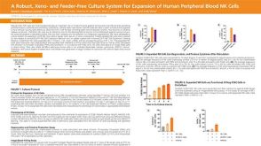 科学海报A Robust, Xeno- and Feeder-Free Culture System for Expansion of Human Peripheral Blood NK Cells

 沪公网安备31010102008431号