
 EasySep™小鼠TIL(CD45)正选试剂盒


搜索结果: 'methocult media formulations for human hematopoietic cells serum containing'
-
 抗人CD45RO抗体,clone UCHL1 小鼠Monoclonal IgG2a抗体,抗人、黑猩猩、普通狨猴CD45RO
 -
 抗人CD56抗体,clone HCD56 小鼠Monoclonal IgG1抗体,抗人CD56 (NCAM)
 -
 抗人CD68抗体,clone Y1/82A 小鼠Monoclonal IgG2b抗体,抗人CD68
 -
 抗人CD83抗体,clone HB15e 抗人、恒河猴、食蟹猴CD83的小鼠Monoclonal IgG1抗体
 -
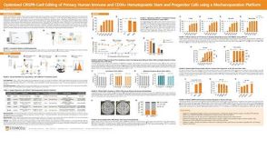 科学海报Optimized CRISPR-Cas9 Editing of Primary Human Immune and CD34+ Hematopoietic Stem and Progenitor Cells using a Mechanoporation Platform
产品类型:
Conference:
ASCGT 2025
产品号#:
产品名:
-
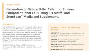 技术公告Generation of Natural Killer Cells from Human Pluripotent Stem Cells Using STEMdiff™ and StemSpan™ Media and Supplements
产品类型:
细胞类型:
NK细胞,多能干细胞
产品号#:
产品名:
发布日期: 07/30/2021 -

产品类型:
产品号#:
18757
18757RF
产品名:
EasySep™小鼠CD117(cKIT)正选试剂盒
RoboSep™ 小鼠CD117(cKIT)正选试剂盒含滤芯吸头
-
产品类型:
产品号#:
19756
19756RF
产品名:
-
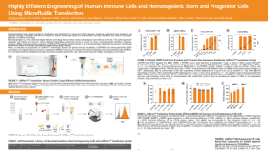 科学海报Highly Efficient Engineering of Human Immune Cells and Hematopoietic Stem and Progenitor Cells Using Microfluidic Transfection
产品类型:
Conference:
AAI 2025
产品号#:
产品名:
-
产品类型:
产品号#:
04434
04444
产品名:
MethoCult™ H4434 Classic
MethoCult™ H4434 Classic
-
产品类型:
产品号#:
19055
19055RF
产品名:
EasySep™人NK细胞富集试剂盒
RoboSep™ 人NK细胞富集试剂盒含滤芯吸头
-
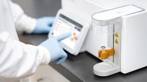 实验方案Gene Editing Human CD34+ Hematopoietic Stem and Progenitor Cells with the CellPore™ Transfection System
产品类型:
研究方向:
免疫学,细胞治疗开发
产品号#:
产品名:

 沪公网安备31010102008431号