D. Park et al. (may 2019)
Scientific reports 9 1 7094
Differences in the molecular signatures of mucosal-associated invariant T cells and conventional T cells.
Mucosal-associated invariant T (MAIT) cells exhibit different characteristics from those of TCRalpha7.2- conventional T cells. They play important roles in various inflammatory diseases,including rheumatoid arthritis and inflammatory bowel disease. MAIT cells express a single T cell receptor alpha chain,TCRalpha7.2 segment associated with Jalpha33 and CDR3 with fixed length,which recognizes bacteria-derived vitamin B metabolites. However,the characteristics of MAIT cells and TCRalpha7.2+ CD161- T cells have never been compared. Here,we performed RNA sequencing to compare the properties of MAIT cells,TCRalpha7.2- conventional T cells and TCRalpha7.2+ CD161- T cells. Genome-wide transcriptomes of MAIT cells,TCRalpha7.2- conventional T cells,and TCRalpha7.2+ CD161- T cells were compared and analyzed using causal network analysis. This is the first report comparing the transcriptomes of MAIT cells,TCRalpha7.2- conventional T cells and TCRalpha7.2+ CD161- T cells. We also identified the predominant signaling pathways of MAIT cells,which differed from those of TCRalpha7.2- conventional T cells and TCRalpha7.2+ CD161- T cells,through a gene set enrichment test and upstream regulator analysis and identified the genes responsible for the characteristic MAIT cell phenotypes. Our study advances the complete understanding of MAIT biology.
View Publication
L. Starck et al. ( 2014)
The Journal of Immunology 192 206-213
Immunotherapy with TCR-Redirected T Cells: Comparison of TCR-Transduced and TCR-Engineered Hematopoietic Stem Cell-Derived T Cells
Redirecting Ag specificity by transfer of TCR genes into PBLs is an attractive method to generate large numbers of cytotoxic T cells for immunotherapy of cancer and viral diseases. However,transferred TCR chains can pair with endogenous TCR chains,resulting in the formation of mispaired TCR dimers and decreased or unspecific reactivity. TCR gene transfer into hematopoietic stem cells (HSCs) is an alternative to create T cells with desired Ag specificity,because in this case expression of endogenous TCR chains is then less likely owing to allelic exclusion. We generated TCR-transduced T cells from peripheral T cells using the lymphocytic choriomeningitis virus-specific P14 TCR. After transfer of the P14 TCR genes into HSCs and subsequent reconstitution of irradiated mice,TCR-engineered HSC-derived T cells were produced. We then compared the Ag-specific T cell populations with P14 TCR-transgenic T cells for their therapeutic efficiency in three in vivo models. In this study,we demonstrate that TCR-transduced T cells and TCR-engineered HSC-derived T cells are comparable in controlling lymphocytic choriomeningitis virus infection in mice and suppress growth of B16 tumor cells expressing the cognate Ag in a comparable manner.
View Publication
Booty MG et al. (FEB 2016)
Journal of Immunology 196 4 1822--31
Multiple Inflammatory Cytokines Converge To Regulate CD8+ T Cell Expansion and Function during Tuberculosis.
The differentiation of effector CD8(+) T cells is a dynamically regulated process that varies during different infections and is influenced by the inflammatory milieu of the host. In this study,we define three signals regulating CD8(+) T cell responses during tuberculosis by focusing on cytokines known to affect disease outcome: IL-12,type I IFN,and IL-27. Using mixed bone marrow chimeras,we compared wild-type and cytokine receptor knockout CD8(+) T cells within the same mouse following aerosol infection with Mycobacterium tuberculosis. Four weeks postinfection,IL-12,type 1 IFN,and IL-27 were all required for efficient CD8(+) T cell expansion in the lungs. We next determined if these cytokines directly promote CD8(+) T cell priming or are required only for expansion in the lungs. Using retrogenic CD8(+) T cells specific for the M. tuberculosis Ag TB10.4 (EsxH),we observed that IL-12 is the dominant cytokine driving both CD8(+) T cell priming in the lymph node and expansion in the lungs; however,type I IFN and IL-27 have nonredundant roles supporting pulmonary CD8(+) T cell expansion. Thus,IL-12 is a major signal promoting priming in the lymph node,but a multitude of inflammatory signals converge in the lung to promote continued expansion. Furthermore,these cytokines regulate the differentiation and function of CD8(+) T cells during tuberculosis. These data demonstrate distinct and overlapping roles for each of the cytokines examined and underscore the complexity of CD8(+) T cell regulation during tuberculosis.
View Publication
产品类型:
产品号#:
19853
19853RF
产品名:
EasySep™小鼠CD8+ T细胞分选试剂盒
RoboSep™ 小鼠CD8+ T细胞分选试剂盒
Smalls-Mantey A et al. ( 2013)
PloS one 8 9 e74858
Comparative efficiency of HIV-1-infected T cell killing by NK cells, monocytes and neutrophils.
HIV-1 infected cells are eliminated in infected individuals by a variety of cellular mechanisms,the best characterized of which are cytotoxic T cell and NK cell-mediated killing. An additional antiviral mechanism is antibody-dependent cellular cytotoxicity. Here we use primary CD4(+) T cells infected with the BaL clone of HIV-1 as target cells and autologous NK cells,monocytes,and neutrophils as effector cells,to quantify the cytotoxicity mediated by the different effectors. This was carried out in the presence or absence of HIV-1-specific antiserum to assess antibody-dependent cellular cytotoxicity. We show that at the same effector to target ratio,NK cells and monocytes mediate similar levels of both antibody-dependent and antibody-independent killing of HIV-1-infected T cells. Neutrophils mediated significant antibody-dependent killing of targets,but were less effective than monocytes or NK cells. These data have implications for acquisition and control of HIV-1 in natural infection and in the context of vaccination.
View Publication
Jeffery LE et al. (NOV 2009)
Journal of immunology (Baltimore,Md. : 1950) 183 9 5458--67
1,25-Dihydroxyvitamin D3 and IL-2 combine to inhibit T cell production of inflammatory cytokines and promote development of regulatory T cells expressing CTLA-4 and FoxP3.
The active form of vitamin D,1,25-dihydroxyvitamin D(3) (1,25(OH)(2)D(3)),has potent immunomodulatory properties that have promoted its potential use in the prevention and treatment of infectious disease and autoimmune conditions. A variety of immune cells,including macrophages,dendritic cells,and activated T cells express the intracellular vitamin D receptor and are responsive to 1,25(OH)(2)D(3.) Despite this,how 1,25(OH)(2)D(3) regulates adaptive immunity remains unclear and may involve both direct and indirect effects on the proliferation and function of T cells. To further clarify this issue,we have assessed the effects of 1,25(OH)(2)D(3) on human CD4(+)CD25(-) T cells. We observed that stimulation of CD4(+)CD25(-) T cells in the presence of 1,25(OH)(2)D(3) inhibited production of proinflammatory cytokines including IFN- gamma,IL-17,and IL-21 but did not substantially affect T cell division. In contrast to its inhibitory effects on inflammatory cytokines,1,25(OH)(2)D(3) stimulated expression of high levels of CTLA-4 as well as FoxP3,the latter requiring the presence of IL-2. T cells treated with 1,25(OH)(2)D(3) could suppress proliferation of normally responsive T cells,indicating that they possessed characteristics of adaptive regulatory T cells. Our results suggest that 1,25(OH)(2)D(3) and IL-2 have direct synergistic effects on activated T cells,acting as potent anti-inflammatory agents and physiologic inducers of adaptive regulatory T cells.
View Publication
产品类型:
产品号#:
14052
产品名:
Drake A et al. ( 2016)
PloS one 11 11 e0166280
Interleukins 7 and 15 Maintain Human T Cell Proliferative Capacity through STAT5 Signaling.
T lymphocytes require signals from self-peptides and cytokines,most notably interleukins 7 and 15 (IL-7,IL-15),for survival. While mouse T cells die rapidly if IL-7 or IL-15 is withdrawn,human T cells can survive prolonged withdrawal of IL-7 and IL-15. Here we show that IL-7 and IL-15 are required to maintain human T cell proliferative capacity through the STAT5 signaling pathway. T cells from humanized mice proliferate better if stimulated in the presence of human IL-7 or IL-15 or if T cells are exposed to human IL-7 or IL-15 in mice. Freshly isolated T cells from human peripheral blood lose proliferative capacity if cultured for 24 hours in the absence of IL-7 or IL-15. We further show that phosphorylation of STAT5 correlates with proliferation and inhibition of STAT5 reduces proliferation. These results reveal a novel role of IL-7 and IL-15 in maintaining human T cell function,provide an explanation for T cell dysfunction in humanized mice,and have significant implications for in vitro studies with human T cells.
View Publication
产品类型:
产品号#:
17951
17951RF
19851
19851RF
15624
15664
100-0695
产品名:
EasySep™人T细胞分选试剂盒
RoboSep™ 人T细胞分选试剂盒
EasySep™小鼠T细胞分选试剂盒
RoboSep™ 小鼠T细胞分选试剂盒
RosetteSep™人粒细胞去除抗体混合物
RosetteSep™人粒细胞去除抗体混合物
EasySep™人T细胞分选试剂盒
Engelhardt BG et al. (MAR 2011)
Bone marrow transplantation 46 3 436--42
Regulatory T cell expression of CLA or α(4)β(7) and skin or gut acute GVHD outcomes.
Regulatory T cells (Tregs) are a suppressive subset of CD4(+) T lymphocytes implicated in the prevention of acute GVHD (aGVHD) after allo-SCT (ASCT). To determine whether increased frequency of Tregs with a skin-homing (cutaneous lymphocyte Ag,CLA(+)) or a gut-homing (α(4)β(7)(+)) phenotype is associated with reduced risk of skin or gut aGVHD,respectively,we quantified circulating CLA(+) or α(4)β(7)(+) on Tregs at the time of neutrophil engraftment in 43 patients undergoing ASCT. Increased CLA(+) Tregs at engraftment was associated with the prevention of skin aGVHD (2.6 vs 1.7%; P=0.038 (no skin aGVHD vs skin aGVHD)),and increased frequencies of CLA(+) and α(4)β(7)(+) Tregs were negatively correlated with severity of skin aGVHD (odds ratio (OR),0.67; 95% confidence interval (CI),0.46-0.98; P=0.041) or gut aGVHD (OR,0.93; 95% CI,0.88-0.99; P=0.031),respectively. This initial report suggests that Treg tissue-homing subsets help to regulate organ-specific risk and severity of aGVHD after human ASCT. These results need to be validated in a larger,multicenter cohort.
View Publication
Maldonado RA et al. (APR 2009)
The Journal of experimental medicine 206 4 877--92
Control of T helper cell differentiation through cytokine receptor inclusion in the immunological synapse.
The antigen recognition interface formed by T helper precursors (Thps) and antigen-presenting cells (APCs),called the immunological synapse (IS),includes receptors and signaling molecules necessary for Thp activation and differentiation. We have recently shown that recruitment of the interferon-gamma receptor (IFNGR) into the IS correlates with the capacity of Thps to differentiate into Th1 effector cells,an event regulated by signaling through the functionally opposing receptor to interleukin-4 (IL4R). Here,we show that,similar to IFN-gamma ligation,TCR stimuli induce the translocation of signal transducer and activator of transcription 1 (STAT1) to IFNGR1-rich regions of the membrane. Unexpectedly,STAT1 is preferentially expressed,is constitutively serine (727) phosphorylated in Thp,and is recruited to the IS and the nucleus upon TCR signaling. IL4R engagement controls this process by interfering with both STAT1 recruitment and nuclear translocation. We also show that in cells with deficient Th1 or constitutive Th2 differentiation,the IL4R is recruited to the IS. This observation suggest that the IL4R is retained outside the IS,similar to the exclusion of IFNGR from the IS during IL4R signaling. This study provides new mechanistic cues for the regulation of lineage commitment by mutual immobilization of functionally antagonistic membrane receptors.
View Publication


 EasySep™小鼠TIL(CD45)正选试剂盒






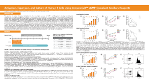 科学海报Activation, Expansion, and Culture of Human T Cells Using ImmunoCult™ cGMP-Compliant Ancillary Reagents
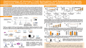 科学海报Optimization of Human T Cell Activation and Expansion Protocols Improves Efficiency of Genetic Modification and Overall Cell Yield
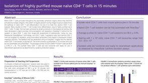 科学海报Immunomagnetic Cell Isolation of Highly Purified Mouse Naïve CD4+ T Cells in 15 Minutes
 沪公网安备31010102008431号