Nocodazole treatment decreases expression of pluripotency markers nanog and Oct4 in human embryonic stem cells
Nocodazole is a known destabiliser of microtubule dynamics and arrests cell-cycle at the G2/M phase. In the context of the human embryonic stem cell (hESC) it is important to understand how this arrest influences the pluripotency of cells. Here we report for the first time the changes in the expression of transcription markers Nanog and Oct4 as well as SSEA-3 and SSEA-4 in human embryonic cells after their treatment with nocodazole. Multivariate permeabilised-cell flow cytometry was applied for characterising the expression of Nanog and Oct4 during different cell cycle phases. Among untreated hESC we detected Nanog-expressing cells,which also expressed Oct4,SSEA-3 and SSEA-4. We also found another population expressing SSEA-4,but without Nanog,Oct4 and SSEA-3 expression. Nocodazole treatment resulted in a decrease of cell population positive for all four markers Nanog,Oct4,SSEA-3,SSEA-4. Nocodazole-mediated cell-cycle arrest was accompanied by higher rate of apoptosis and upregulation of p53. Twenty-four hours after the release from nocodazole block,the cell cycle of hESC normalised,but no increase in the expression of transcription markers Nanog and Oct4 was detected. In addition,the presence of ROCK-2 inhibitor Y-27632 in the medium had no effect on increasing the expression of pluripotency markers Nanog and Oct4 or decreasing apoptosis or the level of p53. The expression of SSEA-3 and SSEA-4 increased in Nanog-positive cells after wash-out of nocodazole in the presence and in the absence of Y-27632. Our data show that in hESC nocodazole reversible blocks cell cycle,which is accompanied by irreversible loss of expression of pluripotency markers Nanog and Oct4.
View Publication
产品类型:
产品号#:
05850
05857
05870
05875
85850
85857
85870
85875
产品名:
mTeSR™1
mTeSR™1
Cox JL et al. (AUG 2011)
Journal of Cell Science 124 Pt 15 2654--65
Banf1 is required to maintain the self-renewal of both mouse and human embryonic stem cells.
Self-renewal is a complex biological process necessary for maintaining the pluripotency of embryonic stem cells (ESCs). Recent studies have used global proteomic techniques to identify proteins that associate with the master regulators Oct4,Nanog and Sox2 in ESCs or in ESCs during the early stages of differentiation. Through an unbiased proteomic screen,Banf1 was identified as a Sox2-associated protein. Banf1 has been shown to be essential for worm and fly development but,until now,its role in mammalian development and ESCs has not been explored. In this study,we examined the effect of knocking down Banf1 on ESCs. We demonstrate that the knockdown of Banf1 promotes the differentiation of mouse ESCs and decreases the survival of both mouse and human ESCs. For mouse ESCs,we demonstrate that knocking down Banf1 promotes their differentiation into cells that exhibit markers primarily associated with mesoderm and trophectoderm. Interestingly,knockdown of Banf1 disrupts the survival of human ESCs without significantly reducing the expression levels of the master regulators Sox2,Oct4 and Nanog or inducing the expression of markers of differentiation. Furthermore,we determined that the knockdown of Banf1 alters the cell cycle distribution of both human and mouse ESCs by causing an uncharacteristic increase in the proportion of cells in the G2-M phase of the cell cycle.
View Publication
产品类型:
产品号#:
05850
05857
05870
05875
85850
85857
85870
85875
产品名:
mTeSR™1
mTeSR™1
Tan Y et al. (JAN 2012)
Journal of biomechanics 45 1 123--8
Probing the mechanobiological properties of human embryonic stem cells in cardiac differentiation by optical tweezers.
Human embryonic stem cells (hESC) and hESC-derived cardiomyocytes (hESC-CM) hold great promise for the treatment of cardiovascular diseases. However the mechanobiological properties of hESC and hESC-CM remains elusive. In this paper,we examined the dynamic and static micromechanical properties of hESC and hESC-CM,by manipulating via optical tweezers at the single-cell level. Theoretical approaches were developed to model the dynamic and static mechanical responses of cells during optical stretching. Our experiments showed that the mechanical stiffness of differentiated hESC-CM increased after cardiac differentiation. Such stiffening could associate with increasingly organized myofibrillar assembly that underlines the functional characteristics of hESC-CM. In summary,our findings lay the ground work for using hESC-CMs as models to study mechanical and contractile defects in heart diseases.
View Publication
产品类型:
产品号#:
05850
05857
05870
05875
85850
85857
85870
85875
产品名:
mTeSR™1
mTeSR™1
Moschidou D et al. (OCT 2012)
Molecular therapy : the journal of the American Society of Gene Therapy 20 10 1953--67
Valproic acid confers functional pluripotency to human amniotic fluid stem cells in a transgene-free approach.
Induced pluripotent stem cells (iPSCs) with potential for therapeutic applications can be derived from somatic cells via ectopic expression of a set of limited and defined transcription factors. However,due to risks of random integration of the reprogramming transgenes into the host genome,the low efficiency of the process,and the potential risk of virally induced tumorigenicity,alternative methods have been developed to generate pluripotent cells using nonintegrating systems,albeit with limited success. Here,we show that c-KIT+ human first-trimester amniotic fluid stem cells (AFSCs) can be fully reprogrammed to pluripotency without ectopic factors,by culture on Matrigel in human embryonic stem cell (hESC) medium supplemented with the histone deacetylase inhibitor (HDACi) valproic acid (VPA). The cells share 82% transcriptome identity with hESCs and are capable of forming embryoid bodies (EBs) in vitro and teratomas in vivo. After long-term expansion,they maintain genetic stability,protein level expression of key pluripotency factors,high cell-division kinetics,telomerase activity,repression of X-inactivation,and capacity to differentiate into lineages of the three germ layers,such as definitive endoderm,hepatocytes,bone,fat,cartilage,neurons,and oligodendrocytes. We conclude that AFSC can be utilized for cell banking of patient-specific pluripotent cells for potential applications in allogeneic cellular replacement therapies,pharmaceutical screening,and disease modeling.
View Publication
产品类型:
产品号#:
05850
05857
05870
05875
85850
85857
85870
85875
产品名:
mTeSR™1
mTeSR™1
Deng Y et al. (NOV 2013)
Acta Biomaterialia 9 11 8840--8850
Long-term self-renewal of human pluripotent stem cells on peptide-decorated poly(OEGMA-co-HEMA) brushes under fully defined conditions
Realization of the full potential of human induced pluripotent stem cells (hiPSC) in clinical applications requires the development of well-defined culture conditions for their long-term growth and directed differentiation. This paper describes a novel fully defined synthetic peptide-decorated substrate that supports self-renewal of hiPSC in commercially available xeno-free,chemically defined medium. The Au surface was deposited by a poly(OEGMA-co-HEMA) film,using the surface-initiated polymerization method (SIP) with the further step of carboxylation. The hiPSC generated from umbilical cord mesenchymal cells were successfully cultured for 10 passages on the peptide-tethered poly(OEGMA-co-HEMA) brushes for the first time. Cells maintained their characteristic morphology,proliferation and expressed high levels of markers of pluripotency,similar to the cells cultured on Matrigel???. Moreover,the cell adhesion could be tuned by the pattern and peptide concentration on the substrate. This well-defined,xeno-free and safe substrate,which supports long-term proliferation and self-renewal of hiPSC,will not only help to accelerate the translational perspectives of hiPSC,but also provide a platform to elucidate the underlying molecular mechanisms that regulate stem cell proliferation and differentiation via SIP technology. ?? 2013 Acta Materialia Inc. Published by Elsevier Ltd. All rights reserved.
View Publication
Minimum Transendothelial Electrical Resistance Thresholds for the Study of Small and Large Molecule Drug Transport in a Human in Vitro Blood-Brain Barrier Model.
A human cell-based in vitro model that can accurately predict drug penetration into the brain as well as metrics to assess these in vitro models are valuable for the development of new therapeutics. Here,human induced pluripotent stem cells (hPSCs) are differentiated into a polarized monolayer that express blood-brain barrier (BBB)-specific proteins and have transendothelial electrical resistance (TEER) values greater than 2500 Ωtextperiodcenteredcm(2). By assessing the permeabilities of several known drugs,a benchmarking system to evaluate brain permeability of drugs was established. Furthermore,relationships between TEER and permeability to both small and large molecules were established,demonstrating that different minimum TEER thresholds must be achieved to study the brain transport of these two classes of drugs. This work demonstrates that this hPSC-derived BBB model exhibits an in vivo-like phenotype,and the benchmarks established here are useful for assessing functionality of other in vitro BBB models.
View Publication
Tateno H et al. (FEB 2014)
Scientific reports 4 4069
A medium hyperglycosylated podocalyxin enables noninvasive and quantitative detection of tumorigenic human pluripotent stem cells.
While human pluripotent stem cells are attractive sources for cell-replacement therapies,a major concern remains regarding their tumorigenic potential. Thus,safety assessment of human pluripotent stem cell-based products in terms of tumorigenicity is critical. Previously we have identified a pluripotent stem cell-specific lectin probe rBC2LCN recognizing hyperglycosylated podocalyxin as a cell surface ligand. Here we demonstrate that hyperglycosylated podocalyxin is secreted from human pluripotent stem cells into cell culture supernatants. We establish a sandwich assay system,named the GlycoStem test,targeting the soluble hyperglycosylated podocalyxin using rBC2LCN. The GlycoStem test is sufficiently sensitive and quantitative to detect residual human pluripotent stem cells. This work provides a proof of concept for the noninvasive and quantitative detection of tumorigenic human pluripotent stem cells using cell culture supernatants. The developed method should increase the safety of human pluripotent stem cell-based cell therapies.
View Publication
产品类型:
产品号#:
05850
05857
05870
05875
85850
85857
85870
85875
产品名:
mTeSR™1
mTeSR™1
Richter A et al. (MAR 2014)
Stem Cells 32 3 636--648
BMP4 promotes EMT and mesodermal commitment in human embryonic stem cells via SLUG and MSX2
Bone morphogenetic proteins (BMPs) initiate differentiation in human embryonic stem cells (hESCs) but the exact mechanisms have not been fully elucidated. We demonstrate here that SLUG and MSX2,transcription factors involved in epithelial-mesenchymal transitions,essential features of gastrulation in development and tumor progression,are important mediators of BMP4-induced differentiation in hESCs. Phosphorylated Smad1/5/8 colocalized with the SLUG protein at the edges of hESC colonies where differentiation takes place. The upregulation of the BMP target SLUG was direct as shown by the binding of phosphorylated Smad1/5/8 to its promoter,which interrupted the formation of adhesion proteins,resulting in migration. Knockdown of SLUG by short hairpin RNA blocked these changes,confirming an important role for SLUG in BMP-mediated mesodermal differentiation. Furthermore,BMP4-induced MSX2 expression leads to mesoderm formation and then preferential differentiation toward the cardiovascular lineage.
View Publication
产品类型:
产品号#:
05850
05857
05870
05875
85850
85857
85870
85875
产品名:
mTeSR™1
mTeSR™1
Takahashi K et al. (APR 2014)
Nature communications 5 3678
Induction of pluripotency in human somatic cells via a transient state resembling primitive streak-like mesendoderm.
During mammalian embryonic development,the primitive streak initiates the differentiation of pluripotent epiblast cells into germ layers. Pluripotency can be reacquired in committed somatic cells using a combination of a handful of transcription factors,such as OCT3/4,SOX2,KLF4 and c-MYC (hereafter referred to as OSKM),albeit with low efficiency. Here we show that during OSKM-induced reprogramming towards pluripotency in human cells,intermediate cells transiently show gene expression profiles resembling mesendoderm,which is a major component of the primitive streak. Based on these findings,we discover that forkhead box H1 (FOXH1),a transcription factor required for anterior primitive streak specification during early development,significantly enhances the reprogramming efficiency of human fibroblasts by promoting their maturation,including mesenchymal to epithelial transition and the activation of late pluripotency markers. These results demonstrate that during the reprogramming process,human somatic cells go through a transient state that resembles mesendoderm.
View Publication

 EasySep™小鼠TIL(CD45)正选试剂盒


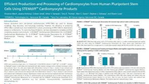 科学海报Efficient Production and Processing of Cardiomyocytes from Human Pluripotent Stem Cells Using STEMdiff™ Cardiomyocyte Products
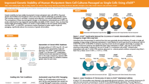

 沪公网安备31010102008431号