Forward RNAi screens in primary human hematopoietic stem/progenitor cells.
The mechanisms regulating key fate decisions such as self-renewal and differentiation in hematopoietic stem and progenitor cells (HSPC) remain poorly understood. We report here a screening strategy developed to assess modulators of human hematopoiesis using a lentiviral short hairpin RNA (shRNA) library transduced into cord blood-derived stem/progenitor cells. To screen for modifiers of self-renewal/differentiation,we used the limited persistence of HSPCs under ex vivo culture conditions as a baseline for functional selection of shRNAs conferring enhanced maintenance or expansion of the stem/progenitor potential. This approach enables complex,pooled screens in large numbers of cells. Functional selection identified novel specific gene targets (exostoses 1) or shRNA constructs capable of altering human hematopoietic progenitor differentiation or stem cell expansion,respectively,thereby demonstrating the potential of this forward screening approach in primary human stem cell populations.
View Publication
A 3D sphere culture system containing functional polymers for large-scale human pluripotent stem cell production
Utilizing human pluripotent stem cells (hPSCs) in cell-based therapy and drug discovery requires large-scale cell production. However,scaling up conventional adherent cultures presents challenges of maintaining a uniform high quality at low cost. In this regard,suspension cultures are a viable alternative,because they are scalable and do not require adhesion surfaces. 3D culture systems such as bioreactors can be exploited for large-scale production. However,the limitations of current suspension culture methods include spontaneous fusion between cell aggregates and suboptimal passaging methods by dissociation and reaggregation. 3D culture systems that dynamically stir carrier beads or cell aggregates should be refined to reduce shearing forces that damage hPSCs. Here,we report a simple 3D sphere culture system that incorporates mechanical passaging and functional polymers. This setup resolves major problems associated with suspension culture methods and dynamic stirring systems and may be optimal for applications involving large-scale hPSC production. ?? 2014 The Authors.
View Publication
Hawley RG et al. (JAN 2006)
Methods in enzymology 419 149--79
Hematopoietic stem cells.
Hematopoietic stem cells (HSCs) have the capacity to self-renew and the potential to differentiate into all of the mature blood cell types. The ability to prospectively identify and isolate HSCs has been the subject of extensive investigation since the first transplantation studies implying their existence almost 50 years ago. Despite significant advances in enrichment protocols,the continuous in vitro propagation of human HSCs has not yet been achieved. This chapter describes current procedures used to phenotypically and functionally characterize candidate human HSCs and initial efforts to derive permanent human HSC lines.
View Publication
产品类型:
产品号#:
01700
01705
01702
产品名:
ALDEFLUOR™ 试剂盒
ALDEFLUOR™ DEAB试剂, 1.5 mM, 1 mL
ALDEFLUOR™检测缓冲液
Gentry T et al. (JAN 2007)
Cytotherapy 9 3 259--74
Simultaneous isolation of human BM hematopoietic, endothelial and mesenchymal progenitor cells by flow sorting based on aldehyde dehydrogenase activity: implications for cell therapy.
BACKGROUND: ALDH(br) cells express high aldehyde dehydrogenase (ALDH) activity and have progenitor cell activity in several contexts. We characterized human BM ALDH(br) cells to determine whether cell sorting based on ALDH activity isolates potentially useful populations for cell therapy. METHOD: We measured the expression of ALDH and cell-surface Ag by flow cytometry and compared the ability of sorted ALDH(br),and BM populations remaining after ALDH(br) cells were removed (ALDH(dim) populations),to develop into several cell lineages in culture. RESULTS: The ALDH(br) population comprised 1.2+/-0.8% (mean+/-SD,n=30) nucleated cells and was enriched in cells expressing CD34,CD117,CD105,CD127,CD133 and CD166,and in primitive CD34(+) CD38(-) and CD34(+) CD133(+) progenitors. Most of the CD34(+) and CD133(+) cells were ALDH(dim). ALDH(br) populations had 144-fold more hematopoietic colony-forming activity than ALDH(dim) cells and included all megakaryocyte progenitors. ALDH(br) populations readily established endothelial cell monolayers in cultures. Cells generating endothelial colonies in 7 days were 435-fold more frequent in ALDH(br) than ALDH(dim) populations. CFU-F were 9.5-fold more frequent in ALDH(br) than ALDH(dim) cells,and ALDH(br) cells gave rise to multipotential mesenchymal cell cultures that could be driven to develop into adipocytes,osteoblasts and chondrocytes. DISCUSSION: Hematopoietic,endothelial and mesenchymal progenitor cells can be isolated simultaneously from human BM by cell sorting based on ALDH activity. BM ALDH(br) populations may be useful in several cell therapy applications.
View Publication

 EasySep™小鼠TIL(CD45)正选试剂盒


 实验方案Transitioning from Feeder-Free Media to mTeSR™ Plus for Human Pluripotent Stem Cell Culture
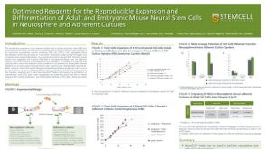 科学海报Optimized Reagents for the Reproducible Expansion and Differentiation of Adult and Embryonic Mouse Neural Stem Cells in Neurosphere and Adherent Cultures
 31:39
线上讲座Serum- and Feeder-Free Differentiation of Erythroid Progenitor Cells from hPSCs发布日期: 05/21/2021
31:39
线上讲座Serum- and Feeder-Free Differentiation of Erythroid Progenitor Cells from hPSCs发布日期: 05/21/2021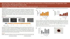 科学海报Efficient Differentiation of Human Pluripotent Stem Cells to Hematopoietic Progenitor Cells in Serum-Free Culture Conditions
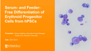

 沪公网安备31010102008431号