Cardiac malformations and disease are the leading causes of death in the United States in live-born infants and adults,respectively. In both of these cases,a decrease in the number of functional cardiomyocytes often results in improper growth of heart tissue,wound healing complications,and poor tissue repair. The field of cardiac tissue engineering seeks to address these concerns by developing cardiac patches created from a variety of biomaterial scaffolds to be used in surgical repair of the heart. These scaffolds should be fully degradable biomaterial systems with tunable properties such that the materials can be altered to meet the needs of both in vitro culture (e.g. disease modeling) and in vivo application (e.g. cardiac patch). Current platforms do not utilize both structural anisotropy and proper cell-matrix contacts to promote functional cardiac phenotypes and thus there is still a need for critically sized scaffolds that mimic both the structural and adhesive properties of native tissue. To address this need,we have developed a silk-based scaffold platform containing cardiac tissue-derived extracellular matrix (cECM). These silk-cECM composite scaffolds have tunable architectures,degradation rates,and mechanical properties. Subcutaneous implantation in rats demonstrated that addition of the cECM to aligned silk scaffold led to 99% endogenous cell infiltration and promoted vascularization of a critically sized scaffold (10 × 5 × 2.5 mm) after 4 weeks in vivo. In vitro,silk-cECM scaffolds maintained the HL-1 atrial cardiomyocytes and human embryonic stem cell-derived cardiomyocytes and promoted a more functional phenotype in both cell types. This class of hybrid silk-cECM anisotropic scaffolds offers new opportunities for developing more physiologically relevant tissues for cardiac repair and disease modeling.
View Publication
产品类型:
产品号#:
05850
05857
05870
05875
85850
85857
85870
85875
产品名:
mTeSR™1
mTeSR™1
Dobo I et al. (JAN 2001)
The hematology journal : the official journal of the European Haematology Association / EHA 2 6 396--403
Comparison of four serum-free, cytokine-free media for analysis of endogenous erythroid colony growth in polycythemia vera and essential thrombocythemia.
INTRODUCTION: The assay of endogenous erythroid colony formation (EEC),a characteristic of polycythemia vera and essential thrombocythemia,is not standardized. In this multicentric study,we tested four semisolid,serum-free,cytokine-free media based on either methylcellulose (M1,M2) or collagen (C1,C2) commercialized for the EEC assay. MATERIALS AND METHODS: Bone marrow mononuclear cells (BMMC) from 73 individuals (62 patients with either polycythemia vera (26),essential thrombocythemia (19),secondary polyglobuly (17) or chronic myeloid leukemia (2) and 11 healthy donors) were grown in parallel in the four media without,or with 0.01 U/ml erythropoietin (EPo). RESULTS: In all four media EEC formation was specific,as it was not observed in cultures of patients with secondary polyglobuly or chronic myeloid leukemia,nor of healthy donors. Analysis of fresh or MGG-stained collagen gel cultures allowed detection of EEC formation significantly more frequently than methylcellulose-based media; addition of 0.01 U/ml of EPo had little or no effect on EEC formation. Collagen-based medium C1 gave better results than the other media tested: the 'C1' EEC assay was positive for 68.2% of polycythemia vera cultures with significantly higher median EEC numbers (6.5/10(5) BMMC for patients with one major criteria of polycythemia vera and 19 and 21/10(5) BMMC for patients with two or three major criteria,respectively). Medium C1 was also better for essential thrombocythemia cultures with 47.4% of positive results but with a low median EEC number (6.7/10(5) BMMC). When associated with the ELISA dosage of serum EPo,the 'C1' EEC assay allowed confirmation or elimination of the diagnosis of polycythemia vera for 91% (20/22) of polyglobulic patients. CONCLUSION: We propose that serum-free collagen-based culture systems be considered to standardize the EEC assay,now part of the new criteria of polycythemia vera.
View Publication
产品类型:
产品号#:
04961
04965
04962
04915
04807
04809
04906
04913
04803
04804
04905
04850
04974
04902
04960
04900
04901
04963
04970
04971
产品名:
MegaCult™-C胶原和含细胞因子培养基
MegaCult™-C CFU-Mk染色试剂盒
MegaCult-C 10% BSA, 6mL
MegaCult-C Human Serum, 6mL
Alkaline Phosphatase Substrate Tabs, pk
Biotin/Conjugate Goat Anti-Mu lgG, 125uL
MegaCult-C Evans Blue Stain, 5mL
Primary Ab, Anti-HuAnti-GPIIb/IIIa 360uL
MegaCult-C Control Antibody, 100 µL
Avidin-Alk Phosphatase Conjugate, 200 uL
MegaCult™-C含脂质培养基
MegaCult™-C胶原和含脂质培养基
胶原蛋白溶液
MegaCult™-C胶原和无细胞因子培养基
MegaCult™-C无细胞因子培养基
MegaCult™-C含细胞因子培养基
双室载玻片套件
MegaCult™-C无细胞因子全套试剂盒
MegaCult™-C含细胞因子全套试剂盒
Ghiaur G et al. (APR 2008)
Blood 111 7 3313--21
Rac1 is essential for intraembryonic hematopoiesis and for the initial seeding of fetal liver with definitive hematopoietic progenitor cells.
Definitive hematopoietic stem and progenitor cells (HSCs/Ps) originating from the yolk sac and/or para-aorta-splanchno-pleura/aorta-gonad-mesonephros are hypothesized to colonize the fetal liver,but mechanisms involved are poorly defined. The Rac subfamily of Rho GTPases has been shown to play essential roles in HSC/P localization to the bone marrow following transplantation. Here,we study the role of Rac1 in HSC/P migration during ontogeny and seeding of fetal liver. Using a triple-transgenic approach,we have deleted Rac1 in HSCs/Ps during very early embryonic development. Without Rac1,there was a decrease in circulating HSCs/Ps in the blood of embryonic day (E) 10.5 embryos,while yolk sac definitive hematopoiesis was quantitatively normal. Intraembryonic hematopoiesis was significantly impaired in Rac1-deficient embryos,culminating with absence of intra-aortic clusters and fetal liver hematopoiesis. At E10.5,Rac1-deficient HSCs/Ps displayed decreased transwell migration and impaired inter-action with the microenvironment in migration-dependent assays. These data suggest that Rac1 plays an important role in HSC/P migration during embryonic development and is essential for the emergence of intraembryonic hematopoiesis.
View Publication
Leung CG et al. (JUL 2007)
The Journal of experimental medicine 204 7 1603--11
Requirements for survivin in terminal differentiation of erythroid cells and maintenance of hematopoietic stem and progenitor cells.
Survivin,which is the smallest member of the inhibitor of apoptosis protein (IAP) family,is a chromosomal passenger protein that mediates the spindle assembly checkpoint and cytokinesis,and also functions as an inhibitor of apoptosis. Frequently overexpressed in human cancers and not expressed in most adult tissues,survivin has been proposed as an attractive target for anticancer therapies and,in some cases,has even been touted as a cancer-specific gene. Survivin is,however,expressed in proliferating adult cells,including human hematopoietic stem cells,T-lymphocytes,and erythroid cells throughout their maturation. Therefore,it is unclear how survivin-targeted anticancer therapies would impact steady-state blood development. To address this question,we used a conditional gene-targeting strategy and abolished survivin expression from the hematopoietic compartment of mice. We show that inducible deletion of survivin leads to ablation of the bone marrow,with widespread loss of hematopoietic progenitors and rapid mortality. Surprisingly,heterozygous deletion of survivin causes defects in erythropoiesis in a subset of the animals,with a dramatic reduction in enucleated erythrocytes and the presence of immature megaloblastic erythroblasts. Our studies demonstrate that survivin is essential for steady-state hematopoiesis and survival of the adult,and further,that a high level of survivin expression is critical for proper erythroid differentiation.
View Publication
产品类型:
产品号#:
19756
19756RF
产品名:
Zhang CC et al. (FEB 2006)
Proceedings of the National Academy of Sciences of the United States of America 103 7 2184--9
Prion protein is expressed on long-term repopulating hematopoietic stem cells and is important for their self-renewal.
Although the wild-type prion protein (PrP) is abundant and widely expressed in various types of tissues and cells,its physiological function(s) remain unknown,and PrP knockout mice do not exhibit overt and undisputed phenotypes. Here we showed that PrP is expressed on the surface of several bone marrow cell populations successively enriched in long-term (LT) hematopoietic stem cells (HSCs) using flow cytometry analysis. Affinity purification of the PrP-positive and -negative fractions from these populations,followed by competitive bone marrow reconstitution assays,shows that all LT HSCs express PrP. HSCs from PrP-null bone marrow exhibited impaired self-renewal in serial transplantation of lethally irradiated mouse recipients both in the presence and absence of competitors. When treated with a cell cycle-specific myelotoxic agent,the animals reconstituted with PrP-null HSCs exhibit increased sensitivity to hematopoietic cell depletion. Ectopic expression of PrP in PrP-null bone marrow cells by retroviral infection rescued the defective hematopoietic engraftment during serial transplantation. Therefore,PrP is a marker for HSCs and supports their self-renewal.
View Publication
产品类型:
产品号#:
03630
03434
03444
09600
09650
28600
产品名:
MethoCult™ M3630
MethoCult™ GF M3434
MethoCult™ GF M3434
StemSpan™ SFEM
StemSpan™ SFEM
L-Calc™有限稀释软件
Phondeechareon T et al. (OCT 2016)
Annals of hematology 95 10 1617--1625
Generation of induced pluripotent stem cells as a potential source of hematopoietic stem cells for transplant in PNH patients.
Paroxysmal nocturnal hemoglobinuria (PNH) is an acquired hemolytic anemia caused by lack of CD55 and CD59 on blood cell membrane leading to increased sensitivity of blood cells to complement. Hematopoietic stem cell transplantation (HSCT) is the only curative therapy for PNH,however,lack of HLA-matched donors and post-transplant complications are major concerns. Induced pluripotent stem cells (iPSCs) derived from patients are an attractive source for generating autologous HSCs to avoid adverse effects resulting from allogeneic HSCT. The disease involves only HSCs and their progeny; therefore,other tissues are not affected by the mutation and may be used to produce disease-free autologous HSCs. This study aimed to derive PNH patient-specific iPSCs from human dermal fibroblasts (HDFs),characterize and differentiate to hematopoietic cells using a feeder-free protocol. Analysis of CD55 and CD59 expression was performed before and after reprogramming,and hematopoietic differentiation. Patients' dermal fibroblasts expressed CD55 and CD59 at normal levels and the normal expression remained after reprogramming. The iPSCs derived from PNH patients had typical pluripotent properties and differentiation capacities with normal karyotype. After hematopoietic differentiation,the differentiated cells expressed early hematopoietic markers (CD34 and CD43) with normal CD59 expression. The iPSCs derived from HDFs of PNH patients have normal levels of CD55 and CD59 expression and hold promise as a potential source of HSCs for autologous transplantation to cure PNH patients.
View Publication
Defects in osteoblast function but no changes in long-term repopulating potential of hematopoietic stem cells in a mouse chronic inflammatory arthritis model.
Recent studies support the notion that there is an intricate relationship between hematopoiesis and bone homeostasis in normal steady states. Using mice undergoing chronic inflammatory arthritis,we investigated the relationship between hematopoiesis and bone homeostasis in pathologic conditions. We demonstrate that mice undergoing chronic inflammatory arthritis displayed osteoporosis resulting from a severe defect in osteoblast function. Despite the defective osteoblast function,however,the hematopoietic stem cells from these mice exhibited normal properties in either long-term repopulation or cell cycling. Therefore,the bone-forming capacity of osteoblasts is distinct from their ability to maintain hematopoietic stem cells in chronic inflammatory conditions.
View Publication
beta-Catenin expression in the bone marrow microenvironment is required for long-term maintenance of primitive hematopoietic cells.
Hematopoiesis is dependent upon the bone marrow microenvironment,which is comprised of multiple mesenchymal cell types,including fibroblasts,endothelial cells,osteoblasts,and stroma progenitors. The canonical Wnt signaling pathway,which relies on the beta-catenin protein to mediate its signal,is necessary for the normal development of mesenchymal tissue. We hypothesized that canonical Wnt signaling regulates the cellular composition and function of the bone marrow microenvironment. We observed that a beta-catenin-deficient bone marrow microenvironment maintained hematopoietic stem cells but exhibited a decreased capacity to support primitive hematopoietic cells. These results correlated with decreased numbers of osteoblasts and with decreased production of basic fibroblast growth factor,stem cell factor,and vascular cell adhesion molecule-1. From these data,we propose a model in which beta-catenin in the microenvironment is required noncell autonomously for long-term maintenance of hematopoietic progenitors.
View Publication
产品类型:
产品号#:
05150
产品名:
MyeloCult™ H5100
Finstad SL et al. (JUL 2007)
Journal of virology 81 13 7274--9
Diminished potential for B-lymphoid differentiation after murine leukemia virus infection in vivo and in EML hematopoietic progenitor cells.
Infection with a recombinant murine-feline gammaretrovirus,MoFe2,or with the parent virus,Moloney murine leukemia virus,caused significant reduction in B-lymphoid differentiation of bone marrow at 2 to 8 weeks postinfection. The suppression was selective,in that myeloid potential was significantly increased by infection. Analysis of cell surface markers and immunoglobulin H gene rearrangements in an in vitro model demonstrated normal B-lymphoid differentiation after infection but significantly reduced viability of differentiating cells. This reduction in viability may confer a selective advantage on undifferentiated lymphoid progenitors in the bone marrow of gammaretrovirus-infected animals and thereby contribute to the establishment of a premalignant state.
View Publication
产品类型:
产品号#:
03630
03434
03444
产品名:
MethoCult™ M3630
MethoCult™ GF M3434
MethoCult™ GF M3434
Yu S et al. (FEB 2011)
Blood 117 7 2166--78
GABP controls a critical transcription regulatory module that is essential for maintenance and differentiation of hematopoietic stem/progenitor cells.
Maintaining a steady pool of self-renewing hematopoietic stem cells (HSCs) is critical for sustained production of multiple blood lineages. Many transcription factors and molecules involved in chromatin and epigenetic modifications have been found to be critical for HSC self-renewal and differentiation; however,their interplay is less understood. The transcription factor GA binding protein (GABP),consisting of DNA-binding subunit GABPα and transactivating subunit GABPβ,is essential for lymphopoiesis as shown in our previous studies. Here we demonstrate cell-intrinsic,absolute dependence on GABPα for maintenance and differentiation of hematopoietic stem/progenitor cells. Through genome-wide mapping of GABPα binding and transcriptomic analysis of GABPα-deficient HSCs,we identified Zfx and Etv6 transcription factors and prosurvival Bcl-2 family members including Bcl-2,Bcl-X(L),and Mcl-1 as direct GABP target genes,underlying its pivotal role in HSC survival. GABP also directly regulates Foxo3 and Pten and hence sustains HSC quiescence. Furthermore,GABP activates transcription of DNA methyltransferases and histone acetylases including p300,contributing to regulation of HSC self-renewal and differentiation. These systematic analyses revealed a GABP-controlled gene regulatory module that programs multiple aspects of HSC biology. Our studies thus constitute a critical first step in decoding how transcription factors are orchestrated to regulate maintenance and multipotency of HSCs.
View Publication


 EasySep™小鼠TIL(CD45)正选试剂盒





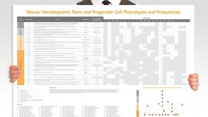 挂图Mouse Hematopoietic Stem and Progenitor Cell Phenotyping Overview of mouse HSPC subset surface markers and frequencies发布日期: 08/10/2018
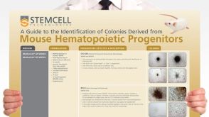 挂图Identification of Colonies Derived from Mouse Hematopoietic Progenitors Representative colony images and tips for identifying progenitor subtypes in CFU assays
 沪公网安备31010102008431号