Cottler-Fox MH et al. (JAN 2003)
Hematology / the Education Program of the American Society of Hematology. American Society of Hematology. Education Program 419--37
Stem cell mobilization.
Successful blood and marrow transplant (BMT),both autologous and allogeneic,requires the infusion of a sufficient number of hematopoietic progenitor/stem cells (HPCs) capable of homing to the marrow cavity and regenerating a full array of hematopoietic cell lineages in a timely fashion. At present,the most commonly used surrogate marker for HPCs is the cell surface marker CD34,identified in the clinical laboratory by flow cytometry. Clinical studies have shown that infusion of at least 2 x 10(6) CD34(+) cells/kg recipient body weight results in reliable engraftment as measured by recovery of adequate neutrophil and platelet counts approximately 14 days after transplant. Recruitment of HPCs from the marrow into the blood is termed mobilization,or,more commonly,stem cell mobilization. In Section I,Dr. Tsvee Lapidot and colleagues review the wide range of factors influencing stem cell mobilization. Our current understanding focuses on chemokines,proteolytic enzymes,adhesion molecules,cytokines and stromal cell-stem cell interactions. On the basis of this understanding,new approaches to mobilization have been designed and are now starting to undergo clinical testing. In Section II,Dr. Michele Cottler-Fox describes factors predicting the ability to mobilize the older patient with myeloma. In addition,clinical approaches to improving collection by individualizing the timing of apheresis and adjusting the volume of blood processed to achieve a desired product are discussed. Key to this process is the daily enumeration of blood CD34(+) cells. Newer methods of enumerating and mobilizing autologous blood HPCs are discussed. In Section III,Dr. John DiPersio and colleagues provide data on clinical results of mobilizing allogeneic donors with G-CSF,GM-CSF and the combination of both as relates to the number and type of cells collected by apheresis. Newer methods of stem cell mobilization as well as the relationship of graft composition on immune reconstitution and GVHD are discussed.
View Publication
产品类型:
产品号#:
01700
01705
01702
产品名:
ALDEFLUOR™ 试剂盒
ALDEFLUOR™ DEAB试剂, 1.5 mM, 1 mL
ALDEFLUOR™检测缓冲液
Guan BX et al. (MAY 2014)
IEEE/ACM transactions on computational biology and bioinformatics / IEEE,ACM 11 3 604--611
Bio-Driven Cell Region Detection in Human Embryonic Stem Cell Assay.
This paper proposes a bio-driven algorithm that detects cell regions automatically in the human embryonic stem cell (hESC) images obtained using a phase contrast microscope. The algorithm uses both statistical intensity distributions of foreground/hESCs and background/substrate as well as cell property for cell region detection. The intensity distributions of foreground/hESCs and background/substrate are modeled as a mixture of two Gaussians. The cell property is translated into local spatial information. The algorithm is optimized by parameters of the modeled distributions and cell regions evolve with the local cell property. The paper validates the method with various videos acquired using different microscope objectives. In comparison with the state-of-the-art methods,the proposed method is able to detect the entire cell region instead of fragmented cell regions. It also yields high marks on measures such as Jacard similarity,Dice coefficient,sensitivity and specificity. Automated detection by the proposed method has the potential to enable fast quantifiable analysis of hESCs using large data sets which are needed to understand dynamic cell behaviors.
View Publication
产品类型:
产品号#:
05850
05857
05870
05875
85850
85857
85870
85875
产品名:
mTeSR™1
mTeSR™1
Kumar S et al. ( 2016)
Stem Cells International 2016 1--20
Utility of Lymphoblastoid Cell Lines for Induced Pluripotent Stem Cell Generation
A large number of EBV immortalized LCLs have been generated and maintained in genetic/epidemiological studies as a perpetual source of DNA and as a surrogate in vitro cell model. Recent successes in reprograming LCLs into iPSCs have paved the way for generating more relevant in vitro disease models using this existing bioresource. However,the overall reprogramming efficiency and success rate remain poor and very little is known about the mechanistic changes that take place at the transcriptome and cellular functional level during LCL-to-iPSC reprogramming. Here,we report a new optimized LCL-to-iPSC reprogramming protocol using episomal plasmids encoding pluripotency transcription factors and mouse p53DD (p53 carboxy-terminal dominant-negative fragment) and commercially available reprogramming media. We achieved a consistently high reprogramming efficiency and 100% success rate using this optimized protocol. Further,we investigated the transcriptional changes in mRNA and miRNA levels,using FC-abs ≥ 2.0 and FDR ≤ 0.05 cutoffs; 5,228 mRNAs and 77 miRNAs were differentially expressed during LCL-to-iPSC reprogramming. The functional enrichment analysis of the upregulated genes and activation of human pluripotency pathways in the reprogrammed iPSCs showed that the generated iPSCs possess transcriptional and functional profiles very similar to those of human ESCs.
View Publication
BACKGROUND: Multiple measures of endothelial progenitor cells (EPCs) have been described,but there has been limited study of the comparability of these assays. We sought to determine the reproducibility of and correlation between alternative EPC assay methodologies. METHODS: We simultaneously assessed EPC numbers in 140 patients undergoing cardiac catheterization using the 2 most commonly used culture techniques: endothelial cell outgrowth and colony-forming unit (CFU). In the final 77 patients,EPCs were also identified on the basis of cell surface marker expression (CD133,CD34,and vascular endothelial growth factor receptor-2 [VEGFR-2]) and aldehyde dehydrogenase (ALDH) activity. RESULTS: Endothelial progenitor cell enumeration based on fluorescence activated cell sorting was more precise than culture assays. There was limited correlation between EPC numbers determined using the 2 common culture-based assays; however,endothelial CFUs correlated with VEGFR-2 and CD34/VEGFR-2-expressing cells. Endothelial progenitor cells defined by expression of CD133,CD34,CD133/CD34,and ALDH activity correlated with each other,but not with VEGFR-2(+) cells. CONCLUSIONS: Endothelial progenitor cells can be broadly classified into 2 classes: VEGFR-2-expressing cells,which give rise to endothelial CFUs,and CD133/CD34 or ALDH(br) cells. These observations underscore the need for better assay standardization and a more precise definition of EPCs in cell therapy research.
View Publication
Polisetti N et al. (JAN 2016)
Stem cells (Dayton,Ohio) 34 1 203--219
Cell Adhesion Molecules and Stem Cell-Niche-Interactions in the Limbal Stem Cell Niche.
Interactions between stem cells and their microenvironment are critical for regulation and maintenance of stem cell function. To elucidate the molecular interactions within the human limbal epithelial stem/progenitor cell (LEPC) niche,which is essential for maintaining corneal transparency and vision,we performed a comprehensive expression analysis of cell adhesion molecules (CAMs) using custom-made quantitative real-time polymerase chain reaction (qRT-PCR) arrays and laser capture-microdissected LEPC clusters,comprising LEPCs,melanocytes,mesenchymal cells,and transmigrating immune cells. We show that LEPCs are anchored to their supporting basement membrane by the laminin receptors $\$3$\$1 and $\$6$\$4 integrin and the dystroglycan complex,while intercellular contacts between LEPCs and melanocytes are mediated by N-,P-,and E-cadherin together with L1-CAM,a member of the immunoglobulin superfamily (Ig)CAMs. In addition to the LEPC-associated heparan sulfate proteoglycans syndecan-2,glypican-3,and glypican-4,the IgCAM members ICAM-1 and VCAM-1 were found to be variably expressed on LEPCs and associated niche cells and to be dynamically regulated in response to chemokines such as interferon-$\$ enhance interactions with immune cells. Moreover,junctional adhesion molecule JAM-C accumulating in the subepithelial limbal matrix,appeared to be involved in recruitment of immune cells,while mesenchymal stromal cells appeared to use the nephronectin receptor integrin $\$8 for approaching the limbal basement membrane. In summary,we identified a novel combination of cell surface receptors that may regulate both stable and dynamic cell-matrix and cell-cell interactions within the limbal niche. The findings provide a solid foundation for further functional studies and for advancement of our current therapeutic strategies for ocular surface reconstruction.
View Publication
产品类型:
产品号#:
05850
05857
05870
05875
27215
27250
27216
27217
27260
27270
85850
85857
85870
85875
产品名:
37µm可逆滤筛,小 (15 mL)
37µm可逆滤筛,大 (50 mL)
70µm可逆滤筛,小 (15 mL)
100µm可逆滤筛,小 (15 mL)
70µm可逆滤筛,大 (50 mL)
100µm可逆滤筛,大 (50 mL)
mTeSR™1
mTeSR™1
Zhao W et al. (MAY 2012)
Molecules (Basel,Switzerland) 17 6 6196--6236
Embryonic stem cell markers.
Embryonic stem cell (ESC) markers are molecules specifically expressed in ES cells. Understanding of the functions of these markers is critical for characterization and elucidation for the mechanism of ESC pluripotent maintenance and self-renewal,therefore helping to accelerate the clinical application of ES cells. Unfortunately,different cell types can share single or sometimes multiple markers; thus the main obstacle in the clinical application of ESC is to purify ES cells from other types of cells,especially tumor cells. Currently,the marker-based flow cytometry (FCM) technique and magnetic cell sorting (MACS) are the most effective cell isolating methods,and a detailed maker list will help to initially identify,as well as isolate ESCs using these methods. In the current review,we discuss a wide range of cell surface and generic molecular markers that are indicative of the undifferentiated ESCs. Other types of molecules,such as lectins and peptides,which bind to ESC via affinity and specificity,are also summarized. In addition,we review several markers that overlap with tumor stem cells (TSCs),which suggest that uncertainty still exists regarding the benefits of using these markers alone or in various combinations when identifying and isolating cells.
View Publication


 EasySep™小鼠TIL(CD45)正选试剂盒




 II型胶原酶 用于消化天然胶原纤维
 挂图Pluripotent Stem Cell Biology Nuclear transfer, reprogramming and cell fusion to induce pluripotency in somatic cells
 III型胶原酶 用于消化天然胶原纤维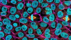

 沪公网安备31010102008431号