Wong JCY et al. (AUG 2003)
Human molecular genetics 12 16 2063--76
Targeted disruption of exons 1 to 6 of the Fanconi Anemia group A gene leads to growth retardation, strain-specific microphthalmia, meiotic defects and primordial germ cell hypoplasia.
Fanconi Anemia (FA) is an autosomal recessive disorder characterized by cellular hypersensitivity to DNA cross-linking agents. Recent studies suggest that FA proteins share a common pathway with BRCA proteins. To study the in vivo role of the FA group A gene (Fanca),gene-targeting techniques were used to generate Fanca(tm1Hsc) mice in which Fanca exons 1-6 were replaced by a beta-galactosidase reporter construct. Fanca(tm1.1Hsc) mice were generated by Cre-mediated removal of the neomycin cassette in Fanca(tm1Hsc) mice. Fanca(tm1.1Hsc) homozygotes display FA-like phenotypes including growth retardation,microphthalmia and craniofacial malformations that are not found in other Fanca mouse models,and the genetic background affects manifestation of certain phenotypes. Both male and female mice homozygous for Fanca mutation exhibit hypogonadism,and homozygous females demonstrate premature reproductive senescence and an increased incidence of ovarian cysts. We showed that fertility defects in Fanca(tm1.1Hsc) homozygotes might be related to a diminished population of primordial germ cells (PGCs) during migration into the gonadal ridges. We also found a high level of Fanca expression in pachytene spermatocytes. Fanca(tm1Hsc) homozygous males exhibited an elevated frequency of mispaired meiotic chromosomes and increased apoptosis in germ cells,implicating a role for Fanca in meiotic recombination. However,the localization of Rad51,Brca1,Fancd2 and Mlh1 appeared normal on Fanca(tm1Hsc) homozygous meiotic chromosomes. Taken together,our results suggest that the FA pathway plays a role in the maintenance of reproductive germ cells and in meiotic recombination.
View Publication
产品类型:
产品号#:
03434
03444
产品名:
MethoCult™ GF M3434
MethoCult™ GF M3434
Visus C et al. (NOV 2007)
Cancer research 67 21 10538--45
Identification of human aldehyde dehydrogenase 1 family member A1 as a novel CD8+ T-cell-defined tumor antigen in squamous cell carcinoma of the head and neck.
Few epitopes are available for vaccination therapy of patients with squamous cell carcinoma of the head and neck (SCCHN). Using a tumor-specific CTL,aldehyde dehydrogenase 1 family member A1 (ALDH1A1) was identified as a novel tumor antigen in SCCHN. Mass spectral analysis of peptides in tumor-derived lysates was used to determine that the CTL line recognized the HLA-A*0201 (HLA-A2) binding ALDH1A1(88-96) peptide. Expression of ALDH1A1 in established SCCHN cell lines,normal mucosa,and primary keratinocytes was studied by quantitative reverse transcription-PCR and immunostaining. Protein expression was further defined by immunoblot analysis,whereas ALDH1A1 activity was measured using ALDEFLUOR. ALDH1A1(88-96) peptide was identified as an HLA-A2-restricted,naturally presented,CD8(+) T-cell-defined tumor peptide. ALDH1A1(88-96) peptide-specific CD8(+) T cells recognized only HLA-A2(+) SCCHN cell lines,which overexpressed ALDH1A1,as well as targets transfected with ALDH1A1 cDNA. Target recognition was blocked by anti-HLA class I and anti-HLA-A2 antibodies. SCCHN cell lines overexpressing ALDH1 had high enzymatic activity. ALDH1A1 protein was expressed in 12 of 17 SCCHN,and 30 of 40 dysplastic mucosa samples,but not in normal mucosa. ALDH1A1 expression levels in target cells correlated with their recognition by ALDH1A1(88-96) peptide-specific CD8(+) T cells. Our findings identify ALDH1A1,a metabolic antigen,as a potential target for vaccination therapy in the cohort of SCCHN subjects with tumors overexpressing this protein. A smaller cohort of subjects with SCCHN,whose tumors express little to no ALDH1A1,and thus are deficient in conversion of retinal to retinoic acid,could benefit from chemoprevention therapy.
View Publication
产品类型:
产品号#:
01700
01705
01702
产品名:
ALDEFLUOR™ 试剂盒
ALDEFLUOR™ DEAB试剂, 1.5 mM, 1 mL
ALDEFLUOR™检测缓冲液
Řeboun M et al. ( 2016)
Folia biologica 62 2 82--89
X-Chromosome Inactivation Analysis in Different Cell Types and Induced Pluripotent Stem Cells Elucidates the Disease Mechanism in a Rare Case of Mucopolysaccharidosis Type II in a Female.
Mucopolysaccharidosis type II (MPS II) is an X-linked lysosomal storage disorder resulting from deficiency of iduronate-2-sulphatase activity. The disease manifests almost exclusively in males; only 16 symptomatic heterozygote girls have been reported so far. We describe the results of X-chromosome inactivation analysis in a 5-year-old girl with clinically severe disease and heterozygous mutation p.Arg468Gln in the IDS gene. X inactivation analysed at three X-chromosome loci showed extreme skewing (96/4 to 99/1) in two patient's cell types. This finding correlated with exclusive expression of the mutated allele. Induced pluripotent stem cells (iPSC) generated from the patient's peripheral blood demonstrated characteristic pluripotency markers,deficiency of enzyme activity,and mutation in the IDS gene. These cells were capable of differentiation into other cell types (cardiomyocytes,neurons). In MPS II iPSC clones,the X inactivation ratio remained highly skewed in culture conditions that led to partial X inactivation reset in Fabry disease iPSC clones. Our data,in accordance with the literature,suggest that extremely skewed X inactivation favouring the mutated allele is a crucial condition for manifestation of MPS II in females. This suggests that the X inactivation status and enzyme activity have a prognostic value and should be used to evaluate MPS II in females. For the first time,we show generation of iPSC from a symptomatic MPS II female patient that can serve as a cellular model for further research of the pathogenesis and treatment of this disease.
View Publication
产品类型:
产品号#:
05850
05857
05870
05875
07920
85850
85857
85870
85875
07922
产品名:
ACCUTASE™
mTeSR™1
mTeSR™1
ACCUTASE™
Pirson L et al. (JUL 2006)
Stem cells (Dayton,Ohio) 24 7 1814--21
Despite inhibition of hematopoietic progenitor cell growth in vitro, the tyrosine kinase inhibitor imatinib does not impair engraftment of human CD133+ cells into NOD/SCIDbeta2mNull mice.
There is potential interest for combining allogeneic hematopoietic cell transplantation (HCT),and particularly allogeneic HCT with a nonmyeloablative regimen,to the tyrosine kinase inhibitor imatinib (Glivec; Novartis,Basel,Switzerland,http://www.novartis.com) in order to maximize anti-leukemic activity against Philadelphia chromosome-positive leukemias. However,because imatinib inhibits c-kit,the stem cell factor receptor,it could interfere with bone marrow engraftment. In this study,we examined the impact of imatinib on normal progenitor cell function. Imatinib decreased the colony-forming capacity of mobilized peripheral blood human CD133(+) cells but not that of long-term culture-initiating cells. Imatinib also decreased the proliferation of cytokine-stimulated CD133(+) cells but did not induce apoptosis of these cells. Expression of very late antigen (VLA)-4,VLA-5,and CXCR4 of CD133(+) cells was not modified by imatinib,but imatinib decreased the ability of CD133(+) cells to migrate. Finally,imatinib did not decrease engraftment of CD133(+) cells into irradiated nonobese diabetic/severe combined immunodeficient/beta2m(null) mice conditioned with 3 or 1 Gy total body irradiation. In summary,our results suggest that,despite inhibition of hematopoietic progenitor cell growth in vitro,imatinib does not interfere with hematopoietic stem cell engraftment.
View Publication
产品类型:
产品号#:
05150
04435
04445
04960
04902
04900
04961
04901
04963
04962
04970
04971
产品名:
MyeloCult™ H5100
MethoCult™ H4435 Enriched
MethoCult™ H4435 Enriched
MegaCult™-C胶原和无细胞因子培养基
胶原蛋白溶液
MegaCult™-C无细胞因子培养基
MegaCult™-C胶原和含细胞因子培养基
MegaCult™-C含细胞因子培养基
双室载玻片套件
MegaCult™-C CFU-Mk染色试剂盒
MegaCult™-C无细胞因子全套试剂盒
MegaCult™-C含细胞因子全套试剂盒
Kaewkhaw R et al. (DEC 2015)
Stem cells (Dayton,Ohio) 33 12 3504--3518
Transcriptome Dynamics of Developing Photoreceptors in Three-Dimensional Retina Cultures Recapitulates Temporal Sequence of Human Cone and Rod Differentiation Revealing Cell Surface Markers and Gene Networks.
The derivation of three-dimensional (3D) stratified neural retina from pluripotent stem cells has permitted investigations of human photoreceptors. We have generated a H9 human embryonic stem cell subclone that carries a green fluorescent protein (GFP) reporter under the control of the promoter of cone-rod homeobox (CRX),an established marker of postmitotic photoreceptor precursors. The CRXp-GFP reporter replicates endogenous CRX expression in vitro when the H9 subclone is induced to form self-organizing 3D retina-like tissue. At day 37,CRX+ photoreceptors appear in the basal or middle part of neural retina and migrate to apical side by day 67. Temporal and spatial patterns of retinal cell type markers recapitulate the predicted sequence of development. Cone gene expression is concomitant with CRX,whereas rod differentiation factor neural retina leucine zipper protein (NRL) is first observed at day 67. At day 90,robust expression of NRL and its target nuclear receptor NR2E3 is evident in many CRX+ cells,while minimal S-opsin and no rhodopsin or L/M-opsin is present. The transcriptome profile,by RNA-seq,of developing human photoreceptors is remarkably concordant with mRNA and immunohistochemistry data available for human fetal retina although many targets of CRX,including phototransduction genes,exhibit a significant delay in expression. We report on temporal changes in gene signatures,including expression of cell surface markers and transcription factors; these expression changes should assist in isolation of photoreceptors at distinct stages of differentiation and in delineating coexpression networks. Our studies establish the first global expression database of developing human photoreceptors,providing a reference map for functional studies in retinal cultures.
View Publication
产品类型:
产品号#:
05850
05857
05870
05875
07921
85850
85857
85870
85875
产品名:
ACCUMAX™
mTeSR™1
mTeSR™1
Barruet E et al. (AUG 2016)
Stem cell research & therapy 7 1 115
The ACVR1 R206H mutation found in fibrodysplasia ossificans progressiva increases human induced pluripotent stem cell-derived endothelial cell formation and collagen production through BMP-mediated SMAD1/5/8 signaling.
BACKGROUND The Activin A and bone morphogenetic protein (BMP) pathways are critical regulators of the immune system and of bone formation. Inappropriate activation of these pathways,as in conditions of congenital heterotopic ossification,are thought to activate an osteogenic program in endothelial cells. However,if and how this occurs in human endothelial cells remains unclear. METHODS We used a new directed differentiation protocol to create human induced pluripotent stem cell (hiPSC)-derived endothelial cells (iECs) from patients with fibrodysplasia ossificans progressiva (FOP),a congenital disease of heterotopic ossification caused by an activating R206H mutation in the Activin A type I receptor (ACVR1). This strategy allowed the direct assay of the cell-autonomous effects of ACVR1 R206H in the endogenous locus without the use of transgenic expression. These cells were challenged with BMP or Activin A ligand,and tested for their ability to activate osteogenesis,extracellular matrix production,and differential downstream signaling in the BMP/Activin A pathways. RESULTS We found that FOP iECs could form in conditions with low or absent BMP4. These conditions are not normally permissive in control cells. FOP iECs cultured in mineralization media showed increased alkaline phosphatase staining,suggesting formation of immature osteoblasts,but failed to show mature osteoblastic features. However,FOP iECs expressed more fibroblastic genes and Collagen 1/2 compared to control iECs,suggesting a mechanism for the tissue fibrosis seen in early heterotopic lesions. Finally,FOP iECs showed increased SMAD1/5/8 signaling upon BMP4 stimulation. Contrary to FOP hiPSCs,FOP iECs did not show a significant increase in SMAD1/5/8 phosphorylation upon Activin A stimulation,suggesting that the ACVR1 R206H mutation has a cell type-specific effect. In addition,we found that the expression of ACVR1 and type II receptors were different in hiPSCs and iECs,which could explain the cell type-specific SMAD signaling. CONCLUSIONS Our results suggest that the ACVR1 R206H mutation may not directly increase the formation of mature chondrogenic or osteogenic cells by FOP iECs. Our results also show that BMP can induce endothelial cell dysfunction,increase expression of fibrogenic matrix proteins,and cause differential downstream signaling of the ACVR1 R206H mutation. This iPSC model provides new insight into how human endothelial cells may contribute to the pathogenesis of heterotopic ossification.
View Publication
产品类型:
产品号#:
05850
05857
05870
05875
85850
85857
85870
85875
产品名:
mTeSR™1
mTeSR™1
Baksh D et al. (NOV 2005)
Blood 106 9 3012--9
Soluble factor cross-talk between human bone marrow-derived hematopoietic and mesenchymal cells enhances in vitro CFU-F and CFU-O growth and reveals heterogeneity in the mesenchymal progenitor cell compartment.
The homeostatic adult bone marrow (BM) is a complex tissue wherein physical and biochemical interactions serve to maintain a balance between the hematopoietic and nonhematopoietic compartments. To focus on soluble factor interactions occurring between mesenchymal and hematopoietic cells,a serum-free adhesion-independent culture system was developed that allows manipulation of the growth of both mesenchymal and hematopoietic human BM-derived progenitors and the balance between these compartments. Factorial experiments demonstrated a role for stem cell factor (SCF) and interleukin 3 (IL-3) in the concomitant growth of hematopoietic (CD45+) and nonhematopoietic (CD45-) cells,as well as their derivatives. Kinetic tracking of IL-3alpha receptor (CD123) and SCF receptor (CD117) expression on a sorted CD45- cell population revealed the emergence of CD45-CD123+ cells capable of osteogenesis. Of the total fibroblast colony-forming units (CFU-Fs) and osteoblast colony-forming units (CFU-O),approximately 24% of CFU-Fs and about 22% of CFU-Os were recovered from this population. Cell-sorting experiments demonstrated that the CD45+ cell population secreted soluble factors that positively affect the survival and proliferation of CFU-Fs and CFU-Os generated from the CD45- cells. Together,our results provide insight into the intercellular cytokine network between hematopoietic and mesenchymal cells and provide a strategy to mutually culture both mesenchymal and hematopoietic cells in a defined scalable bioprocess.
View Publication
产品类型:
产品号#:
09850
产品名:
Ueno NT et al. (NOV 2003)
Blood 102 10 3829--36
Rapid induction of complete donor chimerism by the use of a reduced-intensity conditioning regimen composed of fludarabine and melphalan in allogeneic stem cell transplantation for metastatic solid tumors.
We evaluated the feasibility and efficacy of a reduced-intensity conditioning (RIC) regimen of fludarabine and melphalan to achieve rapid complete donor chimerism after allogeneic stem cell transplantation (SCT) in patients with metastatic solid tumors. Between January 1999 and January 2003,8 patients with metastatic breast cancer (BC) and 15 with metastatic renal cell carcinoma (RCC) underwent allogeneic SCT after an RIC regimen of 5 days of fludarabine and 2 days of melphalan. Filgrastim-mobilized stem cells from HLA-identical related or unrelated donors were infused. Prophylaxis for graft-versus-host disease (GVHD) consisted of tacrolimus and methotrexate. All 22 evaluable patients had 100% donor chimerism at day 30 and at all measurement times thereafter. One patient died 19 days after SCT. Nine patients (39%) had grades II to IV acute GVHD and 10 patients (43%) had chronic GVHD. Five patients (22%) died of nonrelapse treatment-related complications. Treatment-related disease response was seen in 10 patients (45%),with 3 complete responses,2 partial responses,and 5 minor responses. Fludarabine-melphalan is a feasible and effective RIC regimen for allogeneic SCT in metastatic BC and RCC. It induces rapid complete donor chimerism without the need for donor lymphocyte infusion. Tumor regression associated with GVHD is consistent with graft-versus-tumor effect.
View Publication


 EasySep™小鼠TIL(CD45)正选试剂盒





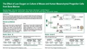 科学海报The Effect of Low Oxygen on Culture of Mouse and Human Mesenchymal Progenitor Cells from Bone Marrow
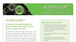 产品手册ALDEFLUOR™: For Identification and Isolation of Viable Stem & Progenitor Cells
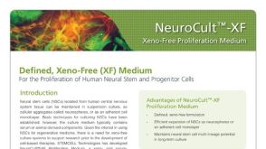 产品手册NeuroCult™-XF: Xeno-Free Culture Medium for the Proliferation of Human Neural Stem Cells
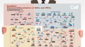 挂图Directed Differentiation of Pluripotent Stem Cells Strategies for differentiating ES and iPS cells into ectoderm, endoderm or mesoderm
 沪公网安备31010102008431号