Hudson JE et al. (JAN 2011)
Stem cells and development 20 1 77--87
A defined medium and substrate for expansion of human mesenchymal stromal cell progenitors that enriches for osteo- and chondrogenic precursors.
Human mesenchymal stromal cells (hMSCs) have generated significant interest due to their potential use in clinical applications. hMSCs are present at low frequency in vivo,but after isolation can be expanded considerably,generating clinically useful numbers of cells. In this study,we demonstrate the use of a defined embryonic stem cell expansion medium,mTeSR (Stem Cell Technologies),for the expansion of bone-marrow-derived hMSCs. The hMSCs grow at comparable rates,demonstrate tri-lineage differentiation potential,and show similar surface marker profiles (CD29(+),CD44(+),CD49a(+),CD73(+),CD90(+),CD105(+),CD146(+),CD166(+),CD34(-),and CD45(-)) in both the fetal bovine serum (FBS)-supplemented medium and mTeSR. However,expression of early differentiation transcription factors runt-related transcription factor 2,sex-determining region Y box 9,and peroxisome proliferator-activated receptor gamma changed significantly. Both runt-related transcription factor 2 and sex-determining region Y box 9 were upregulated,whereas peroxisome proliferator-activated receptor gamma was downregulated in mTeSR compared with FBS. Although osteogenic and chondrogenic differentiation was comparable in cells grown in mTeSR compared to FBS,adipogenic differentiation was significantly decreased in mTeSR-expanded cells,both in terms of gene expression and absolute numbers of adipocytes. The removal of the FBS from the medium and the provision of a defined medium with disclosed composition make mTeSR a superior study platform for hMSC biology in a controlled environment. Further,this provides a key step toward generating a clinical-grade medium for expansion of hMSCs for clinical applications that rely on osteo- and chondroinduction of MSCs,such as bone repair and cartilage generation.
View Publication
产品类型:
产品号#:
05850
05857
05870
05875
85850
85857
85870
85875
产品名:
mTeSR™1
mTeSR™1
Hu K et al. (APR 2017)
Stem cell research 20 115--117
Generation of an induced pluripotent stem cell line from a Loeys-Dietz syndrome patient with transforming growth factor-beta receptor-2 gene mutation.
Loeys-Dietz syndrome (LDS) is an autosomal-dominant connective tissue disorder,commonly caused by genetic mutation of transforming growth factor-beta receptor (TGFBR)-1 or TGFBR2. This study describes the generation of human induced pluripotent stem cells (hiPSCs) from peripheral blood mononuclear cells obtained from an LDS patient with TGFBR2 mutation (R193W). Analysis confirmed the cells had a normal karyotype,expressed typical pluripotency markers,had the ability to differentiate into all three germ layers in vivo,and retained the TGFBR2 mutation from the derived hiPSCs. This iPSC line represents a potentially useful tool for investigating LDS disease mechanisms.
View Publication
Molecular decoy to the Y-box binding protein-1 suppresses the growth of breast and prostate cancer cells whilst sparing normal cell viability.
The Y-box binding protein-1 (YB-1) is an oncogenic transcription/translation factor that is activated by phosphorylation at S102 whereby it induces the expression of growth promoting genes such as EGFR and HER-2. We recently illustrated by an in vitro kinase assay that a novel peptide to YB-1 was highly phosphorylated by the serine/threonine p90 S6 kinases RSK-1 and RSK-2,and to a lesser degree PKCα and AKT. Herein,we sought to develop this decoy cell permeable peptide (CPP) as a cancer therapeutic. This 9-mer was designed as an interference peptide that would prevent endogenous YB-1(S102) phosphorylation based on molecular docking. In cancer cells,the CPP blocked P-YB-1(S102) and down-regulated both HER-2 and EGFR transcript level and protein expression. Further,the CPP prevented YB-1 from binding to the EGFR promoter in a gel shift assay. Notably,the growth of breast (SUM149,MDA-MB-453,AU565) and prostate (PC3,LNCap) cancer cells was inhibited by ∼90% with the CPP. Further,treatment with this peptide enhanced sensitivity and overcame resistance to trastuzumab in cells expressing amplified HER-2. By contrast,the CPP had no inhibitory effect on the growth of normal immortalized breast epithelial (184htert) cells,primary breast epithelial cells,nor did it inhibit differentiation of hematopoietic progenitors. These data collectively suggest that the CPP is a novel approach to suppressing the growth of cancer cells while sparing normal cells and thereby establishes a proof-of-concept that blocking YB-1 activation is a new course of cancer therapeutics.
View Publication
产品类型:
产品号#:
05601
18056
18056RF
04435
04445
产品名:
EpiCult™-B 人培养基
MethoCult™ H4435 Enriched
MethoCult™ H4435 Enriched
Varga E et al. (OCT 2016)
Stem cell research 17 3 482--484
Generation of Mucopolysaccharidosis type II (MPS II) human induced pluripotent stem cell (iPSC) line from a 1-year-old male with pathogenic IDS mutation.
Peripheral blood was collected from a 1-year-old male patient with an X-linked recessive mutation of Iduronate 2-sulfatase (IDS) gene (NM000202.7(IDS):c.85CtextgreaterT) causing MPS II (OMIM 309900). Peripheral blood mononuclear cells (PBMCs) were reprogrammed by lentiviral delivery of a self-silencing hOKSM polycistronic vector. The pluripotency of the iPSC line was confirmed by the expression of pluripotency-associated markers and in vitro spontaneous differentiation towards the 3 germ layers. The iPSC line showed normal karyotype. The cell line offers a good platform to study MPS II pathophysiology,for drug testing,early biomarker discovery and gene therapy studies.
View Publication
产品类型:
产品号#:
05850
05857
05870
05875
85850
85857
85870
85875
产品名:
mTeSR™1
mTeSR™1
Ma D et al. (JAN 2017)
Stem cell research 18 51--53
Development of a human induced pluripotent stem cell (iPSC) line from a Parkinson's disease patient carrying the N551K variant in LRRK2 gene.
Peripheral blood mononuclear cells (PBMCs) were collected from a clinically diagnosed 64-year old male Parkinson's disease (PD) patient with N551K variant in the LRRK2 gene. The PMBCs were reprogrammed with the human OSKM transcription factors using the Sendai-virus reprogramming system. The transgene-free iPSC showed pluripotency confirmed by immunofluorescent staining for pluripotency markers and differentiated into the 3 germ layers in vivo. The iPSC line also showed normal karyotype. This cellular model can complement in vivo PD models for pathophysiological studies and drug screening.
View Publication
产品类型:
产品号#:
05850
05857
05870
05875
85850
85857
85870
85875
产品名:
mTeSR™1
mTeSR™1
Kurita R et al. (SEP 2006)
Stem cells (Dayton,Ohio) 24 9 2014--22
Tal1/Scl gene transduction using a lentiviral vector stimulates highly efficient hematopoietic cell differentiation from common marmoset (Callithrix jacchus) embryonic stem cells.
The development of embryonic stem cell (ESC) therapies requires the establishment of efficient methods to differentiate ESCs into specific cell lineages. Here,we report the in vitro differentiation of common marmoset (CM) (Callithrix jacchus) ESCs into hematopoietic cells after exogenous gene transfer using vesicular stomatitis virus-glycoprotein-pseudotyped lentiviral vectors. We transduced hematopoietic genes,including tal1/scl,gata1,gata2,hoxB4,and lhx2,into CM ESCs. By immunochemical and morphological analyses,we demonstrated that overexpression of tal1/scl,but not the remaining genes,dramatically increased hematopoiesis of CM ESCs,resulting in multiple blood-cell lineages. Furthermore,flow cytometric analysis demonstrated that CD34,a hematopoietic stem/progenitor cell marker,was highly expressed in tal1/scl-overexpressing embryoid body cells. Similar results were obtained from three independent CM ESC lines. These results suggest that transduction of exogenous tal1/scl cDNA into ESCs is a promising method to induce the efficient differentiation of CM ESCs into hematopoietic stem/progenitor cells.
View Publication
产品类型:
产品号#:
03434
03444
04435
04445
产品名:
MethoCult™ GF M3434
MethoCult™ GF M3434
MethoCult™ H4435 Enriched
MethoCult™ H4435 Enriched
Grandy RA et al. (FEB 2015)
Molecular and Cellular Biology 36 December MCB.00877--15
Genome-wide Studies Reveal that H3K4me3 Modification in Bivalent Genes is Dynamically Regulated During the Pluripotent Cell Cycle and Stabilized Upon Differentiation
textlessptextgreaterStem cell phenotypes are reflected by post-translational histone modifications,and this chromatin-related memory must be mitotically inherited to maintain cell identity through proliferative expansion. In human embryonic stem cells (hESCs),bivalent genes with both activating (H3K4me3) and repressive (H3K27me3) histone modifications are essential to sustain pluripotency. Yet the molecular mechanisms by which this epigenetic landscape is transferred to progeny cells remains to be established. By mapping genomic enrichment of H3K4me3/H3K27me3 in pure populations of hESCs in G2,mitotic,and G1 phases of the cell cycle,we found striking variations in the levels of H3K4me3 through the G2-M-G1 transition. Analysis of a representative set of bivalent genes revealed that chromatin modifiers involved in H3K4 methylation/demethylation are recruited to bivalent gene promoters in a cell cycle–dependent fashion. Interestingly,bivalent genes enriched with H3K4me3 exclusively during mitosis undergo the strongest upregulation after induction of differentiation. Furthermore,the histone-modification signature of genes that remain bivalent in differentiated cells resolves into a cell cycle–independent pattern after lineage commitment. These results establish a new dimension of chromatin regulation important in maintenance of pluripotency.textless/ptextgreater
View Publication
产品类型:
产品号#:
05850
05857
05870
05875
85850
85857
85870
85875
产品名:
mTeSR™1
mTeSR™1
Perin EC et al. (JUN 2011)
American heart journal 161 6 1078--87.e3
A randomized study of transendocardial injection of autologous bone marrow mononuclear cells and cell function analysis in ischemic heart failure (FOCUS-HF).
BACKGROUND Autologous bone marrow mononuclear cell (ABMMNC) therapy has shown promise in patients with heart failure (HF). Cell function analysis may be important in interpreting trial results. METHODS In this prospective study,we evaluated the safety and efficacy of the transendocardial delivery of ABMMNCs in no-option patients with chronic HF. Efficacy was assessed by maximal myocardial oxygen consumption,single photon emission computed tomography,2-dimensional echocardiography,and quality-of-life assessment (Minnesota Living with Heart Failure and Short Form 36). We also characterized patients' bone marrow cells by flow cytometry,colony-forming unit,and proliferative assays. RESULTS Cell-treated (n = 20) and control patients (n = 10) were similar at baseline. The procedure was safe; adverse events were similar in both groups. Canadian Cardiovascular Society angina score improved significantly (P = .001) in cell-treated patients,but function was not affected. Quality-of-life scores improved significantly at 6 months (P = .009 Minnesota Living with Heart Failure and P = .002 physical component of Short Form 36) over baseline in cell-treated but not control patients. Single photon emission computed tomography data suggested a trend toward improved perfusion in cell-treated patients. The proportion of fixed defects significantly increased in control (P = .02) but not in treated patients (P = .16). Function of patients' bone marrow mononuclear cells was severely impaired. Stratifying cell results by age showed that younger patients (%60 years) had significantly more mesenchymal progenitor cells (colony-forming unit fibroblasts) than patients<60 years (20.16 ± 14.6 vs 10.92 ± 7.8,P = .04). Furthermore,cell-treated younger patients had significantly improved maximal myocardial oxygen consumption (15 ± 5.8,18.6 ± 2.7,and 17 ± 3.7 mL/kg per minute at baseline,3 months,and 6 months,respectively) compared with similarly aged control patients (14.3 ± 2.5,13.7 ± 3.7,and 14.6 ± 4.7 mL/kg per minute,P = .04). CONCLUSIONS ABMMNC therapy is safe and improves symptoms,quality of life,and possibly perfusion in patients with chronic HF.
View Publication
产品类型:
产品号#:
70001
70001.1
70001.2
70001.3
70001.4
产品名:
Singbrant S et al. (JUN 2010)
Blood 115 23 4689--98
Canonical BMP signaling is dispensable for hematopoietic stem cell function in both adult and fetal liver hematopoiesis, but essential to preserve colon architecture.
Numerous publications have described the importance of bone morphogenetic protein (BMP) signaling in the specification of hematopoietic tissue in developing embryos. Here we investigate the full role of canonical BMP signaling in both adult and fetal liver hematopoiesis using conditional knockout strategies because conventional disruption of components of the BMP signaling pathway result in early death of the embryo. By targeting both Smad1 and Smad5,we have generated a double-knockout mouse with complete disruption of canonical BMP signaling. Interestingly,concurrent deletion of Smad1 and Smad5 results in death because of extrahematopoietic pathologic changes in the colon. However,Smad1/Smad5-deficient bone marrow cells can compete normally with wild-type cells and display unaffected self-renewal and differentiation capacity when transplanted into lethally irradiated recipients. Moreover,although BMP receptor expression is increased in fetal liver,fetal liver cells deficient in both Smad1 and Smad5 remain competent to long-term reconstitute lethally irradiated recipients in a multilineage manner. In conclusion,canonical BMP signaling is not required to maintain either adult or fetal liver hematopoiesis,despite its crucial role in the initial patterning of hematopoiesis in early embryonic development.
View Publication
产品类型:
产品号#:
03231
09600
09650
产品名:
MethoCult™ M3231
StemSpan™ SFEM
StemSpan™ SFEM
Spaggiari GM et al. (FEB 2006)
Blood 107 4 1484--90
Mesenchymal stem cell-natural killer cell interactions: evidence that activated NK cells are capable of killing MSCs, whereas MSCs can inhibit IL-2-induced NK-cell proliferation.
In recent years,mesenchymal stem cells (MSCs) have been shown to inhibit T-lymphocyte proliferation induced by alloantigens or mitogens. However,no substantial information is available regarding their effect on natural killer (NK) cells. Here we show that MSCs sharply inhibit IL-2-induced proliferation of resting NK cells,whereas they only partially affect the proliferation of activated NK cells. In addition,we show that IL-2-activated NK cells (but not freshly isolated NK cells) efficiently lyse autologous and allogeneic MSCs. The activating NK receptors NKp30,NKG2D,and DNAM-1 represented the major receptors responsible for the induction of NK-mediated cytotoxicity against MSCs. Accordingly,MSCs expressed the known ligands for these activating NK receptors-ULBPs,PVR,and Nectin-2. Moreover,NK-mediated lysis was inhibited when IFN-gamma-exposed MSCs were used as target cells as a consequence of the up-regulation of HLA class I molecules at the MSC surface. The interaction between NK cells and MSCs resulted not only in the lysis of MSCs but also in cytokine production by NK cells. These results should be taken into account when evaluating the possible use of MSCs in novel therapeutic strategies designed to improve engraftment or to suppress graft-versus-host disease (GVHD) in bone marrow transplantation.
View Publication
产品类型:
产品号#:
05401
05402
05411
产品名:
MesenCult™ MSC 基础培养基(人)
MesenCult™ MSC刺激添加物(人)
MesenCult™ 增殖试剂盒(人)
Liberski AR et al. (JUL 2013)
Journal of Proteome Research 12 7 3233--3245
Adaptation of a Commonly Used, Chemically Defined Medium for Human Embryonic Stem Cells to Stable Isotope Labeling with Amino Acids in Cell Culture
Metabolic labeling with stable isotopes is a prominent technique for comparative quantitative proteomics,and stable isotope labeling with amino acids in cell culture (SILAC) is the most commonly used approach. SILAC is,however,traditionally limited to simple tissue culture regimens and only rarely employed in the context of complex culturing conditions as those required for human embryonic stem cells (hESCs). Classic hESC culture is based on the use of mouse embryonic fibroblasts (MEFs) as a feeder layer,and as a result,possible xenogeneic contamination,contribution of unlabeled amino acids by the feeders,interlaboratory variability of MEF preparation,and the overall complexity of the culture system are all of concern in conjunction with SILAC. We demonstrate a feeder-free SILAC culture system based on a customized version of a commonly used,chemically defined hESC medium developed by Ludwig et al. and commercially available as mTeSR1 [mTeSR1 is a trade mark of WiCell (Madison,WI) licensed to STEMCELL Technologies (Vancouver,Canada)]. This medium,together with adjustments to the culturing protocol,facilitates reproducible labeling that is easily scalable to the protein amounts required by proteomic work flows. It greatly enhances the usability of quantitative proteomics as a tool for the study of mechanisms underlying hESCs differentiation and self-renewal. Associated data have been deposited to the ProteomeXchange with the identifier PXD000151.
View Publication


 EasySep™小鼠TIL(CD45)正选试剂盒





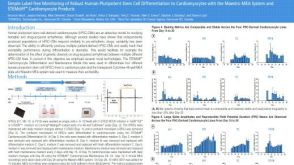 科学海报Simple Label-free Monitoring of Robust Human Pluripotent Stem Cell Differentiation to Cardiomyocytes with the Maestro MEA System and STEMdiff™ Cardiomyocyte Products
 沪公网安备31010102008431号