Sheridan SD et al. (OCT 2011)
PLoS ONE 6 10 e26203
Epigenetic characterization of the FMR1 gene and aberrant neurodevelopment in human induced pluripotent stem cell models of fragile X syndrome
Fragile X syndrome (FXS) is the most common inherited cause of intellectual disability. In addition to cognitive deficits,FXS patients exhibit hyperactivity,attention deficits,social difficulties,anxiety,and other autistic-like behaviors. FXS is caused by an expanded CGG trinucleotide repeat in the 5' untranslated region of the Fragile X Mental Retardation (FMR1) gene leading to epigenetic silencing and loss of expression of the Fragile X Mental Retardation protein (FMRP). Despite the known relationship between FMR1 CGG repeat expansion and FMR1 silencing,the epigenetic modifications observed at the FMR1 locus,and the consequences of the loss of FMRP on human neurodevelopment and neuronal function remain poorly understood. To address these limitations,we report on the generation of induced pluripotent stem cell (iPSC) lines from multiple patients with FXS and the characterization of their differentiation into post-mitotic neurons and glia. We show that clones from reprogrammed FXS patient fibroblast lines exhibit variation with respect to the predominant CGG-repeat length in the FMR1 gene. In two cases,iPSC clones contained predominant CGG-repeat lengths shorter than measured in corresponding input population of fibroblasts. In another instance,reprogramming a mosaic patient having both normal and pre-mutation length CGG repeats resulted in genetically matched iPSC clonal lines differing in FMR1 promoter CpG methylation and FMRP expression. Using this panel of patient-specific,FXS iPSC models,we demonstrate aberrant neuronal differentiation from FXS iPSCs that is directly correlated with epigenetic modification of the FMR1 gene and a loss of FMRP expression. Overall,these findings provide evidence for a key role for FMRP early in human neurodevelopment prior to synaptogenesis and have implications for modeling of FXS using iPSC technology. By revealing disease-associated cellular phenotypes in human neurons,these iPSC models will aid in the discovery of novel therapeutics for FXS and other autism-spectrum disorders sharing common pathophysiology.
View Publication
产品类型:
产品号#:
05850
05857
05870
05875
85850
85857
85870
85875
产品名:
mTeSR™1
mTeSR™1
Close JL et al. (MAR 2017)
Neuron 93 5 1035--1048.e5
Single-Cell Profiling of an In Vitro Model of Human Interneuron Development Reveals Temporal Dynamics of Cell Type Production and Maturation.
GABAergic interneurons are essential for neural circuit function,and their loss or dysfunction is implicated in human neuropsychiatric disease. In vitro methods for interneuron generation hold promise for studying human cellular and functional properties and,ultimately,for therapeutic cell replacement. Here we describe a protocol for generating cortical interneurons from hESCs and analyze the properties and maturation time course of cell types using single-cell RNA-seq. We find that the cell types produced mimic in vivo temporal patterns of neuron and glial production,with immature progenitors and neurons observed early and mature cortical neurons and glial cell types produced late. By comparing the transcriptomes of immature interneurons to those of more mature neurons,we identified genes important for human interneuron differentiation. Many of these genes were previously implicated in neurodevelopmental and neuropsychiatric disorders.
View Publication
产品类型:
产品号#:
05850
05857
05870
05875
85850
85857
85870
85875
产品名:
mTeSR™1
mTeSR™1
Zhang S et al. (MAR 2017)
Stem cell research 19 34--36
Characterization of human induced pluripotent stem cell (iPSC) line from a 72year old male patient with later onset Alzheimer's disease.
Peripheral blood was collected from a clinically diagnosed 72-year old male patient with later onset Alzheimer's disease. Peripheral blood mononuclear cells (PBMCs) were reprogrammed with the Yamanaka KMOS reprogramming factors using the Sendai-virus reprogramming system. The transgene-free iPSC line showed pluripotency verified by immunofluorescent staining for pluripotency markers,and the iPSC line was able to differentiate into the 3 germ layers in vivo. The iPSC line also showed normal karyotype. This in vitro cellular model will be useful for studying the pathological mechanism of Alzheimer's disease.
View Publication
Galat V et al. (MAY 2016)
Stem cells and development 25 14 1060--1072
Transgene Reactivation in Induced Pluripotent Stem Cell Derivatives and Reversion to Pluripotency of Induced Pluripotent Stem Cell-Derived Mesenchymal Cells.
Induced pluripotent stem cells (iPSCs) have enormous potential in regenerative medicine and disease modeling. It is now felt that clinical trials should be performed with iPSCs derived with non-integrative constructs. Numerous studies,however,including those describing disease models,are still being published using cells derived from iPSCs generated with integrative constructs. Our experimental work presents the first evidence of spontaneous transgene reactivation in vitro in several cellular types. Our results show that the transgenes were predominantly silent in parent iPSCs,but in mesenchymal and endothelial iPSC derivatives,the transgenes experienced random up-regulation of Nanog and c-Myc. Additionally,we provide evidence of spontaneous secondary reprogramming and reversion to pluripotency in mesenchymal stem cells derived from iPSCs. These findings strongly suggest that the studies,which utilize cellular products derived from iPSCs generated with retro- or lentiviruses,should be evaluated with consideration of the possibility of transgene reactivation. The in vitro model described here provides insight into the earliest events of culture transformation and suggests the hypothesis that reversion to pluripotency may be responsible for the development of tumors in cell replacement experiments. The main goal of this work,however,is to communicate the possibility of transgene reactivation in retro- or lenti- iPSC derivatives and the associated loss of cellular fidelity in vitro,which may impact the outcomes of disease modeling and related experimentation.
View Publication
产品类型:
产品号#:
05850
05857
05870
05875
85850
85857
85870
85875
产品名:
mTeSR™1
mTeSR™1
Wang H et al. (APR 2016)
The Journal of biological chemistry 291 16 8644--8652
Germ Cell Nuclear Factor (GCNF) Represses Oct4 Expression and Globally Modulates Gene Expression in Human Embryonic Stem (hES) Cells.
Oct4 is considered a key transcription factor for pluripotent stem cell self-renewal. It binds to specific regions within target genes to regulate their expression and is downregulated upon induction of differentiation of pluripotent stem cells; however,the mechanisms that regulate the levels of human Oct4 expression remain poorly understood. Here we show that expression of human Oct4 is directly repressed by germ cell nuclear factor (GCNF),an orphan nuclear receptor,in hES cells. Knockdown of GCNF by siRNA resulted in maintenance of Oct4 expression during RA-induced hES cell differentiation. While overexpression of GCNF promoted repression of Oct4 expression in both undifferentiated and differentiated hES cells. The level of Oct4 repression was dependent on the level of GCNF expression in a dose-dependent manner. mRNA microarray analysis demonstrated that overexpression of GCNF globally regulates gene expression in undifferentiated and differentiated hES cells. Within the group of altered genes,GCNF down-regulated 36% of the genes,and up-regulated 64% in undifferentiated hES cells. In addition,GCNF also showed a regulatory gene pattern that is different from RA treatment during hES cell differentiation. These findings increase our understanding of the mechanisms that maintain hES cell pluripotency and regulate gene expression during the differentiation process.
View Publication
产品类型:
产品号#:
05850
05857
05870
05875
85850
85857
85870
85875
产品名:
mTeSR™1
mTeSR™1
Mateizel I et al. (JAN 2012)
Methods in molecular biology (Clifton,N.J.) 873 81--112
Establishment of hESC lines from the inner cell mass of blastocyst-stage embryos and single blastomeres of 4-cell stage embryos.
More than 600 human embryonic stem cell (hESC) lines have been reported today at the human European Embryonic Stem Cell Registry ( http://www.hescreg.eu/ ). Despite these high numbers,there are currently no general protocols for derivation,culture,and characterization of hESC. Moreover,data on the culture of the embryo used for the derivation (medium,day of ICM isolation) are usually not available but can have an impact on the derivation rate. We present here the protocols for derivation,culture and characterization as we applied them for the 22 hESC lines (named VUB-hESC) in our laboratory.
View Publication
产品类型:
产品号#:
05854
05855
05850
05857
05870
05875
85850
85857
85870
85875
产品名:
mFreSR™
mFreSR™
mTeSR™1
mTeSR™1
Lim MN et al. (MAY 2012)
Molecular vision 18 1289--300
Ex vivo expanded SSEA-4+ human limbal stromal cells are multipotent and do not express other embryonic stem cell markers.
PURPOSE: The presence of multipotent human limbal stromal cells resembling mesenchymal stromal cells (MSC) provides new insights to the characteristic of these cells and its therapeutic potential. However,little is known about the expression of stage-specific embryonic antigen 4 (SSEA-4) and the embryonic stem cell (ESC)-like properties of these cells. We studied the expression of SSEA-4 surface protein and the various ESC and MSC markers in the ex vivo cultured limbal stromal cells. The phenotypes and multipotent differentiation potential of these cells were also evaluated.backslashnbackslashnMETHODS: Limbal stromal cells were derived from corneoscleral rims. The SSEA-4(+) and SSEA-4(-) limbal stromal cells were sorted by fluorescence-activated cells sorting (FACS). Isolated cells were expanded and reanalyzed for their expression of SSEA-4. Expression of MSC and ESC markers on these cells were also analyzed by FACS. In addition,expression of limbal epithelial and corneal stromal proteins such as ATP-binding cassette sub-family G member 2 (ABCG2),tumour protein p63 (p63),paired box 6 (Pax6),cytokeratin 3 (AE5),cytokeratin 10,and keratocan sulfate were evaluated either by immunofluorecence staining or reverse transcription polymerase chain reaction. Appropriate induction medium was used to differentiate these cells into adipocytes,osteocytes,and chondrocytes.backslashnbackslashnRESULTS: Expanded limbal stromal cells expressed the majority of mesenchymal markers. These cells were negative for ABCG2,p63,Pax6,AE-5,and keratocan sulfate. After passaged,a subpopulation of these cells showed low expression of SSEA-4 but were negative for other important ESC surface markers such as Tra-1-60,Tra-1-81,and transcription factors like octamer-binding transcription factor 4 (Oct4),SRY(sex determining region Y)-box 2 (Sox2),and Nanog. Early passaged cells when induced were able to differentiate into adipocytes,osteocytes and chondrocytes.backslashnbackslashnCONCLUSIONS: The expanded limbal stromal cells showed features of multipotent MSC. Our study confirmed the expression of SSEA-4 by a subpopulation of cultured limbal stromal cells. However,despite the expression of SSEA-4,these cells did not express any other markers of ESC. Therefore,we conclude that the cells did not show properties of ESC.
View Publication
产品类型:
产品号#:
05850
05857
05870
05875
85850
85857
85870
85875
产品名:
mTeSR™1
mTeSR™1
Tauchmanovà et al. (MAY 2003)
Cancer 97 10 2453--61
Avascular necrosis in long-term survivors after allogeneic or autologous stem cell transplantation: a single center experience and a review.
BACKGROUND: The most debilitating skeletal complication of stem cell transplantation (SCT) is avascular necrosis (AVN). METHODS: Two hundred seven consecutive patients were evaluated prospectively for AVN. They survived disease free for more than 180 days after autologous or allogeneic SCT for hematologic malignancies. The diagnosis of AVN in suspicious cases was confirmed by magnetic resonance imaging. Possible correlations with treatments,bone mineral density (BMD),graft versus host disease (GVHD),and in vitro growth of fibroblast progenitors were investigated. Bone mineral density was evaluated by dual-energy X-ray absorptiometry in 100 transplanted patients,and the in vitro growth of fibroblast progenitors was monitored by a fibroblast colony-forming unit (CFU-F) assay in 30 patients after allogeneic SCT. RESULTS: Twelve patients developed AVN 3-114 months (median,26 months) following SCT: 10 (10%) after allogeneic SCT and 2 (1.9%) after autologous SCT (P = 0.04). Twenty-five joints were affected by AVN. All patients had femoral head involvement,which was managed with hip replacement in six of them. All but one patient who developed AVN after allogeneic SCT suffered from chronic GVHD (cGVHD). Avascular necrosis occurred 1-4 months after exacerbation or progression of cGVHD. Cumulative dose of steroids was similar in both SCT groups (including steroids given pretransplant for the basic disease),whereas treatment duration was significantly longer in the allogeneic SCT group. Avascular necrosis was related to the decreased number of bone marrow CFU-F colonies in vitro,but not to BMD values. CONCLUSIONS: Avascular necrosis is a skeletal complication that occurs more often after allogeneic than after autologous SCT. Occurrence of AVN symptoms after clinical follow-up of cGVHD suggests that cGVHD requiring long-term steroid therapy is one of the main risk factors for AVN. Avascular necrosis may be facilitated by a severe deficit in the repopulating capacity of bone marrow stromal stem cells after SCT.
View Publication
产品类型:
产品号#:
05401
05402
05411
产品名:
MesenCult™ MSC 基础培养基(人)
MesenCult™ MSC刺激添加物(人)
MesenCult™ 增殖试剂盒(人)
Liu C et al. (OCT 2014)
Biochemical and Biophysical Research Communications 452 4 895--900
Synergistic contribution of SMAD signaling blockade and high localized cell density in the differentiation of neuroectoderm from H9 cells
Directed neural differentiation of human embryonic stem cells (ESCs) enables researchers to generate diverse neuronal populations for human neural development study and cell replacement therapy. To realize this potential,it is critical to precisely understand the role of various endogenous and exogenous factors involved in neural differentiation. Cell density,one of the endogenous factors,is involved in the differentiation of human ESCs. Seeding cell density can result in variable terminal cell densities or localized cell densities (LCDs),giving rise to various outcomes of differentiation. Thus,understanding how LCD determines the differentiation potential of human ESCs is important. The aim of this study is to highlight the role of LCD in the differentiation of H9 human ESCs into neuroectoderm (NE),the primordium of the nervous system. We found the initially seeded cells form derived cells with variable LCDs and subsequently affect the NE differentiation. Using a newly established method for the quantitative examination of LCD,we demonstrated that in the presence of induction medium supplemented with or without SMAD signaling blockers,high LCD promotes the differentiation of NE. Moreover,SMAD signaling blockade promotes the differentiation of NE but not non-NE germ layers,which is dependent on high LCDs. Taken together,this study highlights the need to develop innovative strategies or techniques based on LCDs for generating neural progenies from human ESCs.
View Publication
产品类型:
产品号#:
05850
05857
05870
05875
07923
85850
85857
85870
85875
产品名:
Dispase (1 U/mL)
mTeSR™1
mTeSR™1
Jin S et al. (JUL 2012)
Tissue Engineering Part A 18 13-14 1419--30
Porous membrane substrates offer better niches to enhance the Wnt signaling and promote human embryonic stem cell growth and differentiation.
Human embryonic stem cells (hESCs) require specific niches for adhesion,expansion,and lineage-specific differentiation. In this study,we showed that a membrane substrate offers better tissue niches for hESC attachment,spreading,proliferation,and differentiation. The cell doubling time was shortened from 46.3±5.7 h for hESCs grown on solid substrates to 25.6±2.6 h for those on polyester (PE) membrane substrates with pore size of 0.4 μm. In addition,we observed an increase of approximately five- to ninefold of definitive endoderm marker gene expression in hESCs differentiated on PE or polyethylene terephthalate membrane substrates. Global gene expression analysis revealed upregulated expressions of a number of extracellular matrix and cell adhesion molecules in hESCs grown on membrane substrates. Further,an enhanced nuclear translocation of β-catenin was detected in these cells. These observations suggested the augmentation of Wnt signaling in hESCs grown on membrane substrates. These results also demonstrated that a membrane substrate can offer better physicochemical cues for enhancing in vitro hESC attachment,proliferation,and differentiation.
View Publication
产品类型:
产品号#:
05850
05857
05870
05875
07923
85850
85857
85870
85875
产品名:
Dispase (1 U/mL)
mTeSR™1
mTeSR™1
Sun N and Zhao H (MAY 2014)
Biotechnology and Bioengineering 111 5 1048--53
Seamless correction of the sickle cell disease mutation of the HBB gene in human induced pluripotent stem cells using TALENs.
Sickle cell disease (SCD) is the most common human genetic disease which is caused by a single mutation of human β-globin (HBB) gene. The lack of long-term treatment makes the development of reliable cell and gene therapies highly desirable. Disease-specific patient-derived human induced pluripotent stem cells (hiPSCs) have great potential for developing novel cell and gene therapies. With the disease-causing mutations corrected in situ,patient-derived hiPSCs can restore normal cell functions and serve as a renewable autologous cell source for the treatment of genetic disorders. Here we successfully utilized transcription activator-like effector nucleases (TALENs),a recently emerged novel genome editing tool,to correct the SCD mutation in patient-derived hiPSCs. The TALENs we have engineered are highly specific and generate minimal off-target effects. In combination with piggyBac transposon,TALEN-mediated gene targeting leaves no residual ectopic sequences at the site of correction and the corrected hiPSCs retain full pluripotency and a normal karyotype. Our study demonstrates an important first step of using TALENs for the treatment of genetic diseases such as SCD,which represents a significant advance toward hiPSC-based cell and gene therapies.
View Publication


 EasySep™小鼠TIL(CD45)正选试剂盒





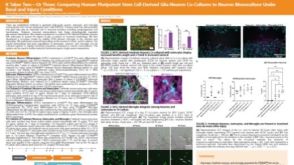 科学海报It Takes Two—Or Three: Comparing Human Pluripotent Stem Cell Derived Glia-Neuron Co-Cultures to Neuron Monoculture Under Basal and Injury Conditions
 沪公网安备31010102008431号