Jaramillo M and Banerjee I (MAR 2012)
Journal of visualized experiments : JoVE 61 2--7
Endothelial cell co-culture mediates maturation of human embryonic stem cell to pancreatic insulin producing cells in a directed differentiation approach.
Embryonic stem cells (ESC) have two main characteristics: they can be indefinitely propagated in vitro in an undifferentiated state and they are pluripotent,thus having the potential to differentiate into multiple lineages. Such properties make ESCs extremely attractive for cell based therapy and regenerative treatment applications. However for its full potential to be realized the cells have to be differentiated into mature and functional phenotypes,which is a daunting task. A promising approach in inducing cellular differentiation is to closely mimic the path of organogenesis in the in vitro setting. Pancreatic development is known to occur in specific stages,starting with endoderm,which can develop into several organs,including liver and pancreas. Endoderm induction can be achieved by modulation of the nodal pathway through addition of Activin A in combination with several growth factors. Definitive endoderm cells then undergo pancreatic commitment by inhibition of sonic hedgehog inhibition,which can be achieved in vitro by addition of cyclopamine. Pancreatic maturation is mediated by several parallel events including inhibition of notch signaling; aggregation of pancreatic progenitors into 3-dimentional clusters; induction of vascularization; to name a few. By far the most successful in vitro maturation of ESC derived pancreatic progenitor cells have been achieved through inhibition of notch signaling by DAPT supplementation. Although successful,this results in low yield of the mature phenotype with reduced functionality. A less studied area is the effect of endothelial cell signaling in pancreatic maturation,which is increasingly being appreciated as an important contributing factor in in-vivo pancreatic islet maturation. The current study explores such effect of endothelial cell signaling in maturation of human ESC derived pancreatic progenitor cells into insulin producing islet-like cells. We report a multi-stage directed differentiation protocol where the human ESCs are first induced towards endoderm by Activin A along with inhibition of PI3K pathway. Pancreatic specification of endoderm cells is achieved by inhibition of sonic hedgehog signaling by Cyclopamine along with retinoid induction by addition of Retinoic Acid. The final stage of maturation is induced by endothelial cell signaling achieved by a co-culture configuration. While several endothelial cells have been tested in the co-culture,herein we present our data with rat heart microvascular endothelial Cells (RHMVEC),primarily for the ease of analysis.
View Publication
产品类型:
产品号#:
05850
05857
05870
05875
85850
85857
85870
85875
产品名:
mTeSR™1
mTeSR™1
Tzeng Y-S et al. (JAN 2011)
Blood 117 2 429--39
Loss of Cxcl12/Sdf-1 in adult mice decreases the quiescent state of hematopoietic stem/progenitor cells and alters the pattern of hematopoietic regeneration after myelosuppression.
The C-X-C-type chemokine Cxcl12,also known as stromal cell-derived factor-1,plays a critical role in hematopoiesis during fetal development. However,the functional requirement of Cxcl12 in the adult hematopoietic stem/progenitor cell (HSPC) regulation was still unclear. In this report,we developed a murine Cxcl12 conditional deletion model in which the target gene can be deleted at the adult stage. We found that loss of stroma-secreted Cxcl12 in the adult led to expansion of the HSPC population as well as a reduction in long-term quiescent stem cells. In Cxcl12-deficient bone marrow,HSPCs were absent along the endosteal surface,and blood cell regeneration occurred predominantly in the perisinusoidal space after 5-fluorouracil myelosuppression challenge. Our results indicate that Cxcl12 is required for HSPC homeostasis regulation and is an important factor for osteoblastic niche organization in adult stage bone marrow.
View Publication
产品类型:
产品号#:
03434
03444
产品名:
MethoCult™ GF M3434
MethoCult™ GF M3434
Nakamura Y et al. (SEP 2010)
Blood 116 9 1422--32
Isolation and characterization of endosteal niche cell populations that regulate hematopoietic stem cells.
The endosteal niche is critical for the maintenance of hematopoietic stem cells (HSCs). However,it consists of a heterogeneous population in terms of differentiation stage and function. In this study,we characterized endosteal cell populations and examined their ability to maintain HSCs. Bone marrow endosteal cells were subdivided into immature mesenchymal cell-enriched ALCAM(-)Sca-1(+) cells,osteoblast-enriched ALCAM(+)Sca-1(-),and ALCAM(-)Sca-1(-) cells. We found that all 3 fractions maintained long-term reconstitution (LTR) activity of HSCs in an in vitro culture. In particular,ALCAM(+)Sca-1(-) cells significantly enhanced the LTR activity of HSCs by the up-regulation of homing- and cell adhesion-related genes in HSCs. Microarray analysis showed that ALCAM(-)Sca-1(+) fraction highly expressed cytokine-related genes,whereas the ALCAM(+)Sca-1(-) fraction expressed multiple cell adhesion molecules,such as cadherins,at a greater level than the other fractions,indicating that the interaction between HSCs and osteoblasts via cell adhesion molecules enhanced the LTR activity of HSCs. Furthermore,we found an osteoblastic marker(low/-) subpopulation in ALCAM(+)Sca-1(-) fraction that expressed cytokines,such as Angpt1 and Thpo,and stem cell marker genes. Altogether,these data suggest that multiple subsets of osteoblasts and mesenchymal progenitor cells constitute the endosteal niche and regulate HSCs in adult bone marrow.
View Publication
产品类型:
产品号#:
03434
03444
产品名:
MethoCult™ GF M3434
MethoCult™ GF M3434
Webb CF et al. (MAR 2011)
Molecular and cellular biology 31 5 1041--53
The ARID family transcription factor bright is required for both hematopoietic stem cell and B lineage development.
Bright/Arid3a has been characterized both as an activator of immunoglobulin heavy-chain transcription and as a proto-oncogene. Although Bright expression is highly B lineage stage restricted in adult mice,its expression in the earliest identifiable hematopoietic stem cell (HSC) population suggests that Bright might have additional functions. We showed that textgreater99% of Bright(-/-) embryos die at midgestation from failed hematopoiesis. Bright(-/-) embryonic day 12.5 (E12.5) fetal livers showed an increase in the expression of immature markers. Colony-forming assays indicated that the hematopoietic potential of Bright(-/-) mice is markedly reduced. Rare survivors of lethality,which were not compensated by the closely related paralogue Bright-derived protein (Bdp)/Arid3b,suffered HSC deficits in their bone marrow as well as B lineage-intrinsic developmental and functional deficiencies in their peripheries. These include a reduction in a natural antibody,B-1 responses to phosphocholine,and selective T-dependent impairment of IgG1 class switching. Our results place Bright/Arid3a on a select list of transcriptional regulators required to program both HSC and lineage-specific differentiation.
View Publication
产品类型:
产品号#:
03434
03444
产品名:
MethoCult™ GF M3434
MethoCult™ GF M3434
Goyama S et al. (DEC 2004)
Blood 104 12 3558--64
The transcriptionally active form of AML1 is required for hematopoietic rescue of the AML1-deficient embryonic para-aortic splanchnopleural (P-Sp) region.
Acute myelogenous leukemia 1 (AML1; runt-related transcription factor 1 [Runx1]) is a member of Runx transcription factors and is essential for definitive hematopoiesis. Although AML1 possesses several subdomains of defined biochemical functions,the physiologic relevance of each subdomain to hematopoietic development has been poorly understood. Recently,the consequence of carboxy-terminal truncation in AML1 was analyzed by the hematopoietic rescue assay of AML1-deficient mouse embryonic stem cells using the gene knock-in approach. Nonetheless,a role for specific internal domains,as well as for mutations found in a human disease,of AML1 remains to be elucidated. In this study,we established an experimental system to efficiently evaluate the hematopoietic potential of AML1 using a coculture system of the murine embryonic para-aortic splanchnopleural (P-Sp) region with a stromal cell line,OP9. In this system,the hematopoietic defect of AML1-deficient P-Sp can be rescued by expressing AML1 with retroviral infection. By analysis of AML1 mutants,we demonstrated that the hematopoietic potential of AML1 was closely related to its transcriptional activity. Furthermore,we showed that other Runx transcription factors,Runx2/AML3 or Runx3/AML2,could rescue the hematopoietic defect of AML1-deficient P-Sp. Thus,this experimental system will become a valuable tool to analyze the physiologic function and domain contribution of Runx proteins in hematopoiesis.
View Publication
Ludwig T et al. (SEP 2007)
Current protocols in stem cell biology Chapter 1 September Unit 1C.2
Defined, Feeder-Independent Medium for Human Embryonic Stem Cell Culture
The developmental potential of human ES cells makes them an important tool in developmental,pharmacological,and clinical research. For human ES cell technology to be fully exploited,however,culture efficiency must be improved,large-scale culture enabled,and safety ensured. Traditional human ES cell culture systems have relied on serum products and mouse feeder layers,which limit the scale,present biological variability,and expose the cells to potential contaminants. Defined,feeder-independent culture systems improve the safety and efficiency of ES cell technology,enabling translational research. The protocols herein are designed with the standard research laboratory in mind. They contain recipes for the formulation of mTeSR (a defined medium for human ES cell culture) and detailed protocols for the culture,transfer,and passage of cells grown in these feeder-independent conditions. They provide a basis for routine feeder-independent culture,and a starting point for additional optimization of culture conditions.
View Publication
产品类型:
产品号#:
05850
05857
05870
05875
85850
85857
85870
85875
产品名:
mTeSR™1
mTeSR™1
Chen X et al. (SEP 2015)
Stem Cell Research 15 2 395--402
OP9-Lhx2 stromal cells facilitate derivation of hematopoietic progenitors both in vitro and in vivo
Generating engraftable hematopoietic stem cells (HSCs) from pluripotent stem cells (PSCs) is an ideal approach for obtaining induced HSCs for cell therapy. However,the path from PSCs to robustly induced HSCs (iHSCs) in vitro remains elusive. We hypothesize that the modification of hematopoietic niche cells by transcription factors facilitates the derivation of induced HSCs from PSCs. The Lhx2 transcription factor is expressed in fetal liver stromal cells but not in fetal blood cells. Knocking out Lhx2 leads to a fetal hematopoietic defect in a cell non-autonomous role. In this study,we demonstrate that the ectopic expression of Lhx2 in OP9 cells (OP9-Lhx2) accelerates the hematopoietic differentiation of PSCs. OP9-Lhx2 significantly increased the yields of hematopoietic progenitor cells via co-culture with PSCs in vitro. Interestingly,the co-injection of OP9-Lhx2 and PSCs into immune deficient mice also increased the proportion of hematopoietic progenitors via the formation of teratomas. The transplantation of phenotypic HSCs from OP9-Lhx2 teratomas but not from the OP9 control supported a transient repopulating capability. The upregulation of Apln gene by Lhx2 is correlated to the hematopoietic commitment property of OP9-Lhx2. Furthermore,the enforced expression of Apln in OP9 cells significantly increased the hematopoietic differentiation of PSCs. These results indicate that OP9-Lhx2 is a good cell line for regeneration of hematopoietic progenitors both in vitro and in vivo.
View Publication
产品类型:
产品号#:
05850
05857
05870
05875
85850
85857
85870
85875
产品名:
mTeSR™1
mTeSR™1
Gallego MJ et al. (JUN 2009)
Stem cells and development 18 5 737--740
Opioid and progesterone signaling is obligatory for early human embryogenesis.
The growth factors that drive the division and differentiation of stem cells during early human embryogenesis are unknown. The secretion of endorphins,progesterone (P(4)),human chorionic gonadotropin,17beta-estradiol,and gonadotropin-releasing hormone by trophoblasts that lie adjacent to the embryoblast in the blastocyst suggests that these pregnancy-associated factors may directly signal the growth and development of the embryoblast. To test this hypothesis,we treated embryoblast-derived human embryonic stem cells (hESCs) with ICI 174,864,a delta-opioid receptor antagonist,and RU-486 (mifepristone),a P(4) receptor competitive antagonist. Both antagonists potently inhibited the differentiation of hESC into embryoid bodies,an in vitro structure akin to the blastocyst containing all three germ layers. Furthermore,these agents prevented the differentiation of hESC aggregates into columnar neuroectodermal cells and their organization into neural tube-like rosettes as determined morphologically. Immunoblot analyses confirmed the obligatory role of these hormones; both antagonists inhibited nestin expression,an early marker of neural precursor cells normally detected during rosette formation. Conversely,addition of P(4) to hESC aggregates induced nestin expression and the formation of neuroectodermal rosettes. These results demonstrate that trophoblast-associated hormones induce blastulation and neurulation during early human embryogenesis.
View Publication
产品类型:
产品号#:
05850
05857
05870
05875
85850
85857
85870
85875
产品名:
mTeSR™1
mTeSR™1
Lin M et al. (AUG 2012)
PLoS ONE 7 8 e44017
Allele-biased expression in differentiating human neurons: implications for neuropsychiatric disorders.
Stochastic processes and imprinting,along with genetic factors,lead to monoallelic or allele-biased gene expression. Stochastic monoallelic expression fine-tunes information processing in immune cells and the olfactory system,and imprinting plays an important role in development. Recent studies suggest that both stochastic events and imprinting may be more widespread than previously considered. We are interested in allele-biased gene expression occurring in the brain because parent-of-origin effects suggestive of imprinting appear to play a role in the transmission of schizophrenia (SZ) and autism spectrum disorders (ASD) in some families. In addition,allele-biased expression could help explain monozygotic (MZ) twin discordance and reduced penetrance. The ability to study allele-biased expression in human neurons has been transformed with the advent of induced pluripotent stem cell (iPSC) technology and next generation sequencing. Using transcriptome sequencing (RNA-Seq) we identified 801 genes in differentiating neurons that were expressed in an allele-biased manner. These included a number of putative SZ and ASD candidates,such as A2BP1 (RBFOX1),ERBB4,NLGN4X,NRG1,NRG3,NRXN1,and NLGN1. Overall,there was a modest enrichment for SZ and ASD candidate genes among those that showed evidence for allele-biased expression (chi-square,p = 0.02). In addition to helping explain MZ twin discordance and reduced penetrance,the capacity to group many candidate genes affecting a variety of molecular and cellular pathways under a common regulatory process - allele-biased expression - could have therapeutic implications.
View Publication
产品类型:
产品号#:
05850
05857
05870
05875
85850
85857
85870
85875
产品名:
mTeSR™1
mTeSR™1
Johansson BM and Wiles MV (JAN 1995)
Molecular and cellular biology 15 1 141--51
Evidence for involvement of activin A and bone morphogenetic protein 4 in mammalian mesoderm and hematopoietic development.
Xenopus in vitro studies have implicated both transforming growth factor beta (TGF-beta) and fibroblast growth factor (FGF) families in mesoderm induction. Although members of both families are present during mouse mesoderm formation,there is little evidence for their functional role in mesoderm induction. We show that mouse embryonic stem cells,which resemble primitive ectoderm,can differentiate to mesoderm in vitro in a chemically defined medium (CDM) in the absence of fetal bovine serum. In CDM,this differentiation is responsive to TGF-beta family members in a concentration-dependent manner,with activin A mediating the formation of dorsoanterior-like mesoderm and bone morphogenetic protein 4 mediating the formation of ventral mesoderm,including hematopoietic precursors. These effects are not observed in CDM alone or when TGF-beta 1,-beta 2,or -beta 3,acid FGF,or basic FGF is added individually to CDM. In vivo,at day 6.5 of mouse development,activin beta A RNA is detectable in the decidua and bone morphogenetic protein 4 RNA is detectable in the egg cylinder. Together,our data strongly implicate the TGF-beta family in mammalian mesoderm development and hematopoietic cell formation.
View Publication
产品类型:
产品号#:
06902
06952
00321
00322
00323
00324
00325
产品名:
Dobo I et al. (AUG 1995)
Journal of hematotherapy 4 4 281--7
Collagen matrix: an attractive alternative to agar and methylcellulose for the culture of hematopoietic progenitors in autologous transplantation products.
Autografts using untreated or in vitro manipulated bone marrow and peripheral blood stem cells represent promising approaches to the treatment of malignant diseases. In this work,the collagen gel culture technique was compared with agar and methylcellulose for its capacity to permit the growth of human granulomonocytic (day 14 CFU-GM; collagen vs agar or MTC) or erythroblastic (day 7 CFU-E and day 14 BFU-E; collagen versus methylcellulose) colonies in autologous transplantation products. Our results show that the collagen culture system always gave as many or more colonies than the other techniques. It also allowed harvesting of gels onto glass slides and subsequent May-Grünwald-Giemsa,cytochemical or immunocytochemical staining. We suggest that the collagen assay represents an interesting alternative to the widely used agar or methylcellulose systems for the culture of hematopoietic progenitors because of the equal or higher number of colonies detected,the easy phenotypical identification of colonies in stained gels,and the ability to store high-quality documentation. This technique is particularly attractive for use in the quality control of autologous bone marrow transplantation procedures.
View Publication

 EasySep™小鼠TIL(CD45)正选试剂盒


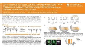 科学海报CRISPR-Cas9 Gene Editing Of CDK5RAP2 In Human Pluripotent Stem Cells, Derivation Of Genetically Stable Clonal Lines And Formation Of Cerebral Organoids
 沪公网安备31010102008431号