
 EasySep™小鼠TIL(CD45)正选试剂盒


搜索结果: 'megacult cytokines cfu'
-
产品类型:
产品号#:
04960
04902
04900
04963
04962
04970
产品名:
MegaCult™-C胶原和无细胞因子培养基
胶原蛋白溶液
MegaCult™-C无细胞因子培养基
双室载玻片套件
MegaCult™-C CFU-Mk染色试剂盒
MegaCult™-C无细胞因子全套试剂盒
-
产品类型:
产品号#:
09850
产品名:
-
产品类型:
产品号#:
产品名:
-
产品类型:
产品号#:
产品名:
-
产品类型:
产品号#:
04564
04534
04544
产品名:
MethoCult™ H4534 Classic 无 EPO 入门试剂盒
MethoCult™ H4534 Classic(不含 EPO)
MethoCult™ H4534 Classic(不含 EPO)
-
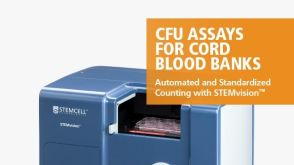 产品手册STEMvision™ 为脐血库提供的自动化和 标准化CFU检测计数
产品类型:
品牌:
STEMvision
产品号#:
27301
27302
04437
04447
22001
22005
22006
22007
22008
22009
22011
22012
22013
27370
27371
产品名:
MethoCult™ Express
MethoCult™ Express
STEMvision™ 人脐带血7-天CFU分析包
STEMvision™ 彩色人脐带血14-天CFU分析包
STEMvision™ 彩色人骨髓14-天CFU分析包
STEMvision™ 彩色人动员外周血14-天CFU分析包
STEMvision™ 小鼠总CFU分析包
STEMvision™ 小鼠髓系CFU分析包
STEMvision™ 小鼠红系CFU分析包
STEMvision™ 小鼠CFU分析包(髓系和红系)
SmartDish™ - 5个
SmartDish™ - 50个
-
产品类型:
产品号#:
04100
产品名:
MethoCult™ H4100
-

-
产品类型:
产品号#:
04961
04965
04962
04915
04807
04809
04906
04913
04803
04804
04905
04850
04974
04902
04960
04900
04901
04963
04970
04971
产品名:
MegaCult™-C胶原和含细胞因子培养基
MegaCult™-C CFU-Mk染色试剂盒
MegaCult-C 10% BSA, 6mL
MegaCult-C Human Serum, 6mL
Alkaline Phosphatase Substrate Tabs, pk
Biotin/Conjugate Goat Anti-Mu lgG, 125uL
MegaCult-C Evans Blue Stain, 5mL
Primary Ab, Anti-HuAnti-GPIIb/IIIa 360uL
MegaCult-C Control Antibody, 100 µL
Avidin-Alk Phosphatase Conjugate, 200 uL
MegaCult™-C含脂质培养基
MegaCult™-C胶原和含脂质培养基
胶原蛋白溶液
MegaCult™-C胶原和无细胞因子培养基
MegaCult™-C无细胞因子培养基
MegaCult™-C含细胞因子培养基
双室载玻片套件
MegaCult™-C无细胞因子全套试剂盒
MegaCult™-C含细胞因子全套试剂盒
-
产品类型:
产品号#:
04960
04902
04900
04961
04901
04963
04962
04970
04971
产品名:
MegaCult™-C胶原和无细胞因子培养基
胶原蛋白溶液
MegaCult™-C无细胞因子培养基
MegaCult™-C胶原和含细胞因子培养基
MegaCult™-C含细胞因子培养基
双室载玻片套件
MegaCult™-C CFU-Mk染色试剂盒
MegaCult™-C无细胞因子全套试剂盒
MegaCult™-C含细胞因子全套试剂盒
-
产品类型:
产品号#:
04960
04902
04900
04961
04901
04963
04962
04970
04971
产品名:
MegaCult™-C胶原和无细胞因子培养基
胶原蛋白溶液
MegaCult™-C无细胞因子培养基
MegaCult™-C胶原和含细胞因子培养基
MegaCult™-C含细胞因子培养基
双室载玻片套件
MegaCult™-C CFU-Mk染色试剂盒
MegaCult™-C无细胞因子全套试剂盒
MegaCult™-C含细胞因子全套试剂盒
-
产品类型:
产品号#:
03234
03434
03444
04960
04902
04900
04961
04901
04963
04962
04970
04971
产品名:
MethoCult™ M3234
MethoCult™ GF M3434
MethoCult™ GF M3434
MegaCult™-C胶原和无细胞因子培养基
胶原蛋白溶液
MegaCult™-C无细胞因子培养基
MegaCult™-C胶原和含细胞因子培养基
MegaCult™-C含细胞因子培养基
双室载玻片套件
MegaCult™-C CFU-Mk染色试剂盒
MegaCult™-C无细胞因子全套试剂盒
MegaCult™-C含细胞因子全套试剂盒

 沪公网安备31010102008431号