Ausubel LJ et al. (JAN 2011)
Methods in molecular biology (Clifton,N.J.) 767 147--159
GMP scale-up and banking of pluripotent stem cells for cellular therapy applications.
Human pluripotent stem cells (PSCs),which include human embryonic stem cells (ESCs) as well as induced pluripotent stem cells (iPSCs),represent an important source of cellular therapies in regenerative medicine and the study of early human development. As such,it is becoming increasingly important to develop methods for the large-scale banking of human PSC lines. There are several well-established methods for the propagation of human PSCs. The key to development of a good manufacturing practice (GMP) bank is to determine a manufacturing method that is amenable to large-scale production using materials that are fully documented. We have developed several banks of hESCs using animal feeder cells,animal-based matrices,or animal-free matrices. Protocols for growing hESCs on mouse embryonic fibroblasts (MEFs) are well established and are very helpful for producing research grade banks of cells. As most human ESCs cultured by research laboratories have been exposed to xenogeneic reagents,it is not imperative that all materials used in the production of a master cell bank be animal-free in origin. Nevertheless,as the field develops,it will no doubt become increasingly important to produce a bank of cells for clinical use without xenogeneic reagents,particularly nonhuman feeder cells which might harbor viruses with potential risk to human health or cell product integrity. Thus,even for cell lines previously exposed to xenogeneic reagents,it is important to minimize any subsequent exposure of the cell lines to additional adventitious agents. We have specifically described procedures for the growth of hESCs on Matrigel,an animal-matrix,and CELLstart,an animal-free matrix,and these can be used to produce hESCs as part of a clinical manufacturing process.
View Publication
产品类型:
产品号#:
05850
05857
05870
05875
85850
85857
85870
85875
产品名:
mTeSR™1
mTeSR™1
Cutler AJ et al. (DEC 2010)
Journal of immunology (Baltimore,Md. : 1950) 185 11 6617--23
Umbilical cord-derived mesenchymal stromal cells modulate monocyte function to suppress T cell proliferation.
Mesenchymal stromal cells (MSCs) may be derived from a variety of tissues,with human umbilical cord (UC) providing an abundant and noninvasive source. Human UC-MSCs share similar in vitro immunosuppressive properties as MSCs obtained from bone marrow and cord blood. However,the mechanisms and cellular interactions used by MSCs to control immune responses remain to be fully elucidated. In this paper,we report that suppression of mitogen-induced T cell proliferation by human UC-,bone marrow-,and cord blood-MSCs required monocytes. Removal of monocytes but not B cells from human adult PBMCs (PBMNCs) reduced the immunosuppressive effects of MSCs on T cell proliferation. There was rapid modulation of a number of cell surface molecules on monocytes when PBMCs or alloantigen-activated PBMNCs were cultured with UC-MSCs. Indomethacin treatment significantly inhibited the ability of UC-MSCs to suppress T cell proliferation,indicating an important role for PGE(2). Monocytes purified from UC-MSC coculture had significantly reduced accessory cell and allostimulatory function when tested in subsequent T cell proliferation assays,an effect mediated in part by UC-MSC PGE(2) production and enhanced by PBMNC alloactivation. Therefore,we identify monocytes as an essential intermediary through which UC-MSCs mediate their suppressive effects on T cell proliferation.
View Publication
Reprogramming of T cells from human peripheral blood.
Vogt-Koyanagi-Harada (VKH) disease (and sympathetic ophthalmia) is an ocular inflammatory disease that is considered to be a cell-mediated autoimmune disease against melanocytes. The purpose of this study was to determine the Ags specific to VKH disease and to develop an animal model of VKH disease. We found that exposure of lymphocytes from patients with VKH disease to peptides (30-mer) derived from the tyrosinase family proteins led to significant proliferation of the lymphocytes. Immunization of these peptides into pigmented rats induced ocular and extraocular changes that highly resembled human VKH disease,and we suggest that an experimental VKH disease was induced in these rats. We conclude that VKH disease is an autoimmune disease against the tyrosinase family proteins.
View Publication
Heinzel K et al. (JAN 2007)
Journal of immunology (Baltimore,Md. : 1950) 178 2 858--68
Bone marrow-derived hemopoietic precursors commit to the T cell lineage only after arrival in the thymic microenvironment.
T lymphocytes develop in the thymus from hemopoietic precursors that commit to the T cell lineage under the influence of Notch signals. In this study,we show by single cell analyses that the most immature hemopoietic precursors in the adult mouse thymus are uncommitted and specify to the T cell lineage only after their arrival in the thymus. These precursors express high levels of surface Notch receptors and rapidly lose B cell potential upon the provision of Notch signals. Using a novel culture system with complexed,soluble Notch ligands that allows the titration of T cell lineage commitment,we find that these precursors are highly sensitive to both Delta and Jagged ligands. In contrast,their phenotypical and functional counterparts in the bone marrow are resistant to Notch signals that efficiently induce T cell lineage commitment in thymic precursors. Mechanistically,this is not due to differences in receptor expression,because early T lineage precursors,bone marrow lineage marker-negative,Sca-1-positive,c-Kit-positive and common lymphoid progenitor cells,express comparable amounts of surface Notch receptors. Our data demonstrate that the sensitivity to Notch-mediated T lineage commitment is stage-dependent and argue against the bone marrow as the site of T cell lineage commitment.
View Publication
产品类型:
产品号#:
03231
产品名:
MethoCult™ M3231
Agosti V et al. (MAR 2004)
The Journal of experimental medicine 199 6 867--78
Critical role for Kit-mediated Src kinase but not PI 3-kinase signaling in pro T and pro B cell development.
The Kit receptor functions in hematopoiesis,lymphocyte development,gastrointestinal tract motility,melanogenesis,and gametogenesis. To investigate the roles of different Kit signaling pathways in vivo,we have generated knock-in mice in which docking sites for PI 3-kinase (KitY719) or Src kinase (KitY567) have been mutated. Whereas steady-state hematopoiesis is normal in KitY719F/Y719F and KitY567F/Y567F mice,lymphopoiesis is affected differentially. The KitY567F mutation,but not the KitY719F mutation,blocks pro T cell and pro B cell development in an age-dependent manner. Thus,the Src family kinase,but not the PI 3-kinase docking site in Kit,mediates a critical signal for lymphocyte development. In agreement with these results,treatment of normal mice with the Kit tyrosine kinase inhibitor imatinib (Gleevec) leads to deficits in pro T and pro B cell development,similar to those seen in KitY567F/Y567F and KitW/W mice. The two mutations do not affect embryonic gametogenesis but the KitY719F mutation blocks spermatogenesis at the spermatogonial stages and in contrast the KitY567F mutation does not affect this process. Therefore,Kit-mediated PI 3-kinase signaling and Src kinase family signaling is highly specific for different cellular contexts in vivo.
View Publication
产品类型:
产品号#:
09600
09650
产品名:
StemSpan™ SFEM
StemSpan™ SFEM
Abdelwahab SF et al. (DEC 2003)
Proceedings of the National Academy of Sciences of the United States of America 100 25 15006--10
HIV-1-suppressive factors are secreted by CD4+ T cells during primary immune responses.
CD4+ T cells are required for immunity against many viral infections,including HIV-1 where a positive correlation has been observed between strong recall responses and low HIV-1 viral loads. Some HIV-1-specific CD4+ T cells are preferentially infected with HIV-1,whereas others escape infection by unknown mechanisms. One possibility is that some CD4+ T cells are protected from infection by the secretion of soluble HIV-suppressive factors,although it is not known whether these factors are produced during primary antigen-specific responses. Here,we show that soluble suppressive factors are produced against CXCR4 and CCR5 isolates of HIV-1 during the primary immune response of human CD4+ T cells. This activity requires antigenic stimulation of naïve CD4+ T cells. One anti-CXCR4 factor is macrophage-derived chemokine (chemokine ligand 22,CCL22),and anti-CCR5 factors include macrophage inflammatory protein-1 alpha (CCL3),macrophage inflammatory protein-1 beta (CCL4),and RANTES (regulated upon activation of normal T cells expressed and secreted) (CCL5). Intracellular staining confirms that CD3+CD4+ T cells are the source of the prototype HIV-1-inhibiting chemokines CCL22 and CCL4. These results show that CD4+ T cells secrete an evolving HIV-1-suppressive activity during the primary immune response and that this activity is comprised primarily of CC chemokines. The data also suggest that production of such factors should be considered in the design of vaccines against HIV-1 and as a mechanism whereby the host can control infections with this virus.
View Publication
产品类型:
产品号#:
09500
09600
09650
19155
19155RF
产品名:
BIT 9500血清替代物
StemSpan™ SFEM
StemSpan™ SFEM
Trzonkowski P et al. (MAR 2009)
Cytometry. Part A : the journal of the International Society for Analytical Cytology 75 3 175--88
Ex vivo expansion of CD4(+)CD25(+) T regulatory cells for immunosuppressive therapy.
Immunosuppressants are powerful drugs,capable of triggering severe adverse effects. Hence,there is tremendous interest in replacing them with less-toxic agents. Adoptive therapy with CD25(+)CD4(+) T regulatory cells (Tregs) holds promise as an alternative to immunosuppressants. Tregs have been described as the most potent immunosuppressive cells in the human body. In a number of experimental models,they have been found to quench autoimmune diseases,maintain allogeneic transplants,and prevent allergic diseases. A major stumbling block in their clinical application is related to Treg phenotype and the very limited number of these cells in the periphery,not exceeding 1-5% of total CD4(+) T cells. Recent progress in multicolor flow cytometry and cell sorting as well as cellular immunology has found ways of overcoming these obstacles,and has opened the doors to the clinical application of Tregs. In the review,we describe Treg sorting and expansion techniques that have been developed in recent years. In the experience of our laboratory,as well as some published reports,Treg adoptive therapy is a promising tool in immunosuppressive therapy,and should be considered for clinical trials.
View Publication
产品类型:
产品号#:
19052
19052RF
产品名:
EasySep™人CD4+ T细胞富集试剂盒
RoboSep™ 人CD4+ T细胞富集试剂盒含滤芯吸头
Esensten JH et al. (JUL 2009)
Journal of immunology (Baltimore,Md. : 1950) 183 1 75--82
T-bet-deficient NOD mice are protected from diabetes due to defects in both T cell and innate immune system function.
The transcription factor T-bet (Tbx21) is critical for Th1 polarization of CD4(+) T cells. Genetic deletion of Tbx21 can cause either exacerbation or attenuation of different autoimmune diseases in animal models. In the nonobese diabetic (NOD) mouse,genetic deletion of the Ifng or the Il12b (IL-12p40) genes,which are both critical Th1 cytokines,does not reduce the incidence of autoimmune diabetes. These results suggest that autoimmune diabetes in the NOD may not be a Th1-driven disease. However,we report that Tbx21 deficiency in the NOD mouse completely blocks insulitis and diabetes due to defects both in the initiation of the anti-islet immune response and in the function of CD4(+) effector T cells. We find defective priming of naive islet-reactive T cells by the innate immune system in Tbx21(-/-) animals. By contrast to naive cells,activated islet-reactive BDC2.5 TCR-transgenic T cells do not require Tbx21 in recipient animals for efficient adoptive transfer of diabetes. However,when these BDC2.5 TCR-transgenic effector cells lack Tbx21,they are less effective at entering the pancreas and promoting diabetes than Tbx21(+/+) cells. Tbx21(-/-) regulatory T cells function normally in vitro and diabetes can be restored in Tbx21(-/-) mice by reducing regulatory T cell numbers. Thus,the absence of diabetes in the NOD.Tbx21(-/-) is due to intrinsic defects in both T cells and cells of the innate immune system paired with the relative preservation of regulatory T cell function.
View Publication
产品类型:
产品号#:
21000
20119
20155
19752
19752RF
产品名:
RoboSep™- S
RoboSep™ 吸头组件抛光剂
RoboSep™分选管套装(9个塑料管)
Crist SA et al. (APR 2008)
Blood 111 7 3553--61
Nuclear factor of activated T cells (NFAT) mediates CD154 expression in megakaryocytes.
Platelets are an abundant source of CD40 ligand (CD154),an immunomodulatory and proinflammatory molecule implicated in the onset and progression of several inflammatory diseases,including systemic lupus erythematosus (SLE),diabetes,and cardiovascular disease. Heretofore considered largely restricted to activated T cells,we initiated studies to investigate the source and regulation of platelet-associated CD154. We found that CD154 is abundantly expressed in platelet precursor cells,megakaryocytes. We show that CD154 is expressed in primary human CD34+ and murine hematopoietic precursor cells only after cytokine-driven megakaryocyte differentiation. Furthermore,using several established megakaryocyte-like cells lines,we performed promoter analysis of the CD154 gene and found that NFAT,a calcium-dependent transcriptional regulator associated with activated T cells,mediated both differentiation-dependent and inducible megakaryocyte-specific CD154 expression. Overall,these data represent the first investigation of the regulation of a novel source of CD154 and suggests that platelet-associated CD154 can be biochemically modulated.
View Publication
产品类型:
产品号#:
09600
09650
产品名:
StemSpan™ SFEM
StemSpan™ SFEM
Brown ME et al. (JAN 2010)
PLoS ONE 5 6 e11373
Derivation of induced pluripotent stem cells from human peripheral blood T lymphocytes
Induced pluripotent stem cells (iPSCs) hold enormous potential for the development of personalized in vitro disease models,genomic health analyses,and autologous cell therapy. Here we describe the generation of T lymphocyte-derived iPSCs from small,clinically advantageous volumes of non-mobilized peripheral blood. These T-cell derived iPSCs (TiPS") retain a normal karyotype and genetic identity to the donor. They share common characteristics with human embryonic stem cells (hESCs) with respect to morphology�
View Publication


 EasySep™小鼠TIL(CD45)正选试剂盒





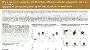 科学海报Stroma-Free, Serum-Free Expansion and Differentiation of Hematopoietic Stem and Progenitor Cells to the T Cell Lineage

 沪公网安备31010102008431号