Prasain N et al. (NOV 2014)
Nature biotechnology 32 11 1151--1157
Differentiation of human pluripotent stem cells to cells similar to cord-blood endothelial colony-forming cells.
The ability to differentiate human pluripotent stem cells into endothelial cells with properties of cord-blood endothelial colony-forming cells (CB-ECFCs) may enable the derivation of clinically relevant numbers of highly proliferative blood vessel-forming cells to restore endothelial function in patients with vascular disease. We describe a protocol to convert human induced pluripotent stem cells (hiPSCs) or embryonic stem cells (hESCs) into cells similar to CB-ECFCs at an efficiency of textgreater10(8) ECFCs produced from each starting pluripotent stem cell. The CB-ECFC-like cells display a stable endothelial phenotype with high clonal proliferative potential and the capacity to form human vessels in mice and to repair the ischemic mouse retina and limb,and they lack teratoma formation potential. We identify Neuropilin-1 (NRP-1)-mediated activation of KDR signaling through VEGF165 as a critical mechanism for the emergence and maintenance of CB-ECFC-like cells.
View Publication
产品类型:
产品号#:
05850
05857
05870
05875
85850
85857
85870
85875
产品名:
mTeSR™1
mTeSR™1
Wang L-S et al. (FEB 2010)
Biomaterials 31 6 1148--57
Injectable biodegradable hydrogels with tunable mechanical properties for the stimulation of neurogenesic differentiation of human mesenchymal stem cells in 3D culture.
We report an injectable hydrogel scaffold system with tunable stiffness for controlling the proliferation rate and differentiation of human mesenchymal stem cells (hMSCs) in a three-dimensional (3D) context in normal growth media. The hydrogels composed of gelatin-hydroxyphenylpropionic acid (Gtn-HPA) conjugate were formed using the oxidative coupling of HPA moieties catalyzed by hydrogen peroxide (H(2)O(2)) and horseradish peroxidase (HRP). The stiffness of the hydrogels was readily tuned by varying the H(2)O(2) concentration without changing the concentration of polymer precursor. We found that the hydrogel stiffness strongly affected the cell proliferation rates. The rate of hMSC proliferation increased with the decrease in the stiffness of the hydrogel. Also,the neurogenesis of hMSCs was controlled by the hydrogel stiffness in a 3D context without the use of any additional biochemical signal. These cells which were cultured in hydrogels with lower stiffness for 3 weeks expressed much more neuronal protein markers compared to those cultured within stiffer hydrogels for the same period of time.
View Publication
产品类型:
产品号#:
05401
05402
05411
产品名:
MesenCult™ MSC 基础培养基(人)
MesenCult™ MSC刺激添加物(人)
MesenCult™ 增殖试剂盒(人)
Carlson AL et al. (AUG 2012)
FASEB journal : official publication of the Federation of American Societies for Experimental Biology 26 8 3240--51
Microfibrous substrate geometry as a critical trigger for organization, self-renewal, and differentiation of human embryonic stem cells within synthetic 3-dimensional microenvironments.
Substrates used to culture human embryonic stem cells (hESCs) are typically 2-dimensional (2-D) in nature,with limited ability to recapitulate in vivo-like 3-dimensional (3-D) microenvironments. We examined critical determinants of hESC self-renewal in poly-d-lysine-pretreated synthetic polymer-based substrates with variable microgeometries,including planar 2-D films,macroporous 3-D sponges,and microfibrous 3-D fiber mats. Completely synthetic 2-D substrates and 3-D macroporous scaffolds failed to retain hESCs or support self-renewal or differentiation. However,synthetic microfibrous geometries made from electrospun polymer fibers were found to promote cell adhesion,viability,proliferation,self-renewal,and directed differentiation of hESCs in the absence of any exogenous matrix proteins. Mechanistic studies of hESC adhesion within microfibrous scaffolds indicated that enhanced cell confinement in such geometries increased cell-cell contacts and altered colony organization. Moreover,the microfibrous scaffolds also induced hESCs to deposit and organize extracellular matrix proteins like laminin such that the distribution of laminin was more closely associated with the cells than the Matrigel treatment,where the laminin remained associated with the coated fibers. The production of and binding to laminin was critical for formation of viable hESC colonies on synthetic fibrous scaffolds. Thus,synthetic substrates with specific 3-D microgeometries can support hESC colony formation,self-renewal,and directed differentiation to multiple lineages while obviating the stringent needs for complex,exogenous matrices. Similar scaffolds could serve as tools for developmental biology studies in 3-D and for stem cell differentiation in situ and transplantation using defined humanized conditions.
View Publication
产品类型:
产品号#:
05850
05857
05870
05875
85850
85857
85870
85875
产品名:
mTeSR™1
mTeSR™1
Chlon TM et al. (OCT 2014)
Journal of virology 88 19 11315--11326
High-risk human papillomavirus E6 protein promotes reprogramming of Fanconi anemia patient cells through repression of p53 but does not allow for sustained growth of induced pluripotent stem cells.
DNA repair plays a crucial role in embryonic and somatic stem cell biology and cell reprogramming. The Fanconi anemia (FA) pathway,which promotes error-free repair of DNA double-strand breaks,is required for somatic cell reprogramming to induced pluripotent stem cells (iPSC). Thus,cells from Fanconi anemia patients,which lack this critical pathway,fail to be reprogrammed to iPSC under standard conditions unless the defective FA gene is complemented. In this study,we utilized the oncogenes of high-risk human papillomavirus 16 (HPV16) to overcome the resistance of FA patient cells to reprogramming. We found that E6,but not E7,recovers FA iPSC colony formation and,furthermore,that p53 inhibition is necessary and sufficient for this activity. The iPSC colonies resulting from each of these approaches stained positive for alkaline phosphatase,NANOG,and Tra-1-60,indicating that they were fully reprogrammed into pluripotent cells. However,FA iPSC were incapable of outgrowth into stable iPSC lines regardless of p53 suppression,whereas their FA-complemented counterparts grew efficiently. Thus,we conclude that the FA pathway is required for the growth of iPSC beyond reprogramming and that p53-independent mechanisms are involved. IMPORTANCE A novel approach is described whereby HPV oncogenes are used as tools to uncover DNA repair-related molecular mechanisms affecting somatic cell reprogramming. The findings indicate that p53-dependent mechanisms block FA cells from reprogramming but also uncover a previously unrecognized defect in FA iPSC proliferation independent of p53.
View Publication
产品类型:
产品号#:
05850
05857
05870
05875
85850
85857
85870
85875
产品名:
mTeSR™1
mTeSR™1
Gerrits A et al. (APR 2010)
Blood 115 13 2610--8
Cellular barcoding tool for clonal analysis in the hematopoietic system.
Clonal analysis is important for many areas of hematopoietic stem cell research,including in vitro cell expansion,gene therapy,and cancer progression and treatment. A common approach to measure clonality of retrovirally transduced cells is to perform integration site analysis using Southern blotting or polymerase chain reaction-based methods. Although these methods are useful in principle,they generally provide a low-resolution,biased,and incomplete assessment of clonality. To overcome those limitations,we labeled retroviral vectors with random sequence tags or barcodes." On integration�
View Publication
产品类型:
产品号#:
09600
09650
产品名:
StemSpan™ SFEM
StemSpan™ SFEM
Qué et al. (JUN 2011)
Blood 117 22 5918--30
Smad4 binds Hoxa9 in the cytoplasm and protects primitive hematopoietic cells against nuclear activation by Hoxa9 and leukemia transformation.
We studied leukemic stem cells (LSCs) in a Smad4(-/-) mouse model of acute myelogenous leukemia (AML) induced either by the HOXA9 gene or by the fusion oncogene NUP98-HOXA9. Although Hoxa9-Smad4 complexes accumulate in the cytoplasm of normal hematopoietic stem cells and progenitor cells (HSPCs) transduced with these oncogenes,there is no cytoplasmic stabilization of HOXA9 in Smad4(-/-) HSPCs,and as a consequence increased levels of Hoxa9 is observed in the nucleus leading to increased immortalization in vitro. Loss of Smad4 accelerates the development of leukemia in vivo because of an increase in transformation of HSPCs. Therefore,the cytoplasmic binding of Hoxa9 by Smad4 is a mechanism to protect Hoxa9-induced transformation of normal HSPCs. Because Smad4 is a potent tumor suppressor involved in growth control,we developed a strategy to modify the subcellular distribution of Smad4. We successfully disrupted the interaction between Hoxa9 and Smad4 to activate the TGF-β pathway and apoptosis,leading to a loss of LSCs. Together,these findings reveal a major role for Smad4 in the negative regulation of leukemia initiation and maintenance induced by HOXA9/NUP98-HOXA9 and provide strong evidence that antagonizing Smad4 stabilization by these oncoproteins might be a promising novel therapeutic approach in leukemia.
View Publication
产品类型:
产品号#:
03434
03444
03236
产品名:
MethoCult™ GF M3434
MethoCult™ GF M3434
MethoCult™ SF M3236
Ortiz-Sá et al. (JAN 2009)
Leukemia 23 1 59--70
Enhanced cytotoxicity of an anti-transferrin receptor IgG3-avidin fusion protein in combination with gambogic acid against human malignant hematopoietic cells: functional relevance of iron, the receptor, and reactive oxygen species.
The human transferrin receptor (hTfR) is a target for cancer immunotherapy due to its overexpression on the surface of cancer cells. We previously developed an antibody-avidin fusion protein that targets hTfR (anti-hTfR IgG3-Av) and exhibits intrinsic cytotoxicity against certain malignant cells. Gambogic acid (GA),a drug that also binds hTfR,induces cytotoxicity in several malignant cell lines. We now report that anti-hTfR IgG3-Av and GA induce cytotoxicity in a new broader panel of hematopoietic malignant cell lines. Our results show that the effect of anti-hTfR IgG3-Av is iron-dependent whereas that of GA is iron-independent in all cells tested. In addition,we observed that GA exerts a TfR-independent cytotoxicity. We also found that GA increases the generation of reactive oxygen species that may play a role in the cytotoxicity induced by this drug. Additive cytotoxicity was observed by simultaneous combination treatment with these drugs and synergy by using anti-hTfR IgG3-Av as a chemosensitizing agent. In addition,we found a concentration of GA that is toxic to malignant hematopoietic cells but not to human hematopoietic progenitor cells. Our results suggest that these two compounds may be effective,alone or in combination,for the treatment of human hematopoietic malignancies.
View Publication
产品类型:
产品号#:
04434
04444
产品名:
MethoCult™ H4434 Classic
MethoCult™ H4434 Classic
Deglincerti A et al. (NOV 2016)
Nature protocols 11 11 2223--2232
Self-organization of human embryonic stem cells on micropatterns.
Fate allocation in the gastrulating embryo is spatially organized as cells differentiate into specialized cell types depending on their positions with respect to the body axes. There is a need for in vitro protocols that allow the study of spatial organization associated with this developmental transition. Although embryoid bodies and organoids can exhibit some spatial organization of differentiated cells,methods that generate embryoid bodies or organoids do not yield consistent and fully reproducible results. Here,we describe a micropatterning approach in which human embryonic stem cells are confined to disk-shaped,submillimeter colonies. After 42 h of BMP4 stimulation,cells form self-organized differentiation patterns in concentric radial domains,which express specific markers associated with the embryonic germ layers,reminiscent of gastrulating embryos. Our protocol takes 3 d; it uses commercial microfabricated slides (from CYTOO),human laminin-521 (LN-521) as extracellular matrix coating,and either conditioned or chemically defined medium (mTeSR). Differentiation patterns within individual colonies can be determined by immunofluorescence and analyzed with cellular resolution. Both the size of the micropattern and the type of medium affect the patterning outcome. The protocol is appropriate for personnel with basic stem cell culture training. This protocol describes a robust platform for quantitative analysis of the mechanisms associated with pattern formation at the onset of gastrulation.
View Publication
产品类型:
产品号#:
05850
05857
05870
05875
85850
85857
85870
85875
产品名:
mTeSR™1
mTeSR™1
Fraga AM et al. (MAR 2011)
Cell Transplantation 20 3 431--40
Establishment of a Brazilian line of human embryonic stem cells in defined medium: implications for cell therapy in an ethnically diverse population.
Pluripotent human embryonic stem (hES) cells are an important experimental tool for basic and applied research,and a potential source of different tissues for transplantation. However,one important challenge for the clinical use of these cells is the issue of immunocompatibility,which may be dealt with by the establishment of hES cell banks to attend different populations. Here we describe the derivation and characterization of a line of hES cells from the Brazilian population,named BR-1,in commercial defined medium. In contrast to the other hES cell lines established in defined medium,BR-1 maintained a stable normal karyotype as determined by genomic array analysis after 6 months in continuous culture (passage 29). To our knowledge,this is the first reported line of hES cells derived in South America. We have determined its genomic ancestry and compared the HLA-profile of BR-1 and another 22 hES cell lines established elsewhere with those of the Brazilian population,finding they would match only 0.011% of those individuals. Our results highlight the challenges involved in hES cell banking for populations with a high degree of ethnic admixture.
View Publication
产品类型:
产品号#:
05850
05857
05870
05875
85850
85857
85870
85875
产品名:
mTeSR™1
mTeSR™1
Liberski AR et al. (JUL 2013)
Journal of Proteome Research 12 7 3233--3245
Adaptation of a Commonly Used, Chemically Defined Medium for Human Embryonic Stem Cells to Stable Isotope Labeling with Amino Acids in Cell Culture
Metabolic labeling with stable isotopes is a prominent technique for comparative quantitative proteomics,and stable isotope labeling with amino acids in cell culture (SILAC) is the most commonly used approach. SILAC is,however,traditionally limited to simple tissue culture regimens and only rarely employed in the context of complex culturing conditions as those required for human embryonic stem cells (hESCs). Classic hESC culture is based on the use of mouse embryonic fibroblasts (MEFs) as a feeder layer,and as a result,possible xenogeneic contamination,contribution of unlabeled amino acids by the feeders,interlaboratory variability of MEF preparation,and the overall complexity of the culture system are all of concern in conjunction with SILAC. We demonstrate a feeder-free SILAC culture system based on a customized version of a commonly used,chemically defined hESC medium developed by Ludwig et al. and commercially available as mTeSR1 [mTeSR1 is a trade mark of WiCell (Madison,WI) licensed to STEMCELL Technologies (Vancouver,Canada)]. This medium,together with adjustments to the culturing protocol,facilitates reproducible labeling that is easily scalable to the protein amounts required by proteomic work flows. It greatly enhances the usability of quantitative proteomics as a tool for the study of mechanisms underlying hESCs differentiation and self-renewal. Associated data have been deposited to the ProteomeXchange with the identifier PXD000151.
View Publication
产品类型:
产品号#:
05850
05857
05870
05875
07923
85850
85857
85870
85875
产品名:
Dispase (1 U/mL)
mTeSR™1
mTeSR™1
Sumitomo A et al. (OCT 2010)
Molecular and cellular biology 30 20 4818--27
The transcriptional mediator subunit MED1/TRAP220 in stromal cells is involved in hematopoietic stem/progenitor cell support through osteopontin expression.
MED1/TRAP220,a subunit of the transcriptional Mediator/TRAP complex,is crucial for various biological events through its interaction with distinct activators,such as nuclear receptors and GATA family activators. In hematopoiesis,MED1 plays a pivotal role in optimal nuclear receptor-mediated myelomonopoiesis and GATA-1-induced erythropoiesis. In this study,we present evidence that MED1 in stromal cells is involved in supporting hematopoietic stem and/or progenitor cells (HSPCs) through osteopontin (OPN) expression. We found that the proliferation of bone marrow (BM) cells cocultured with MED1 knockout (Med1(-/-)) mouse embryonic fibroblasts (MEFs) was significantly suppressed compared to the control. Furthermore,the number of long-term culture-initiating cells (LTC-ICs) was attenuated for BM cells cocultured with Med1(-/-) MEFs. The vitamin D receptor (VDR)- and Runx2-mediated expression of OPN,as well as Mediator recruitment to the Opn promoter,was specifically attenuated in the Med1(-/-) MEFs. Addition of OPN to these MEFs restored the growth of cocultured BM cells and the number of LTC-ICs,both of which were attenuated by the addition of the anti-OPN antibody to Med1(+/+) MEFs and to BM stromal cells. Consequently,MED1 in niche appears to play an important role in supporting HSPCs by upregulating VDR- and Runx2-mediated transcription on the Opn promoter.
View Publication

 EasySep™小鼠TIL(CD45)正选试剂盒


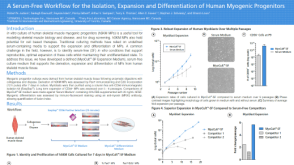 科学海报A Serum-Free Workflow for the Isolation, Expansion and Differentiation of Human Myogenic Progenitors
 沪公网安备31010102008431号