Diekmann U et al. (JAN 2015)
Stem cells and development 24 2 190--204
A reliable and efficient protocol for human pluripotent stem cell differentiation into the definitive endoderm based on dispersed single cells.
Differentiation of pluripotent cells into endoderm-related cell types initially requires in vitro gastrulation into the definitive endoderm (DE). Most differentiation protocols are initiated from colonies of pluripotent cells complicating their adaption due to insufficiently defined starting conditions. The protocol described here was initiated from a defined cell number of dispersed single cells and tested on three different human embryonic stem cell lines and one human induced pluripotent stem cell line. Combined activation of ActivinA/Nodal signaling and GSK3 inhibition for the first 24 h,followed by ActivinA/Nodal signaling efficiently induced the DE state. Activation of ActivinA/Nodal signaling alone was not effective. Efficient GSK3 inhibition allowed the reduction of the ActivinA concentration during the entire protocol. A feeder-independent cultivation of pluripotent cells was preferred to achieve the high efficiency and robustness since feeder cells hindered the differentiation process. Additionally,inhibition of the phosphatidylinositol 3-kinase (PI3K) signaling pathway was not required,nonetheless yielding high cell numbers efficiently committed toward the DE. Finally,the endoderm generated could be differentiated further into PDX1-positive pan-pancreatic cells and NGN3-positive endocrine progenitors. Thus,this efficient and robust DE differentiation protocol is a step forward toward better reproducibility due to the well-defined conditions based on dispersed single cells from feeder-free-cultivated human pluripotent cells.
View Publication
产品类型:
产品号#:
05850
05857
05870
05875
07923
07174
85850
85857
85870
85875
100-0485
100-1077
产品名:
Dispase (1 U/mL)
mTeSR™1
mTeSR™1
温和细胞解离试剂
ReLeSR™
Liu Y et al. (MAR 2015)
Journal of Biomedical Materials Research - Part A 103 3 1053--1059
Native nucleus pulposus tissue matrix promotes notochordal differentiation of human induced pluripotent stem cells with potential for treating intervertebral disc degeneration
Native porcine nucleus pulposus (NP) tissue harbors a number of notochordal cells (NCs). Whether the native NP matrix supports the homeostasis of notochordal cells is poorly understood. We hypothesized the NP matrix alone may contain sufficient regulatory factors and can serve as stimuli to generate notochordal cells (NCs) from human pluripotent stem cells. NCs are a promising cell sources for cell-based therapy to treat some types of intervertebral disc (IVD) degeneration. One major limitation of this emerging technique is the lack of available NCs as a potential therapeutic cell source. Human pluripotent stem cells derived from reprogramming or somatic cell nuclear transfer technique may yield stable and unlimited source for therapeutic use. We devised a new method to use porcine NP matrix to direct notochordal differentiation of human induced pluripotent stem cells (hiPSCs). The results showed that hiPSCs successfully differentiated into NC-like cells under the influence of devitalized porcine NP matrix. The NC-like cells expressed typical notochordal marker genes including brachyury (T),cytokeratin-8 (CK-8) and cytokeratin-18 (CK-18),and they displayed the ability to generate NP-like tissue in vitro,which was rich in aggrecan and collagen type II. These findings demonstrated the proof of concept for using native NP matrix to direct notochordal differentiation of hiPSCs. It provides a foundation for further understanding the biology of NCs,and eventually towards regenerative therapies for disc degeneration.
View Publication
Bone HK et al. (JUN 2011)
Journal of cell science 124 Pt 12 1992--2000
A novel chemically directed route for the generation of definitive endoderm from human embryonic stem cells based on inhibition of GSK-3.
The use of small molecules to 'chemically direct' differentiation represents a powerful approach to promote specification of embryonic stem cells (ESCs) towards particular functional cell types for use in regenerative medicine and pharmaceutical applications. Here,we demonstrate a novel route for chemically directed differentiation of human ESCs (hESCs) into definitive endoderm (DE) exploiting a selective small-molecule inhibitor of glycogen synthase kinase 3 (GSK-3). This GSK-3 inhibitor,termed 1m,when used as the only supplement to a chemically defined feeder-free culture system,effectively promoted differentiation of ESC lines towards primitive streak (PS),mesoderm and DE. This contrasts with the role of GSK-3 in murine ESCs,where GSK-3 inhibition promotes pluripotency. Interestingly,1m-mediated induction of differentiation involved transient NODAL expression and Nodal signalling. Prolonged treatment of hESCs with 1m resulted in the generation of a population of cells displaying hepatoblast characteristics,that is expressing α-fetoprotein and HNF4α. Furthermore,1m-induced DE had the capacity to mature and generate hepatocyte-like cells capable of producing albumin. These findings describe,for the first time,the utility of GSK-3 inhibition,in a chemically directed approach,to a method of DE generation that is robust,potentially scalable and applicable to different hESC lines.
View Publication
产品类型:
产品号#:
05850
05857
05870
05875
85850
85857
85870
85875
产品名:
mTeSR™1
mTeSR™1
Hu Y-L et al. (SEP 2010)
Nucleic acids research 38 16 5472--8
HOXA9 regulates miR-155 in hematopoietic cells.
HOXA9-mediated up-regulation of miR-155 was noted during an array-based analysis of microRNA expression in Hoxa9(-/-)bone marrow (BM) cells. HOXA9 induction of miR-155 was confirmed in these samples,as well as in wild-type versus Hoxa9-deficient marrow,using northern analysis and qRT-PCR. Infection of wild-type BM with HOXA9 expressing or GFP(+) control virus further confirmed HOXA9-mediated regulation of miR-155. miR-155 expression paralleled Hoxa9 mRNA expression in fractionated BM progenitors,being highest in the stem cell enriched pools. HOXA9 capacity to induce myeloid colony formation was blunted in miR-155-deficient BM cells,indicating that miR-155 is a downstream mediator of HOXA9 function in blood cells. Pu.1,an important regulator of myelopoiesis,was identified as a putative down stream target for miR-155. Although miR-155 was shown to down-regulate the Pu.1 protein,HOXA9 did not appear to modulate Pu.1 expression in murine BM cells.
View Publication
产品类型:
产品号#:
03434
03444
产品名:
MethoCult™ GF M3434
MethoCult™ GF M3434
Yang L et al. ( 2014)
1114 245--267
CRISPR-cas-mediated targeted genome editing in human cells
The clustered regularly interspaced short palindromic repeats (CRISPR) and CRISPR-associated (Cas) systems have evolved as an adaptive surveillance and defense mechanism in bacteria and archaea that uses short RNAs to direct degradation of foreign genetic elements. Here,we present our protocol for utilizing the S. pyogenes type II bacterial CRISPR system to achieve sequence-specific genome alterations in human cells. In principle,any genomic sequence of the form N(19)NGG can be targeted with the generation of custom guide RNA (gRNA) which functions to direct the Cas9 protein to genomic targets and induce DNA cleavage. Here,we describe our methods for designing and generating gRNA expression constructs either singly or in a multiplexed manner,as well as optimized protocols for the delivery of Cas9-gRNA components into human cells. Genomic alterations at the target site are then introduced either through nonhomologous end joining (NHEJ) or through homologous recombination (HR) in the presence of an appropriate donor sequence. This RNA-guided editing tool offers greater ease of customization and synthesis in comparison to existing sequence-specific endonucleases and promises to become a highly versatile and multiplexable human genome engineering platform.
View Publication
产品类型:
产品号#:
05850
05857
05870
05875
85850
85857
85870
85875
产品名:
mTeSR™1
mTeSR™1
Sun Y et al. (MAR 2010)
Blood 115 9 1709--17
Slug deficiency enhances self-renewal of hematopoietic stem cells during hematopoietic regeneration.
Both extrinsic and intrinsic mechanisms tightly govern hematopoietic stem cell (HSC) decisions of self-renewal and differentiation. However,transcription factors that can selectively regulate HSC self-renewal division after stress remain to be identified. Slug is an evolutionarily conserved zinc-finger transcription factor that is highly expressed in primitive hematopoietic cells and is critical for the radioprotection of these key cells. We studied the effect of Slug in the regulation of HSCs in Slug-deficient mice under normal and stress conditions using serial functional assays. Here,we show that Slug deficiency does not disturb hematopoiesis or alter HSC homeostasis and differentiation in bone marrow but increases the numbers of primitive hematopoietic cells in the extramedullary spleen site. Deletion of Slug enhances HSC repopulating potential but not its homing and differentiation ability. Furthermore,Slug deficiency increases HSC proliferation and repopulating potential in vivo after myelosuppression and accelerates HSC expansion during in vitro culture. Therefore,we propose that Slug is essential for controlling the transition of HSCs from relative quiescence under steady-state condition to rapid proliferation under stress conditions. Our data suggest that inhibition of Slug in HSCs may present a novel strategy for accelerating hematopoietic recovery,thus providing therapeutic benefits for patients after clinical myelosuppressive treatment.
View Publication
产品类型:
产品号#:
09600
09650
28600
产品名:
StemSpan™ SFEM
StemSpan™ SFEM
L-Calc™有限稀释软件
Tropel P et al. (MAY 2017)
Stem cells and development
CpG island methylation correlates with the use of alternative promoter for USP44 gene expression in human pluripotent stem cells and testis.
Deubiquitinating enzymes may play a major regulatory role in pluripotent stem cells (PSCs) but few studies have investigated this topic. Within this family of enzymes,we found that the ubiquitin specific peptidase,USP44,is highly expressed in embryonic stem cells,induced PSCs and testes as compared to differentiated progenies and somatic organs. Analysis by qPCR and 5'RACE showed that alternate promoters are responsible for expression in PSCs and organs. We noticed 7 regions of transcription initiation,some of them with cell- or tissue-specific activity. Close analysis showed that one of the promoters involved in stem cell and testis-specific activity is differentially regulated in those tissues. At the epigenetic level,USP44 transcription was correlated with DNA methylation of a CpG island close to the main promoter region. These data imply a complex picture where regulating factors like OCT4 may interact with other epigenetic mechanisms to regulate USP44 expression in PSCs and testes.
View Publication
产品类型:
产品号#:
05850
05857
05870
05875
85850
85857
85870
85875
产品名:
mTeSR™1
mTeSR™1
Baksh D et al. (NOV 2005)
Blood 106 9 3012--9
Soluble factor cross-talk between human bone marrow-derived hematopoietic and mesenchymal cells enhances in vitro CFU-F and CFU-O growth and reveals heterogeneity in the mesenchymal progenitor cell compartment.
The homeostatic adult bone marrow (BM) is a complex tissue wherein physical and biochemical interactions serve to maintain a balance between the hematopoietic and nonhematopoietic compartments. To focus on soluble factor interactions occurring between mesenchymal and hematopoietic cells,a serum-free adhesion-independent culture system was developed that allows manipulation of the growth of both mesenchymal and hematopoietic human BM-derived progenitors and the balance between these compartments. Factorial experiments demonstrated a role for stem cell factor (SCF) and interleukin 3 (IL-3) in the concomitant growth of hematopoietic (CD45+) and nonhematopoietic (CD45-) cells,as well as their derivatives. Kinetic tracking of IL-3alpha receptor (CD123) and SCF receptor (CD117) expression on a sorted CD45- cell population revealed the emergence of CD45-CD123+ cells capable of osteogenesis. Of the total fibroblast colony-forming units (CFU-Fs) and osteoblast colony-forming units (CFU-O),approximately 24% of CFU-Fs and about 22% of CFU-Os were recovered from this population. Cell-sorting experiments demonstrated that the CD45+ cell population secreted soluble factors that positively affect the survival and proliferation of CFU-Fs and CFU-Os generated from the CD45- cells. Together,our results provide insight into the intercellular cytokine network between hematopoietic and mesenchymal cells and provide a strategy to mutually culture both mesenchymal and hematopoietic cells in a defined scalable bioprocess.
View Publication
产品类型:
产品号#:
09850
产品名:
Huber BC et al. (NOV 2013)
STEM CELLS 31 11 2354--2363
Costimulation-adhesion blockade is superior to Cyclosporine A and prednisone immunosuppressive therapy for preventing rejection of differentiated human embryonic stem cells following transplantation
RATIONALE: Human embryonic stem cell (hESC) derivatives are attractive candidates for therapeutic use. The engraftment and survival of hESC derivatives as xenografts or allografts require effective immunosuppression to prevent immune cell infiltration and graft destruction.backslashnbackslashnOBJECTIVE: To test the hypothesis that a short-course,dual-agent regimen of two costimulation-adhesion blockade agents can induce better engraftment of hESC derivatives compared to current immunosuppressive agents.backslashnbackslashnMETHODS AND RESULTS: We transduced hESCs with a double fusion reporter gene construct expressing firefly luciferase (Fluc) and enhanced green fluorescent protein,and differentiated these cells to endothelial cells (hESC-ECs). Reporter gene expression enabled longitudinal assessment of cell engraftment by bioluminescence imaging. Costimulation-adhesion therapy resulted in superior hESC-EC and mouse EC engraftment compared to cyclosporine therapy in a hind limb model. Costimulation-adhesion therapy also promoted robust hESC-EC and hESC-derived cardiomyocyte survival in an ischemic myocardial injury model. Improved hESC-EC engraftment had a cardioprotective effect after myocardial injury,as assessed by magnetic resonance imaging. Mechanistically,costimulation-adhesion therapy is associated with systemic and intragraft upregulation of T-cell immunoglobulin and mucin domain 3 (TIM3) and a reduced proinflammatory cytokine profile.backslashnbackslashnCONCLUSIONS: Costimulation-adhesion therapy is a superior alternative to current clinical immunosuppressive strategies for preventing the post-transplant rejection of hESC derivatives. By extending the window for cellular engraftment,costimulation-adhesion therapy enhances functional preservation following ischemic injury. This regimen may function through a TIM3-dependent mechanism.
View Publication
产品类型:
产品号#:
05850
05857
05870
05875
85850
85857
85870
85875
产品名:
mTeSR™1
mTeSR™1
Wu J et al. (APR 2015)
Stem cells and development 24 7 892--903
Increased culture density is linked to decelerated proliferation, prolonged G1 phase, and enhanced propensity for differentiation of self-renewing human pluripotent stem cells.
Human pluripotent stem cells (hPSCs) display a very short G1 phase and rapid proliferation kinetics. Regulation of the cell cycle,which is linked to pluripotency and differentiation,is dependent on the stem cell environment,particularly on culture density. This link has been so far empirical and central to disparities in the growth rates and fractions of self-renewing hPSCs residing in different cycle phases. In this study,hPSC cycle progression in conjunction with proliferation and differentiation were comprehensively investigated for different culture densities. Cell proliferation decelerated significantly at densities beyond 50×10(4) cells/cm(2). Correspondingly,the G1 fraction increased from 25% up to 60% at densities greater than 40×10(4) cells/cm(2) while still hPSC pluripotency marker expression was maintained. In parallel,expression of the cycle inhibitor CDKN1A (p21) was increased,while that of p27 and p53 did not change significantly. After 4 days of culture in an unconditioned medium,greater heterogeneity was noted in the differentiation outcomes and was limited by reducing the density variation. A quantitative model was constructed for self-renewing and differentiating hPSC ensembles to gain a better understanding of the link between culture density,cycle progression,and stem cell state. Results for multiple hPSC lines and medium types corroborated experimental findings. Media commonly used for maintenance of self-renewing hPSCs exhibited the slowest kinetics of induction of differentiation (kdiff),while BMP4 supplementation led to 14-fold higher kdiff values. Spontaneous differentiation in a growth factor-free medium exhibited the largest variation in outcomes at different densities. In conjunction with the quantitative framework,our findings will facilitate rationalizing the selection of cultivation conditions for the generation of stem cell therapeutics.
View Publication

 EasySep™小鼠TIL(CD45)正选试剂盒


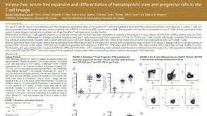 科学海报Stroma-Free, Serum-Free Expansion and Differentiation of Hematopoietic Stem and Progenitor Cells to the T Cell Lineage
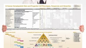 挂图Human Hematopoietic Stem and Progenitor Cell Phenotyping Overview of subset surface markers, frequencies and assays for analysis
 沪公网安备31010102008431号