Arbab AS et al. (SEP 2008)
FASEB journal : official publication of the Federation of American Societies for Experimental Biology 22 9 3234--46
Detection of migration of locally implanted AC133+ stem cells by cellular magnetic resonance imaging with histological findings.
This study investigated the factors responsible for migration and homing of magnetically labeled AC133(+) cells at the sites of active angiogenesis in tumor. AC133(+) cells labeled with ferumoxide-protamine sulfate were mixed with either rat glioma or human melanoma cells and implanted in flank of nude mice. An MRI of the tumors including surrounding tissues was performed. Tumor sections were stained for Prussian blue (PB),platelet-derived growth factor (PDGF),hypoxia-inducible factor-1alpha (HIF-1alpha),stromal cell derived factor-1 (SDF-1),matrix metalloproteinase-2 (MMP-2),vascular endothelial growth factor (VEGF),and endothelial markers. Fresh snap-frozen strips from the central and peripheral parts of the tumor were collected for Western blotting. MRIs demonstrated hypointense regions at the periphery of the tumors where the PB(+)/AC133(+) cells were positive for endothelial cells markers. At the sites of PB(+)/AC133(+) cells,both HIF-1alpha and SDF-1 were strongly positive and PDGF and MMP-2 showed generalized expression in the tumor and surrounding tissues. There was no significant association of PB(+)/AC133(+) cell localization and VEGF expression in tumor cells. Western blot demonstrated strong expression of the SDF-1,MMP-2,and PDGF at the peripheral parts of the tumors. HIF-1alpha was expressed at both the periphery and central parts of the tumor. This work demonstrates that magnetically labeled cells can be used as probes for MRI and histological identification of administered cells.
View Publication
产品类型:
产品号#:
09600
09650
产品名:
StemSpan™ SFEM
StemSpan™ SFEM
Luo M et al. (JAN 2009)
Cancer research 69 2 466--74
Mammary epithelial-specific ablation of the focal adhesion kinase suppresses mammary tumorigenesis by affecting mammary cancer stem/progenitor cells.
Focal adhesion kinase (FAK) has been implicated in the development of cancers,including those of the breast. Nevertheless,the molecular and cellular mechanisms by which FAK promotes mammary tumorigenesis in vivo are not well understood. Here,we show that targeted deletion of FAK in mouse mammary epithelium significantly suppresses mammary tumorigenesis in a well-characterized breast cancer model. Ablation of FAK leads to the depletion of a subset of bipotent cells in the tumor that express both luminal marker keratin 8/18 and basal marker keratin 5. Using mammary stem/progenitor markers,including aldehyde dehydrogenase,CD24,CD29,and CD61,we further revealed that ablation of FAK reduced the pool of cancer stem/progenitor cells in primary tumors of FAK-targeted mice and impaired their self-renewal and migration in vitro. Finally,through transplantation in NOD-SCID mice,we found that cancer stem/progenitor cells isolated from FAK-targeted mice have compromised tumorigenicity and impaired maintenance in vivo. Together,these results show a novel function of FAK in maintaining the mammary cancer stem/progenitor cell population and provide a novel mechanism by which FAK may promote breast cancer development and progression.
View Publication
产品类型:
产品号#:
18556
18556RF
产品名:
Charafe-Jauffret E et al. (FEB 2009)
Cancer research 69 4 1302--13
Breast cancer cell lines contain functional cancer stem cells with metastatic capacity and a distinct molecular signature.
Tumors may be initiated and maintained by a cellular subcomponent that displays stem cell properties. We have used the expression of aldehyde dehydrogenase as assessed by the ALDEFLUOR assay to isolate and characterize cancer stem cell (CSC) populations in 33 cell lines derived from normal and malignant mammary tissue. Twenty-three of the 33 cell lines contained an ALDEFLUOR-positive population that displayed stem cell properties in vitro and in NOD/SCID xenografts. Gene expression profiling identified a 413-gene CSC profile that included genes known to play a role in stem cell function,as well as genes such as CXCR1/IL-8RA not previously known to play such a role. Recombinant interleukin-8 (IL-8) increased mammosphere formation and the ALDEFLUOR-positive population in breast cancer cell lines. Finally,we show that ALDEFLUOR-positive cells are responsible for mediating metastasis. These studies confirm the hierarchical organization of immortalized cell lines,establish techniques that can facilitate the characterization of regulatory pathways of CSCs,and identify potential stem cell markers and therapeutic targets.
View Publication
Gene expression profiling and localization of Hoechst-effluxing CD45- and CD45+ cells in the embryonic mouse lung.
Hoechst-effluxing cells (side population cells) are a rare subset of cells found in adult tissues that are highly enriched for stem and progenitor cell activity. To identify potential stem and progenitor cells during lung development,we generated gene expression profiles for CD45- and CD45+ side population cells in the embryonic day 17.5 lung. We found that side population cells comprise 1% of total embryonic day 17.5 lung cells (55% CD45+,45% CD45-). Gene profiling data demonstrated an overrepresentation of endothelial genes within the CD45- side population. We used expression of several distinct genes to identify two types of CD45- side population cells: 1) von Willebrand factor+/smooth muscle actin+ cells that reside in the muscular layer of select large vessels and 2) von Willebrand factor+/intercellular adhesion molecule+ cells that reside within the endothelial layer of select small vessels. Gene profiling of the CD45+ side population indicated an overrepresentation of genes associated with myeloid cell differentiation. Consistent with this,culturing CD45+ side population cells was associated with induction of mature dendritic markers (CD86). The microarray results suggested that expression of myeloperoxidase and proteinase-3 might be used to identify CD45+ side population cells. By immunohistochemistry,we found that myeloperoxidase+/proteinase-3+ cells represent a small subset of total CD45+ cells in the embryonic day 17.5 lung and that they reside in the mesenchyme and perivascular regions. This is the first detailed information regarding the phenotype and localization of side population cells in a developing organ.
View Publication
产品类型:
产品号#:
09600
09650
产品名:
StemSpan™ SFEM
StemSpan™ SFEM
Cardoso SC et al. (JAN 2011)
PLoS ONE 6 12 e29244
Synchrotron radiation X-ray microfluorescence reveals polarized distribution of atomic elements during differentiation of pluripotent stem cells.
The mechanisms underlying pluripotency and differentiation in embryonic and reprogrammed stem cells are unclear. In this work,we characterized the pluripotent state towards neural differentiated state through analysis of trace elements distribution using the Synchrotron Radiation X-ray Fluorescence Spectroscopy. Naive and neural-stimulated embryoid bodies (EB) derived from embryonic and induced pluripotent stem (ES and iPS) cells were irradiated with a spatial resolution of 20 µm to make elemental maps and qualitative chemical analyses. Results show that these embryo-like aggregates exhibit self-organization at the atomic level. Metallic elements content rises and consistent elemental polarization pattern of P and S in both mouse and human pluripotent stem cells were observed,indicating that neural differentiation and elemental polarization are strongly correlated.
View Publication
Gamma-catenin contributes to leukemogenesis induced by AML-associated translocation products by increasing the self-renewal of very primitive progenitor cells.
Acute myeloid leukemia (AML) is characterized by the block of differentiation,deregulated apoptosis,and an increased self-renewal of hematopoietic precursors. It is unclear whether the self-renewal of leukemic blasts results from the cumulative effects of blocked differentiation and impaired apoptosis or whether there are mechanisms directly increasing self-renewal. The AML-associated translocation products (AATPs) promyelocytic leukemia/retinoic acid receptor alpha (PML/RAR alpha),promyelocytic leukemia zinc finger (PLZF)/RAR alpha (X-RAR alpha),and AML-1/ETO block hematopoietic differentiation. The AATPs activate the Wnt signaling by up-regulating gamma-catenin. Activation of the Wnt signaling augments self-renewal of hematopoietic stem cells (HSCs). Therefore,we investigated how AATPs influence self-renewal of HSCs and evaluated the role of gamma-catenin in the determination of the phenotype of HSCs expressing AATPs. Here we show that the AATPs directly activate the gamma-catenin promoter. The crucial role of gamma-catenin in increasing the self-renewal of HSCs upon expression of AATPs is demonstrated by (i) the abrogation of replating efficiency upon hindrance of gamma-catenin expression through RNA interference,and (ii) the augmentation of replating efficiency of HSCs upon overexpression of gamma-catenin itself. In addition,the inoculation of gamma-catenin-transduced HSCs into irradiated recipient mice establishes the clinical picture of AML. These data provide the first evidence that the aberrant activation of Wnt signaling by the AATP decisively contributes to the pathogenesis of AML.
View Publication
产品类型:
产品号#:
03434
03444
产品名:
MethoCult™ GF M3434
MethoCult™ GF M3434
Baens M et al. (MAY 2006)
Cancer research 66 10 5270--7
Selective expansion of marginal zone B cells in Emicro-API2-MALT1 mice is linked to enhanced IkappaB kinase gamma polyubiquitination.
The translocation t(11;18)(q21;q21) that generates an API2-MALT1 fusion protein is the most common structural abnormality among the genetic defects reported in mucosa-associated lymphoid tissue (MALT)-type lymphomas,and its presence correlates with the apparent lack of further genetic instability or chromosomal imbalances. Hence,constitutive nuclear factor-kappaB (NF-kappaB) activation induced by the API2-MALT1 fusion protein is considered essential for B-cell transformation. To examine its role in B-cell development and lymphomagenesis,Emu-API2-MALT1 transgenic mice were produced. Our data show that expression of the API2-MALT1 fusion protein alone is not sufficient for the development of lymphoma masses within 50 weeks. Nevertheless,API2-MALT1 expression affected B-cell maturation in the bone marrow and triggered the specific expansion of splenic marginal zone B cells. Polyubiquitination of IkappaB kinase gamma (IKKgamma),indicative for enhanced NF-kappaB activation,was increased in splenic lymphocytes and promoted the survival of B cells ex vivo. In addition,we show that the API2-MALT1 fusion resided in the cholesterol- and sphingolipid-enriched membrane microdomains,termed lipid rafts. We provide evidence that association of the MALT1 COOH terminal with the lipid rafts,which is mediated by the API2 portion,is sufficient to trigger NF-kappaB activation via enhanced polyubiquitination of IKKgamma. Taken together,these data support the hypothesis that the API2-MALT1 fusion protein can contribute to MALT lymphoma formation via increased NF-kappaB activation.
View Publication
产品类型:
产品号#:
03630
产品名:
MethoCult™ M3630
Mü et al. (SEP 2012)
Stem Cell Reviews and Reports 8 3 720--740
Ca2+ Activated K Channels-New Tools to Induce Cardiac Commitment from Pluripotent Stem Cells in Mice and Men
Bruserud &O et al. (MAR 2007)
Haematologica 92 3 332--41
Subclassification of patients with acute myelogenous leukemia based on chemokine responsiveness and constitutive chemokine release by their leukemic cells.
BACKGROUND AND OBJECTIVES: Chemokines are soluble mediators involved in angiogenesis,cellular growth control and immunomodulation. In the present study we investigated the effects of various chemokines on proliferation of acute myelogenous leukemia (AML) cells and constitutive chemokine release by primary AML cells. DESIGN AND METHODS: Native human AML cells derived from 68 consecutive patients were cultured in vitro. We investigated AML cell proliferation (3H-thymidine incorporation,colony formation),chemokine receptor expression,constitutive chemokine release and chemotaxis of normal peripheral blood mononuclear cells. RESULTS: Exogenous chemokines usually did not have any effect on AML blast proliferation in the absence of hematopoietic growth factors,but when investigating growth factor-dependent (interleukin 3 + granulocyte-macrophage colony-stimulating factor + stem cell factor) proliferation in suspension cultures the following patient subsets were identified: (i) patients whose cells showed chemokine-induced growth enhancement (8 patients); (ii) divergent effects on proliferation (15 patients); and (iii) no effect (most patients). These patient subsets did not differ in chemokine receptor expression,but,compared to CD34- AML cells,CD34+ cells showed higher expression of several receptors. Chemokines also increased the proliferation of clonogenic AML cells from the first subset of patients. Furthermore,a broad constitutive chemokine release profile was detected for most patients,and the following chemokine clusters could be identified: CCL2-4/CXCL1/8,CCL5/CXCL9-11 (possibly also CCL23) and CCL13/17/22/24/CXCL5 (possibly also CXCL6). Only the CCL2-4/CXCL1/8 cluster showed significant correlations between corresponding mRNA levels and NFkB levels/activation. The chemotaxis of normal immunocompetent cells for patients without constitutive chemokine release was observed to be decreased. INTERPRETATION AND CONCLUSIONS: Differences in chemokine responsiveness as well as chemokine release contribute to patient heterogeneity in AML. Patients with AML can be classified into distinct subsets according to their chemokine responsiveness and chemokine release profile.
View Publication
产品类型:
产品号#:
04434
04444
09600
09650
产品名:
MethoCult™ H4434 Classic
MethoCult™ H4434 Classic
StemSpan™ SFEM
StemSpan™ SFEM
Ma N et al. (MAY 2015)
Journal of Biological Chemistry 290 19 12079--12089
Factor-induced Reprogramming and Zinc Finger Nuclease-aided Gene Targeting Cause Different Genome Instability in $\$-Thalassemia Induced Pluripotent Stem Cells (iPSCs).
The generation of personalized induced pluripotent stem cells (iPSCs) followed by targeted genome editing provides an opportunity for developing customized effective cellular therapies for genetic disorders. However,it is critical to ascertain whether edited iPSCs harbor unfavorable genomic variations before their clinical application. To examine the mutation status of the edited iPSC genome and trace the origin of possible mutations at different steps,we have generated virus-free iPSCs from amniotic cells carrying homozygous point mutations in beta-hemoglobin gene (HBB) that cause severe beta-thalassemia (beta-Thal),corrected the mutations in both HBB alleles by zinc finger nuclease-aided gene targeting,and obtained the final HBB gene-corrected iPSCs by excising the exogenous drug resistance gene with Cre recombinase. Through comparative genomic hybridization and whole-exome sequencing,we uncovered seven copy number variations,five small insertions/deletions,and 64 single nucleotide variations (SNVs) in beta-Thal iPSCs before the gene targeting step and found a single small copy number variation,19 insertions/deletions,and 340 single nucleotide variations in the final gene-corrected beta-Thal iPSCs. Our data revealed that substantial but different genomic variations occurred at factor-induced somatic cell reprogramming and zinc finger nuclease-aided gene targeting steps,suggesting that stringent genomic monitoring and selection are needed both at the time of iPSC derivation and after gene targeting.
View Publication


 EasySep™小鼠TIL(CD45)正选试剂盒





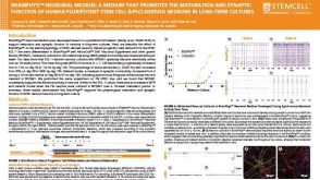 科学海报BrainPhys™ Neuronal Medium: A Medium That Promotes the Maturation and Synaptic Function of Human Pluripotent Stem Cell (hPSC)-Derived Neurons in Long-Term Cultures

 沪公网安备31010102008431号