Chen Y-X et al. (JAN 2006)
Proceedings of the National Academy of Sciences of the United States of America 103 4 1018--23
The tumor suppressor menin regulates hematopoiesis and myeloid transformation by influencing Hox gene expression.
Menin is the product of the tumor suppressor gene Men1 that is mutated in the inherited tumor syndrome multiple endocrine neoplasia type 1 (MEN1). Menin has been shown to interact with SET-1 domain-containing histone 3 lysine 4 (H3K4) methyltransferases including mixed lineage leukemia proteins to regulate homeobox (Hox) gene expression in vitro. Using conditional Men1 knockout mice,we have investigated the requirement for menin in hematopoiesis and myeloid transformation. Men1 excision causes reduction of Hoxa9 expression,colony formation by hematopoietic progenitors,and the peripheral white blood cell count. Menin directly activates Hoxa9 expression,at least in part,by binding to the Hoxa9 locus,facilitating methylation of H3K4,and recruiting the methylated H3K4 binding protein chd1 to the locus. Consistent with signaling downstream of menin,ectopic expression of both Hoxa9 and Meis1 rescues colony formation defects in Men1-excised bone marrow. Moreover,Men1 excision also suppresses proliferation of leukemogenic mixed lineage leukemia-AF9 fusion-protein-transformed myeloid cells and Hoxa9 expression. These studies uncover an important role for menin in both normal hematopoiesis and myeloid transformation and provide a mechanistic understanding of menin's function in these processes that may be used for therapy.
View Publication
产品类型:
产品号#:
03534
产品名:
MethoCult™ GF M3534
Futami M et al. (JUL 2011)
Blood 118 4 1077--86
G-CSF receptor activation of the Src kinase Lyn is mediated by Gab2 recruitment of the Shp2 phosphatase.
Src activation involves the coordinated regulation of positive and negative tyrosine phosphorylation sites. The mechanism whereby receptor tyrosine kinases,cytokine receptors,and integrins activate Src is not known. Here,we demonstrate that granulocyte colony-stimulating factor (G-CSF) activates Lyn,the predominant Src kinase in myeloid cells,through Gab2-mediated recruitment of Shp2. After G-CSF stimulation,Lyn dynamically associates with Gab2 in a spatiotemporal manner. The dephosphorylation of phospho-Lyn Tyr507 was abrogated in Shp2-deficient cells transfected with the G-CSF receptor but intact in cells expressing phosphatase-defective Shp2. Auto-phosphorylation of Lyn Tyr396 was impaired in cells treated with Gab2 siRNA. The constitutively activated Shp2E76A directed the dephosphorylation of phospho-Lyn Tyr507 in vitro. Tyr507 did not undergo dephosphorylation in G-CSF-stimulated cells expressing a mutant Gab2 unable to bind Shp2. We propose that Gab2 forms a complex with Lyn and after G-CSF stimulation,Gab2 recruits Shp2,which dephosphorylates phospho-Lyn Tyr507,leading to Lyn activation.
View Publication
Griswold IJ et al. (AUG 2006)
Molecular and cellular biology 26 16 6082--93
Kinase domain mutants of Bcr-Abl exhibit altered transformation potency, kinase activity, and substrate utilization, irrespective of sensitivity to imatinib.
Kinase domain (KD) mutations of Bcr-Abl interfering with imatinib binding are the major mechanism of acquired imatinib resistance in patients with Philadelphia chromosome-positive leukemia. Mutations of the ATP binding loop (p-loop) have been associated with a poor prognosis. We compared the transformation potency of five common KD mutants in various biological assays. Relative to unmutated (native) Bcr-Abl,the ATP binding loop mutants Y253F and E255K exhibited increased transformation potency,M351T and H396P were less potent,and the performance of T315I was assay dependent. The transformation potency of Y253F and M351T correlated with intrinsic Bcr-Abl kinase activity,whereas the kinase activity of E255K,H396P,and T315I did not correlate with transforming capabilities,suggesting that additional factors influence transformation potency. Analysis of the phosphotyrosine proteome by mass spectroscopy showed differential phosphorylation among the mutants,a finding consistent with altered substrate specificity and pathway activation. Mutations in the KD of Bcr-Abl influence kinase activity and signaling in a complex fashion,leading to gain- or loss-of-function variants. The drug resistance and transformation potency of mutants may determine the outcome of patients on therapy with Abl kinase inhibitors.
View Publication
产品类型:
产品号#:
03236
产品名:
MethoCult™ SF M3236
Wang Y et al. (MAR 2007)
Blood 109 5 2147--55
Adaptive secretion of granulocyte-macrophage colony-stimulating factor (GM-CSF) mediates imatinib and nilotinib resistance in BCR/ABL+ progenitors via JAK-2/STAT-5 pathway activation.
Overcoming imatinib mesylate (IM) resistance and disease persistence in patients with chronic myeloid leukemia (CML) is of considerable importance to the issue of potential cure. Here we asked whether autocrine signaling contributes to survival of BCR/ABL+ cells in the presence of IM and nilotinib (NI; AMN107),a novel,more selective Abl inhibitor. Conditioned media (CM) of IM-resistant LAMA84 cell clones (R-CM) was found to substantially protect IM-naive LAMA cells and primary CML progenitors from IM- or NI-induced cell death. This was due to an increased secretion of the granulocyte-macrophage colony-stimulating factor (GM-CSF),which was identified as the causative factor mediating IM resistance in R-CM. GM-CSF elicited IM and NI drug resistance via a BCR/ABL-independent activation of the janus kinases 2 (JAK-2)/signal transducer and activator of transcription 5 (STAT-5) signaling pathway in GM-CSF receptor alpha receptor (CD116)-expressing cells,including primary CD34+/CD116+ GM progenitors (GMPs). Elevated mRNA and protein levels of GM-CSF were detected in IM-resistant patient samples,suggesting a contribution of GM-CSF secretion for IM and NI resistance in vivo. Importantly,inhibition of JAK-2 with AG490 abrogated GM-CSF-mediated STAT-5 phosphorylation and NI resistance in vitro. Together,adaptive autocrine secretion of GM-CSF mediates BCR/ABL-independent IM and NI resistance via activation of the antiapoptotic JAK-2/STAT-5 pathway. Inhibition of JAK-2 overcomes GM-CSF-induced IM and NI progenitor cell resistance,providing a rationale for the application of JAK-2 inhibitors to eradicate residual disease in CML.
View Publication
产品类型:
产品号#:
04230
产品名:
MethoCult™ H4230
Feng R et al. (MAR 2007)
Blood 109 5 2130--8
SDX-308, a nonsteroidal anti-inflammatory agent, inhibits NF-kappaB activity, resulting in strong inhibition of osteoclast formation/activity and multiple myeloma cell growth.
Multiple myeloma is characterized by increased osteoclast activity that results in bone destruction and lytic lesions. With the prolonged overall patient survival achieved by new treatment modalities,additional drugs are required to inhibit bone destruction. We focused on a novel and more potent structural analog of the nonsteroidal anti-inflammatory drug etodolac,known as SDX-308,and its effects on osteoclastogenesis and multiple myeloma cells. SDX-101 is another structural analog of etodolac that is already used in clinical trials for the treatment of B-cell chronic lymphocytic leukemia (B-CLL). Compared with SDX-101,a 10-fold lower concentration of SDX-308 induced potent (60%-80%) inhibition of osteoclast formation,and a 10- to 100-fold lower concentration inhibited multiple myeloma cell proliferation. Bone resorption was completely inhibited by SDX-308,as determined in dentin-based bone resorption assays. SDX-308 decreased constitutive and RANKL-stimulated NF-kappaB activation and osteoclast formation in an osteoclast cellular model,RAW 264.7. SDX-308 effectively suppressed TNF-alpha-induced IKK-gamma and IkappaB-alpha phosphorylation and degradation and subsequent NF-kappaB activation in human multiple myeloma cells. These results indicate that SDX-308 effectively inhibits multiple myeloma cell proliferation and osteoclast activity,potentially by controlling NF-kappaB activation signaling. We propose that SDX-308 is a promising therapeutic candidate to inhibit multiple myeloma growth and osteoclast activity and that it should receive attention for further study.
View Publication
产品类型:
产品号#:
04434
04444
产品名:
MethoCult™ H4434 Classic
MethoCult™ H4434 Classic
Sjogren A-KM et al. (MAY 2007)
The Journal of clinical investigation 117 5 1294--304
GGTase-I deficiency reduces tumor formation and improves survival in mice with K-RAS-induced lung cancer.
Protein geranylgeranyltransferase type I (GGTase-I) is responsible for the posttranslational lipidation of CAAX proteins such as RHOA,RAC1,and cell division cycle 42 (CDC42). Inhibition of GGTase-I has been suggested as a strategy to treat cancer and a host of other diseases. Although several GGTase-I inhibitors (GGTIs) have been synthesized,they have very different properties,and the effects of GGTIs and GGTase-I deficiency are unclear. One concern is that inhibiting GGTase-I might lead to severe toxicity. In this study,we determined the effects of GGTase-I deficiency on cell viability and K-RAS-induced cancer development in mice. Inactivating the gene for the critical beta subunit of GGTase-I eliminated GGTase-I activity,disrupted the actin cytoskeleton,reduced cell migration,and blocked the proliferation of fibroblasts expressing oncogenic K-RAS. Moreover,the absence of GGTase-I activity reduced lung tumor formation,eliminated myeloproliferative phenotypes,and increased survival of mice in which expression of oncogenic K-RAS was switched on in lung cells and myeloid cells. Interestingly,several cell types remained viable in the absence of GGTase-I,and myelopoiesis appeared to function normally. These findings suggest that inhibiting GGTase-I may be a useful strategy to treat K-RAS-induced malignancies.
View Publication
产品类型:
产品号#:
03234
产品名:
MethoCult™ M3234
Wang J et al. (SEP 2010)
Proceedings of the National Academy of Sciences of the United States of America 107 37 16131--6
CCAAT/enhancer binding protein delta (C/EBPdelta, CEBPD)-mediated nuclear import of FANCD2 by IPO4 augments cellular response to DNA damage.
Maintenance of genomic integrity is an essential cellular function. We previously reported that the transcription factor and tumor suppressor CCAAT/enhancer binding protein δ (C/EBPδ,CEBPD; also known as NFIL-6β") promotes genomic stability. However�
View Publication
产品类型:
产品号#:
03434
03444
产品名:
MethoCult™ GF M3434
MethoCult™ GF M3434
N'jai AU et al. (APR 2011)
Molecular pharmacology 79 4 724--34
Acute disruption of bone marrow hematopoiesis by benzo(a)pyrene is selectively reversed by aryl hydrocarbon receptor-mediated processes.
Bone marrow (BM) hematopoietic cells are selectively sensitive to polycyclic aromatic hydrocarbons (PAH) in vivo. 7,12-Dimethylbenz(a)anthracene (DMBA),but not benzo(a)pyrene (BP),depletes BM hematopoietic cells in C57BL/6 mice. This difference is due to a BP-selective aryl hydrocarbon receptor (AhR)-mediated recovery. Colony-forming unit assays show suppression of lymphoid progenitors by each PAH within 6 h but a subsequent recovery,exclusively after BP treatment. Suppression of myeloid progenitors (6 h) occurs only for DMBA. Each progenitor responded equally to DMBA and BP in congenic mice expressing the PAH-resistant AhR (AhR(d)). AhR,therefore,mediates this BP recovery in each progenitor type. These PAH suppressions depend on Cyp1b1-mediated metabolism. Paradoxically,few genes responded to DMBA,whereas 12 times more responded to BP. Progenitor suppression by DMBA,therefore,occurs with minimal effects on the general BM population. Standard AhR-mediated stimulations (Cyp1a1,Cyp1b1,Ahrr) were similar for each PAH and for the specific agonist 2,3,7,8-tetrachlorodibenzo-p-dioxin but were absent in AhR(d) mice. A group of 12 such AhR responses was sustained from 6 to 24 h. A second,larger set of BP responses (chemokines,cytokines,cyclooxygenase 2) differed in two respects; DMBA responses were low and BP responses declined extensively from 6 to 24 h. A third cluster exhibited BP-induced increases in protective genes (Nqo1,GST-mu) that appeared only after 12 h. Conversion of BP to quinones contributes oxidative signaling not seen with DMBA. We propose that genes in this second cluster,which share oxidative signaling and AhR activation,provide the AhR-dependent protection of hematopoietic progenitors seen for BP.
View Publication

 EasySep™小鼠TIL(CD45)正选试剂盒


 产品手册Registration Form - Fresh Cord Blood Proficiency Testing Program
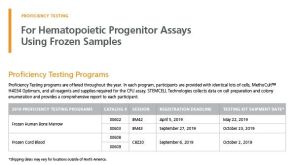 产品手册Registration Form - Proficiency Testing Programs
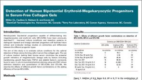 科学海报Detection of Human Bipotential Erythroid-Megakaryocytic Progenitors in Serum-Free Collagen Gels
 沪公网安备31010102008431号