Embryonic stem cells differentiate in vitro to endothelial cells through successive maturation steps.
The mechanisms involved in the regulation of vasculogenesis still remain unclear in mammals. Totipotent embryonic stem (ES) cells may represent a suitable in vitro model to study molecular events involved in vascular development. In this study,we followed the expression kinetics of a relatively large set of endothelial-specific markers in ES-derived embryoid bodies (EBs). Results of both reverse transcription-polymerase chain reaction and/or immunofluorescence analysis show that a spontaneous endothelial differentiation occurs during EBs development. ES-derived endothelial cells express a full range of cell lineage-specific markers: platelet endothelial cell adhesion molecule (PECAM),Flk-1,tie-1,tie-2,vascular endothelial (VE) cadherin,MECA-32,and MEC-14.7. Analysis of the kinetics of endothelial marker expression allows the distinction of successive maturation steps. Flk-1 was the first to be detected; its mRNA is apparent from day 3 of differentiation. PECAM and tie-2 mRNAs were found to be expressed only from day 4,whereas VE-cadherin and tie-1 mRNAs cannot be detected before day 5. Immunofluorescence stainings of EBs with antibodies directed against Flk-1,PECAM,VE-cadherin,MECA-32,and MEC-14.7 confirmed that the expression of these antigens occurs at different steps of endothelial cell differentiation. The addition of an angiogenic growth factor mixture including erythropoietin,interleukin-6,fibroblast growth factor 2,and vascular endothelial growth factor in the EB culture medium significantly increased the development of primitive vascular-like structures within EBs. These results indicate that this in vitro system contains a large part of the endothelial cell differentiation program and constitutes a suitable model to study the molecular mechanisms involved in vasculogenesis.
View Publication
产品类型:
产品号#:
06902
06952
00321
00322
00323
00324
00325
产品名:
Miyoshi H et al. (JAN 1999)
Science (New York,N.Y.) 283 5402 682--6
Transduction of human CD34+ cells that mediate long-term engraftment of NOD/SCID mice by HIV vectors.
Efficient gene transfer into human hematopoietic stem cells (HSCs) is an important goal in the study of the hematopoietic system as well as for gene therapy of hematopoietic disorders. A lentiviral vector based on the human immunodeficiency virus (HIV) was able to transduce human CD34+ cells capable of stable,long-term reconstitution of nonobese diabetic/severe combined immunodeficient (NOD/SCID) mice. High-efficiency transduction occurred in the absence of cytokine stimulation and resulted in transgene expression in multiple lineages of human hematopoietic cells for up to 22 weeks after transplantation.
View Publication
Hwang GH et al. (DEC 2017)
Journal of cellular physiology 232 12 3384--3395
Purification of small molecule-induced cardiomyocytes from human induced pluripotent stem cells using a reporter system.
In order to realize the practical use of human pluripotent stem cell (hPSC)-derived cardiomyocytes for the purpose of clinical use or cardiovascular research,the generation of large numbers of highly purified cardiomyocytes should be achieved. Here,we show an efficient method for cardiac differentiation of human induced pluripotent stem cells (hiPSCs) in chemically defined conditions and purification of hiPSC-derived cardiomyocytes using a reporter system. Regulation of the Wnt/β-catenin signaling pathway is implicated in the induction of the cardiac differentiation of hPSCs. We increased cardiac differentiation efficiency of hiPSCs in chemically defined conditions through combined treatment with XAV939,a tankyrase inhibitor and IWP2,a porcupine inhibitor and optimized concentrations. Although cardiac differentiation efficiency was high (>80%),it was difficult to suppress differentiation into non-cardiac cells,Therefore,we applied a lentiviral reporter system,wherein green fluorescence protein (GFP) and Zeocin-resistant gene are driven by promoter activation of a gene (TNNT2) encoding cardiac troponin T (cTnT),a cardiac-specific protein,to exclude non-cardiomyocytes from differentiated cell populations. We transduced this reporter construct into differentiated cells using a lentiviral vector and then obtained highly purified hiPSC-derived cardiomyocytes by treatment with the lowest effective dose of Zeocin. We significantly increased transgenic efficiency through manipulation of the cells in which the differentiated cells were simultaneously infected with virus and re-plated after single-cell dissociation. Purified cells specifically expressed GFP,cTnT,displayed typical properties of cardiomyocytes. This study provides an efficient strategy for obtaining large quantities of highly purified hPSC-derived cardiomyocytes for application in regenerative medicine and biomedical research.
View Publication
产品类型:
产品号#:
85850
85857
85870
85875
产品名:
mTeSR™1
mTeSR™1
Lu J et al. (APR 2017)
Journal of biomedical materials research. Part A 105 4 1094--1104
Interactions of human embryonic stem cell-derived cardiovascular progenitor cells with immobilized extracellular matrix proteins.
Human embryonic stem cell-derived cardiovascular progenitor cells (hESC-CVPCs) hold great promise for cell-based therapies of heart diseases. However,little is known about their niche microenvironment and in particular the required extracellular matrix (ECM) components. Here we screened combinations of surface-immobilized ECM proteins to identify substrates that support the attachment and survival of hESC-CVPCs. Covalent immobilization of ECM proteins laminin (Lm),fibronectin (Fn),collagen I (CI),collagen III (CIII),and collagen IV (CIV) in multiple combinations and concentrations was achieved by reductive amination on transparent acetaldehyde plasma polymer (AAPP) interlayer coatings. We identified that CI,CIII,CIV,and Fn and their combinations were important for hESC-CVPC attachment and survival,while Lm was dispensable. Moreover,for coatings displaying single ECM proteins,CI and CIII performed better than CIV and Fn,while coatings displaying the combined ECM proteins CIII + CIV and Fn + CIII + CIV at 100 µg/mL were comparable to Matrigel in regard to supporting hESC-CVPC attachment and viability. Our results identify ECM proteins required for hESC-CVPCs and demonstrate that coatings displaying multiple immobilized ECM proteins offer a suitable microenvironment for the attachment and survival of hESC-CVPCs. This knowledge contributes to the development of approaches for maintaining hESC-CVPCs and therefore to advances in cardiovascular regeneration. textcopyright 2017 Wiley Periodicals,Inc. J Biomed Mater Res Part A: 105A: 1094-1104,2017.
View Publication
产品类型:
产品号#:
85850
85857
85870
85875
产品名:
mTeSR™1
mTeSR™1
Wu X et al. (JAN 2018)
Cell 172 3 423--438.e25
Intrinsic Immunity Shapes Viral Resistance of Stem Cells.
Stem cells are highly resistant to viral infection compared to their differentiated progeny; however,the mechanism is mysterious. Here,we analyzed gene expression in mammalian stem cells and cells at various stages of differentiation. We find that,conserved across species,stem cells express a subset of genes previously classified as interferon (IFN) stimulated genes (ISGs) but that expression is intrinsic,as stem cells are refractory to interferon. This intrinsic ISG expression varies in a cell-type-specific manner,and many ISGs decrease upon differentiation,at which time cells become IFN responsive,allowing induction of a broad spectrum of ISGs by IFN signaling. Importantly,we show that intrinsically expressed ISGs protect stem cells against viral infection. We demonstrate the in vivo importance of intrinsic ISG expression for protecting stem cells and their differentiation potential during viral infection. These findings have intriguing implications for understanding stem cell biology and the evolution of pathogen resistance.
View Publication
产品类型:
产品号#:
02691
04434
04444
05110
05711
05712
05872
05873
18056
18056RF
72052
72054
72302
72304
72307
72308
70039
70039.1
70039.2
70039.3
70039.4
70039.5
70039.6
60062
60062BT
60062FI
60062FI.1
60062PE
60062PE.1
60045
60045AZ
60045AZ.1
60045BT
60045FI
60045FI.1
600
产品名:
StemSpan™ CD34+扩增添加物 (10X)
MethoCult™ H4434 Classic
MethoCult™ H4434 Classic
STEMdiff™定型内胚层检测试剂盒
NeuroCult™ SM1 神经添加物
CHIR99021
CHIR99021
Y-27632(二盐酸盐)
Y-27632(二盐酸盐)
Y-27632(二盐酸盐)
Y-27632(二盐酸盐)
抗人SSEA-4抗体,克隆号MC-813-70,生物素
抗人SSEA-4抗体,克隆号MC-813-70,FITC
抗人SSEA-4抗体, 克隆号MC-813-70,FITC
抗人SSEA-4抗体,克隆号MC-813-70,PE
抗人SSEA-4抗体,克隆号MC-813-70,PE
抗人CD90抗体,克隆5E10
抗人CD90抗体,克隆5E10,APC
抗人CD90抗体,克隆5E10,APC
抗人CD90抗体,克隆5E10,Biotin
抗人CD90抗体,克隆5E10,FITC
抗人CD90抗体,克隆5E10,FITC
Bershteyn M et al. (APR 2017)
Cell stem cell 20 4 435--449.e4
Human iPSC-Derived Cerebral Organoids Model Cellular Features of Lissencephaly and Reveal Prolonged Mitosis of Outer Radial Glia.
Classical lissencephaly is a genetic neurological disorder associated with mental retardation and intractable epilepsy,and Miller-Dieker syndrome (MDS) is the most severe form of the disease. In this study,to investigate the effects of MDS on human progenitor subtypes that control neuronal output and influence brain topology,we analyzed cerebral organoids derived from control and MDS-induced pluripotent stem cells (iPSCs) using time-lapse imaging,immunostaining,and single-cell RNA sequencing. We saw a cell migration defect that was rescued when we corrected the MDS causative chromosomal deletion and severe apoptosis of the founder neuroepithelial stem cells,accompanied by increased horizontal cell divisions. We also identified a mitotic defect in outer radial glia,a progenitor subtype that is largely absent from lissencephalic rodents but critical for human neocortical expansion. Our study,therefore,deepens our understanding of MDS cellular pathogenesis and highlights the broad utility of cerebral organoids for modeling human neurodevelopmental disorders.
View Publication
Human Induced Pluripotent Stem Cell-Derived Macrophages Share Ontogeny with MYB-Independent Tissue-Resident Macrophages.
Tissue-resident macrophages,such as microglia,Kupffer cells,and Langerhans cells,derive from Myb-independent yolk sac (YS) progenitors generated before the emergence of hematopoietic stem cells (HSCs). Myb-independent YS-derived resident macrophages self-renew locally,independently of circulating monocytes and HSCs. In contrast,adult blood monocytes,as well as infiltrating,gut,and dermal macrophages,derive from Myb-dependent HSCs. These findings are derived from the mouse,using gene knockouts and lineage tracing,but their applicability to human development has not been formally demonstrated. Here,we use human induced pluripotent stem cells (iPSCs) as a tool to model human hematopoietic development. By using a CRISPR-Cas9 knockout strategy,we show that human iPSC-derived monocytes/macrophages develop in an MYB-independent,RUNX1-,and SPI1 (PU.1)-dependent fashion. This result makes human iPSC-derived macrophages developmentally related to and a good model for MYB-independent tissue-resident macrophages,such as alveolar and kidney macrophages,microglia,Kupffer cells,and Langerhans cells.
View Publication
产品类型:
产品号#:
85850
85857
85870
85875
产品名:
mTeSR™1
mTeSR™1
Chou S-J et al. (APR 2017)
International journal of cardiology 232 255--263
Energy utilization of induced pluripotent stem cell-derived cardiomyocyte in Fabry disease.
BACKGROUND Fabry disease (FD) is a lysosomal storage disease in which glycosphingolipids (GB3) accumulate in organs of the human body,leading to idiopathic hypertrophic cardiomyopathy and target organ damage. Its pathophysiology is still poorly understood. OBJECTIVES We aimed to generate patient-specific induced pluripotent stem cells (iPSC) from FD patients presenting cardiomyopathy to determine whether the model could recapitulate key features of the disease phenotype and to investigate the energy metabolism in Fabry disease. METHODS Peripheral blood mononuclear cells from a 30-year-old Chinese man with a diagnosis of Fabry disease,GLA gene (IVS4+919G>A) mutation were reprogrammed into iPSCs and differentiated into iPSC-CMs and energy metabolism was analyzed in iPSC-CMs. RESULTS The FD-iPSC-CMs recapitulated numerous aspects of the FD phenotype including reduced GLA activity,cellular hypertrophy,GB3 accumulation and impaired contractility. Decreased energy metabolism with energy utilization shift to glycolysis was observed,but the decreased energy metabolism was not modified by enzyme rescue replacement (ERT) in FD-iPSCs-CMs. CONCLUSION This model provided a promising in vitro model for the investigation of the underlying disease mechanism and development of novel therapeutic strategies for FD. This potential remedy for enhancing the energetic network and utility efficiency warrants further study to identify novel therapies for the disease.
View Publication
Sessarego N et al. (MAR 2008)
Haematologica 93 3 339--46
Multipotent mesenchymal stromal cells from amniotic fluid: solid perspectives for clinical application.
BACKGROUND: Mesenchymal stromal cells are multipotent cells considered to be of great promise for use in regenerative medicine. However,the cell dose may be a critical factor in many clinical conditions and the yield resulting from the ex vivo expansion of mesenchymal stromal cells derived from bone marrow may be insufficient. Thus,alternative sources of mesenchymal stromal cells need to be explored. In this study,mesenchymal stromal cells were successfully isolated from second trimester amniotic fluid and analyzed for chromosomal stability to validate their safety for potential utilization as a cell therapy product. DESIGN AND METHODS: Mesenchymal stromal cells were expanded up to the sixth passage starting from amniotic fluid using different culture conditions to optimize large-scale production. RESULTS: The highest number of mesenchymal stromal cells derived from amniotic fluid was reached at a low plating density; in these conditions the expansion of mesenchymal stromal cells from amniotic fluid was significantly greater than that of adult bone marrow-derived mesenchymal stromal cells. Mesenchymal stromal cells from amniotic fluid represent a relatively homogeneous population of immature cells with immunosuppressive properties and extensive proliferative potential. Despite their high proliferative capacity in culture,we did not observe any karyotypic abnormalities or transformation potential in vitro nor any tumorigenic effect in vivo. CONCLUSIONS: Fetal mesenchymal stromal cells can be extensively expanded from amniotic fluid,showing no karyotypic abnormalities or transformation potential in vitro and no tumorigenic effect in vivo. They represent a relatively homogeneous population of immature mesenchymal stromal cells with long telomeres,immunosuppressive properties and extensive proliferative potential. Our results indicate that amniotic fluid represents a rich source of mesenchymal stromal cells suitable for banking to be used when large amounts of cells are required.
View Publication


 EasySep™小鼠TIL(CD45)正选试剂盒





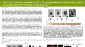 科学海报STEMdiff Cerebral Organoid Kit: A New Tool for the Culture of 3D Brain Organoids Derived from Human Pluripotent Stem Cells
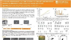 科学海报A Feeder-Independent Culture System to Convert and Maintain Human Pluripotent Stem Cells in a Naïve-Like State
 技术公告Flow Cytometry Methods for Identifying Mouse Hematopoietic Stem and Progenitor Cells
 沪公网安备31010102008431号