Bhadriraju K et al. (JUL 2016)
Stem Cell Research 17 1 122--129
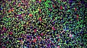
Large-scale time-lapse microscopy of Oct4 expression in human embryonic stem cell colonies
Identification and quantification of the characteristics of stem cell preparations is critical for understanding stem cell biology and for the development and manufacturing of stem cell based therapies. We have developed image analysis and visualization software that allows effective use of time-lapse microscopy to provide spatial and dynamic information from large numbers of human embryonic stem cell colonies. To achieve statistically relevant sampling,we examined textgreater 680 colonies from 3 different preparations of cells over 5 days each,generating a total experimental dataset of 0.9 terabyte (TB). The 0.5 Giga-pixel images at each time point were represented by multi-resolution pyramids and visualized using the Deep Zoom Javascript library extended to support viewing Giga-pixel images over time and extracting data on individual colonies. We present a methodology that enables quantification of variations in nominally-identical preparations and between colonies,correlation of colony characteristics with Oct4 expression,and identification of rare events.
View Publication
产品类型:
产品号#:
05850
05857
05870
05875
05940
85850
85857
85870
85875
产品名:
mTeSR™1
mTeSR™1
Smith GH (JAN 1996)
Breast cancer research and treatment 39 1 21--31
Experimental mammary epithelial morphogenesis in an in vivo model: evidence for distinct cellular progenitors of the ductal and lobular phenotype.
An in vivo transplantation system has been used to evaluate the developmental capacities of specific mouse mammary epithelial cell populations. Specifically,mouse mammary epithelial cells with distinctly limited developmental potentials have been identified using this procedure. Two distinct epithelial cell progenitors have been identified by experiments designed to determine whether basal lobular and ductal phenotypes could develop independently under conditions imposed by a limiting dilution. The prediction that these separate epithelial progenitors must exist was based upon the results from transplantation experiments carried out in epithelium-divested mammary fat pads of syngeneic mice with mammary epithelium from two different transgenic mouse models. The results presented here demonstrate the following points: 1) lobular,i.e. secretory,progenitor cells are present as distinct entities among the mammary epithelial cells found in immature virgin female mice; 2) similarly,ductal epithelial progenitors are present within the same population; 3) lobular progenitors are present in greater numbers,although both cell populations are extremely small; 4) as expected,some inocula produce outgrowths with simultaneous development of both lobular and ductal phenotypes--it is not known whether this indicates cooperative interaction between the two epithelial progenitors or signals the presence of a third progenitor type capable of producing both ductular and lobular committed daughters; 5) these findings have important consequences in the design of experiments aimed at testing the effects of known and putative mammary oncogenes and tumor suppressor genes,using techniques which include cellular transformation in vitro followed by in vivo cultivation and evaluation.
View Publication
产品类型:
产品号#:
01700
01705
05601
05610
05620
01702
产品名:
ALDEFLUOR™ 试剂盒
ALDEFLUOR™ DEAB试剂, 1.5 mM, 1 mL
EpiCult™-B 人培养基
EpiCult™-B 小鼠培养基
MammoCult™ 人源培养基套装
ALDEFLUOR™检测缓冲液
Miyoshi N et al. (JAN 2010)
Proceedings of the National Academy of Sciences of the United States of America 107 1 40--5
Defined factors induce reprogramming of gastrointestinal cancer cells.
Although cancer is a disease with genetic and epigenetic origins,the possible effects of reprogramming by defined factors remain to be fully understood. We studied the effects of the induction or inhibition of cancer-related genes and immature status-related genes whose alterations have been reported in gastrointestinal cancer cells. Retroviral-mediated introduction of induced pluripotent stem (iPS) cell genes was necessary for inducing the expression of immature status-related proteins,including Nanog,Ssea4,Tra-1-60,and Tra-1-80 in esophageal,stomach,colorectal,liver,pancreatic,and cholangiocellular cancer cells. Induced cells,but not parental cells,possessed the potential to express morphological patterns of ectoderm,mesoderm,and endoderm,which was supported by epigenetic studies,indicating methylation of DNA strands and the histone H3 protein at lysine 4 in promoter regions of pluripotency-associated genes such as NANOG. In in vitro analysis induced cells showed slow proliferation and were sensitized to differentiation-inducing treatment,and in vivo tumorigenesis was reduced in NOD/SCID mice. This study demonstrated that pluripotency was manifested in induced cells,and that the induced pluripotent cancer (iPC) cells were distinct from natural cancer cells with regard to their sensitivity to differentiation-inducing treatment. Retroviral-mediated introduction of iPC cells confers higher sensitivity to chemotherapeutic agents and differentiation-inducing treatment.
View Publication
产品类型:
产品号#:
05850
05857
05870
05875
85850
85857
85870
85875
产品名:
mTeSR™1
mTeSR™1
Boheler KR et al. (AUG 2002)
Circulation research 91 3 189--201
Differentiation of pluripotent embryonic stem cells into cardiomyocytes.
Embryonic stem (ES) cells have been established as permanent lines of undifferentiated pluripotent cells from early mouse embryos. ES cells provide a unique system for the genetic manipulation and the creation of knockout strains of mice through gene targeting. By cultivation in vitro as 3D aggregates called embryoid bodies,ES cells can differentiate into derivatives of all 3 primary germ layers,including cardiomyocytes. Protocols for the in vitro differentiation of ES cells into cardiomyocytes representing all specialized cell types of the heart,such as atrial-like,ventricular-like,sinus nodal-like,and Purkinje-like cells,have been established. During differentiation,cardiac-specific genes as well as proteins,receptors,and ion channels are expressed in a developmental continuum,which closely recapitulates the developmental pattern of early cardiogenesis. Exploitation of ES cell-derived cardiomyocytes has facilitated the analysis of early cardiac development and has permitted in vitro gain-of-function" or "loss-of-function" genetic studies. Recently�
View Publication
产品类型:
产品号#:
06902
06952
00321
00322
00323
00324
00325
产品名:
Ichida JK et al. (AUG 2014)
Nature chemical biology 10 8 632--9
Notch inhibition allows oncogene-independent generation of iPS cells.
The reprogramming of somatic cells to pluripotency using defined transcription factors holds great promise for biomedicine. However,human reprogramming remains inefficient and relies either on the use of the potentially dangerous oncogenes KLF4 and CMYC or the genetic inhibition of the tumor suppressor gene p53. We hypothesized that inhibition of signal transduction pathways that promote differentiation of the target somatic cells during development might relieve the requirement for non-core pluripotency factors during induced pluripotent stem cell (iPSC) reprogramming. Here,we show that inhibition of Notch greatly improves the efficiency of iPSC generation from mouse and human keratinocytes by suppressing p21 in a p53-independent manner and thereby enriching for undifferentiated cells capable of long-term self-renewal. Pharmacological inhibition of Notch enabled routine production of human iPSCs without KLF4 and CMYC while leaving p53 activity intact. Thus,restricting the development of somatic cells by altering intercellular communication enables the production of safer human iPSCs.
View Publication
产品类型:
产品号#:
05850
05857
05870
05875
73092
85850
85857
85870
85875
产品名:
DBZ
mTeSR™1
mTeSR™1
Cai J et al. (JAN 2004)
Journal of neurochemistry 88 1 212--26
Membrane properties of rat embryonic multipotent neural stem cells.
We have characterized several potential stem cell markers and defined the membrane properties of rat fetal (E10.5) neural stem cells (NSC) by immunocytochemistry,electrophysiology and microarray analysis. Immunocytochemical analysis demonstrates specificity of expression of Sox1,ABCG2/Bcrp1,and shows that nucleostemin labels both progenitor and stem cell populations. NSCs,like hematopoietic stem cells,express high levels of aldehyde dehydrogenase (ALDH) as assessed by Aldefluor labeling. Microarray analysis of 96 transporters and channels showed that Glucose transporter 1 (Glut1/Slc2a1) expression is unique to fetal NSCs or other differentiated cells. Electrophysiological examination showed that fetal NSCs respond to acetylcholine and its agonists,such as nicotine and muscarine. NSCs express low levels of tetrodotoxin (TTX) sensitive and insensitive sodium channels and calcium channels while expressing at least three kinds of potassium channels. We find that gap junction communication is mediated by connexin (Cx)43 and Cx45,and is essential for NSC survival and proliferation. Overall,our results show that fetal NSCs exhibit a unique signature that can be used to determine their location and assess their ability to respond to their environment.
View Publication
产品类型:
产品号#:
01700
01705
01701
01702
产品名:
ALDEFLUOR™ 试剂盒
ALDEFLUOR™ DEAB试剂, 1.5 mM, 1 mL
ALDEFLUOR™检测缓冲液
Juopperi TA et al. (FEB 2007)
Experimental hematology 35 2 335--41
Isolation of bone marrow-derived stem cells using density-gradient separation.
OBJECTIVE: Our laboratory has established two unique methods to isolate murine hematopoietic stem cells on the basis of functional characteristics such as the ability of stem cells to home to bone marrow and aldehyde dehydrogenase (ALDH) activity. An essential component of both protocols is the separation of whole bone marrow into small-sized cells by counter-flow elutriation. We sought to provide the scientific community with an alternate approach to acquire our stem cells by replacing elutriation with the use of density-gradient centrifugation. METHODS: The elutriated fraction 25 population was characterized based on density using a discontinuous gradient. The long-term reconstituting potential of whole bone marrow cells collected at each density interface was determined by subjecting the fractions to the two-day homing protocol,transplanting them into lethally irradiated recipient mice,and assessing peripheral blood chimerism. We also investigated the ability of high-density bone marrow cells isolated in conjunction with the ALDH protocol to repopulate the hematopoietic system of myeloablated recipients. RESULTS: Bone marrow cells collected at the high-density interface of 1.081/1.087 g/mL (fraction 3) had the capacity for homing to marrow and the ability to provide long-term hematopoietic reconstitution. Fraction three lineage-depleted ALDH-bright cells could also engraft and provide long-term hematopoiesis at limiting dilutions. CONCLUSIONS: Density-gradient centrifugation can be used in conjunction with either of our stem cell isolation protocols to obtain cells with long-term reconstitution ability. We anticipate that this strategy will encourage and enable investigators to study the biology of HSCs isolated using functional characteristics.
View Publication
产品类型:
产品号#:
01700
01705
01701
01702
产品名:
ALDEFLUOR™ 试剂盒
ALDEFLUOR™ DEAB试剂, 1.5 mM, 1 mL
ALDEFLUOR™检测缓冲液
Carmona G et al. (MAR 2008)
Blood 111 5 2640--6
Activation of Epac stimulates integrin-dependent homing of progenitor cells.
Cell therapy is a novel promising option for treatment of ischemic diseases. Administered endothelial progenitor cells (EPCs) are recruited to ischemic regions and improve neovascularization. However,the number of cells that home to ischemic tissues is restricted. The GTPase Rap1 plays an important role in the regulation of adhesion and chemotaxis. We investigated whether pharmacologic activation of Epac1,a nucleotide exchange protein for Rap1,which is directly activated by cAMP,can improve the adhesive and migratory capacity of distinct progenitor cell populations. Stimulation of Epac by a cAMP-analog increased Rap1 activity and stimulated the adhesion of human EPCs,CD34(+) hematopoietic progenitor cells,and mesenchymal stem cells (MSCs). Specifically,short-term stimulation with a specific Epac activator increased the beta2-integrin-dependent adhesion of EPCs to endothelial cell monolayers,and of EPC and CD34(+) cells to ICAM-1. Furthermore,the Epac activator enhanced the beta1-integrin-dependent adhesion of EPCs and MSCs to the matrix protein fibronectin. In addition,Epac1 activation induced the beta1- and beta2-integrin-dependent migration of EPCs on fibronectin and fibrinogen. Interestingly,activation of Epac rapidly increased lateral mobility of beta1- and beta2-integrins,thereby inducing integrin polarization,and stimulated beta1-integrin affinity,whereas the beta2-integrin affinity was not increased. Furthermore,prestimulation of EPCs with the Epac activator increased homing to ischemic muscles and neovascularization-promoting capacity of intravenously injected EPCs in the model of hind limb ischemia. These data demonstrate that activation of Epac1 increases integrin activity and integrin-dependent homing functions of progenitor cells and enhances their in vivo therapeutic potential. These results may provide a platform for the development of novel therapeutic approaches to improve progenitor cell homing.
View Publication
产品类型:
产品号#:
05401
05402
05411
产品名:
MesenCult™ MSC 基础培养基(人)
MesenCult™ MSC刺激添加物(人)
MesenCult™ 增殖试剂盒(人)
Woltjen K et al. (APR 2009)
Nature 458 7239 766--70
piggyBac transposition reprograms fibroblasts to induced pluripotent stem cells.
Transgenic expression of just four defined transcription factors (c-Myc,Klf4,Oct4 and Sox2) is sufficient to reprogram somatic cells to a pluripotent state. The resulting induced pluripotent stem (iPS) cells resemble embryonic stem cells in their properties and potential to differentiate into a spectrum of adult cell types. Current reprogramming strategies involve retroviral,lentiviral,adenoviral and plasmid transfection to deliver reprogramming factor transgenes. Although the latter two methods are transient and minimize the potential for insertion mutagenesis,they are currently limited by diminished reprogramming efficiencies. piggyBac (PB) transposition is host-factor independent,and has recently been demonstrated to be functional in various human and mouse cell lines. The PB transposon/transposase system requires only the inverted terminal repeats flanking a transgene and transient expression of the transposase enzyme to catalyse insertion or excision events. Here we demonstrate successful and efficient reprogramming of murine and human embryonic fibroblasts using doxycycline-inducible transcription factors delivered by PB transposition. Stable iPS cells thus generated express characteristic pluripotency markers and succeed in a series of rigorous differentiation assays. By taking advantage of the natural propensity of the PB system for seamless excision,we show that the individual PB insertions can be removed from established iPS cell lines,providing an invaluable tool for discovery. In addition,we have demonstrated the traceless removal of reprogramming factors joined with viral 2A sequences delivered by a single transposon from murine iPS lines. We anticipate that the unique properties of this virus-independent simplification of iPS cell production will accelerate this field further towards full exploration of the reprogramming process and future cell-based therapies.
View Publication
产品类型:
产品号#:
27845
27945
27840
27865
27940
27965
产品名:
Dani C et al. (JUN 1997)
Journal of cell science 110 ( Pt 1 1279--85
Differentiation of embryonic stem cells into adipocytes in vitro.
Embryonic stem cells,derived from the inner cell mass of murine blastocysts,can be maintained in a totipotent state in vitro. In appropriate conditions embryonic stem cells have been shown to differentiate in vitro into various derivatives of all three primary germ layers. We describe in this paper conditions to induce differentiation of embryonic stem cells reliably and at high efficiency into adipocytes. A prerequisite is to treat early developing embryonic stem cell-derived embryoid bodies with retinoic acid for a precise period of time. Retinoic acid could not be substituted by adipogenic hormones nor by potent activators of peroxisome proliferator-activated receptors. Treatment with retinoic acid resulted in the subsequent appearance of large clusters of mature adipocytes in embryoid body outgrowths. Lipogenic and lipolytic activities as well as high level expression of adipocyte specific genes could be detected in these cultures. Analysis of expression of potential adipogenic genes,such as peroxisome proliferator-activated receptors gamma and delta and CCAAT/enhancer binding protein beta,during differentiation of retinoic acid-treated embryoid bodies has been performed. The temporal pattern of expression of genes encoding these nuclear factors resembled that found during mouse embryogenesis. The differentiation of embryonic stem cells into adipocytes will provide an invaluable model for the characterisation of the role of genes expressed during the adipocyte development programme and for the identification of new adipogenic regulatory genes.
View Publication
产品类型:
产品号#:
06902
06952
72262
72264
00321
00322
00323
00324
00325
100-1045
产品名:
全反式视黄酸
全反式视黄酸
全反式视黄酸
Aanei CM et al. (NOV 2011)
Experimental cell research 317 18 2616--29
Focal adhesion protein abnormalities in myelodysplastic mesenchymal stromal cells.
Direct cell-cell contact between haematopoietic progenitor cells (HPCs) and their cellular microenvironment is essential to maintain 'stemness'. In cancer biology,focal adhesion (FA) proteins are involved in survival signal transduction in a wide variety of human tumours. To define the role of FA proteins in the haematopoietic microenvironment of myelodysplastic syndromes (MDS),CD73-positive mesenchymal stromal cells (MSCs) were immunostained for paxillin,pFAK [Y(397)],and HSP90α/β and p130CAS,and analysed for reactivity,intensity and cellular localisation. Immunofluorescence microscopy allowed us to identify qualitative and quantitative differences,and subcellular localisation analysis revealed that in pathological MSCs,paxillin,pFAK [Y(397)],and HSP90α/β formed nuclear molecular complexes. Increased expression of paxillin,pFAK [Y(397)],and HSP90α/β and enhanced nuclear co-localisation of these proteins correlated with a consistent proliferative advantage in MSCs from patients with refractory anaemia with excess blasts (RAEB) and negatively impacted clonogenicity of HPCs. These results suggest that signalling via FA proteins could be implicated in HPC-MSC interactions. Further,because FAK is an HSP90α/β client protein,these results suggest the utility of HSP90α/β inhibition as a target for adjuvant therapy for myelodysplasia.
View Publication

 EasySep™小鼠TIL(CD45)正选试剂盒


 实验方案Maintenance of Porcine Pluripotent Stem Cells (pPSCs) Using eTeSR™
 沪公网安备31010102008431号