Detection, isolation, and stimulation of quiescent primitive leukemic progenitor cells from patients with acute myeloid leukemia (AML).
Although many acute myeloid leukemia (AML) colony-forming cells (CFCs) and long-term culture-initiating cells (LTC-ICs) directly isolated from patients are actively cycling,quiescent progenitors are present in most samples. In the current study,(3)H-thymidine ((3)H-Tdr) suicide assays demonstrated that most NOD/SCID mouse leukemia-initiating cells (NOD/SL-ICs) are quiescent in 6 of 7 AML samples. AML cells in G(0),G(1),and S/G(2)+M were isolated from 4 of these samples using Hoechst 33342/pyroninY staining and cell sorting. The progenitor content of each subpopulation was consistent with the (3)H-Tdr suicide results,with NOD/SL-ICs found almost exclusively among G(0) cells while the cycling status of AML CFCs and LTC-ICs was more heterogeneous. Interestingly,after 72 hours in serum-free culture with or without Steel factor (SF),Flt-3 ligand (FL),and interleukin-3 (IL-3),most G(0) AML cells entered active cell cycle (percentage of AML cells remaining in G(0) at 72 hours,1.2% to 37%,and 0% to 7.6% in cultures without and with growth factors [GFs],respectively) while G(0) cells from normal lineage-depleted bone marrow remained quiescent in the absence of GF. All 4 AML samples showed evidence of autocrine production of 2 or more of SF,FL,IL-3,and granulocyte-macrophage colony-stimulating factor (GM-CSF). In addition,3 of 4 samples contained an internal tandem duplication of the FLT3 gene. In summary,quiescent leukemic cells,including NOD/SL-ICs,are present in most AML patients. Their spontaneous entry into active cell cycle in short-term culture might be explained by the deregulated GF signaling present in many AMLs.
View Publication
产品类型:
产品号#:
05150
09500
09600
09650
产品名:
MyeloCult™ H5100
BIT 9500血清替代物
StemSpan™ SFEM
StemSpan™ SFEM
Zhang S et al. (APR 2014)
Biomaterials 35 12 3786--3793
The influence of a spatiotemporal 3D environment on endothelial cell differentiation of human induced pluripotent stem cells.
Current EC differentiation protocols are inefficient,and the phenotypes of the differentiated ECs are only briefly stable,which significantly inhibits their utility for basic science research. Here,a remarkably more efficient hiPSC-EC differentiation protocol that incorporates a three-dimensional (3D) fibrin scaffold is presented. With this protocol,up to 45% of the differentiated hiPSCs assumed an EC phenotype,and after purification,greater than 95% of the cells displayed the EC phenotype (based on CD31 expression). The hiPSC-ECs continued to display EC characteristics for 4 weeks invitro. Gene and protein expression levels of CD31,CD144 and von Willebrand factor-8 (vWF-8) were significantly up-regulated in differentiated hiPSC-ECs. hiPSC-ECs also have biological function to up-take Dil-conjugated acetylated LDL (Dil-ac-LDL) and form tubular structures on Matrigel. Collectively,these data demonstrate that a 3D differentiation protocol can efficiently generate ECs from hiPSCs and,furthermore,the differentiated hiPSC-ECs are functional and can maintain EC fate up to 4 weeks invitro. ?? 2014 Elsevier Ltd.
View Publication
产品类型:
产品号#:
05850
05857
05870
05875
85850
85857
85870
85875
产品名:
mTeSR™1
mTeSR™1
Mehta A et al. (NOV 2014)
Biochimica et biophysica acta 1843 11 2394--2402
Phasic modulation of Wnt signaling enhances cardiac differentiation in human pluripotent stem cells by recapitulating developmental ontogeny.
Cardiomyocytes (CMs) derived from human pluripotent stem cells (hPSCs) offer immense value in studying cardiovascular regenerative medicine. However,intrinsic biases and differential responsiveness of hPSCs towards cardiac differentiation pose significant technical and logistic hurdles that hamper human cardiomyocyte studies. Tandem modulation of canonical and non-canonical Wnt signaling pathways may play a crucial role in cardiac development that can efficiently generate cardiomyocytes from pluripotent stem cells. Our Wnt signaling expression profiles revealed that phasic modulation of canonical/non-canonical axis enabled orderly recapitulation of cardiac developmental ontogeny. Moreover,evaluation of 8 hPSC lines showed marked commitment towards cardiac-mesoderm during the early phase of differentiation,with elevated levels of canonical Wnts (Wnt3 and 3a) and Mesp1. Whereas continued activation of canonical Wnts was counterproductive,its discrete inhibition during the later phase of cardiac differentiation was accompanied by significant up-regulation of non-canonical Wnt expression (Wnt5a and 11) and enhanced Nkx2.5(+) (up to 98%) populations. These Nkx2.5(+) populations transited to contracting cardiac troponin T-positive CMs with up to 80% efficiency. Our results suggest that timely modulation of Wnt pathways would transcend intrinsic differentiation biases of hPSCs to consistently generate functional CMs that could facilitate their scalable production for meaningful clinical translation towards personalized regenerative medicine.
View Publication
PDX1 binds and represses hepatic genes to ensure robust pancreatic commitment in differentiating human embryonic stem cells.
Inactivation of the Pancreatic and Duodenal Homeobox 1 (PDX1) gene causes pancreatic agenesis,which places PDX1 high atop the regulatory network controlling development of this indispensable organ. However,little is known about the identity of PDX1 transcriptional targets. We simulated pancreatic development by differentiating human embryonic stem cells (hESCs) into early pancreatic progenitors and subjected this cell population to PDX1 chromatin immunoprecipitation sequencing (ChIP-seq). We identified more than 350 genes bound by PDX1,whose expression was upregulated on day 17 of differentiation. This group included known PDX1 targets and many genes not previously linked to pancreatic development. ChIP-seq also revealed PDX1 occupancy at hepatic genes. We hypothesized that simultaneous PDX1-driven activation of pancreatic and repression of hepatic programs underlie early divergence between pancreas and liver. In HepG2 cells and differentiating hESCs,we found that PDX1 binds and suppresses expression of endogenous liver genes. These findings rebrand PDX1 as a context-dependent transcriptional repressor and activator within the same cell type.
View Publication
产品类型:
产品号#:
05850
05857
05870
05875
85850
85857
85870
85875
产品名:
mTeSR™1
mTeSR™1
Daga A et al. (MAY 2000)
Experimental hematology 28 5 569--74
The retroviral transduction of HOXC4 into human CD34(+) cells induces an in vitro expansion of clonogenic and early progenitors.
OBJECTIVE: +HOX genes are expressed in the hematopoietic system and increasing data point to their involvement in the control of proliferation and/or differentiation. Genes belonging to the C cluster are preferentially expressed in developing and differentiated lymphoid lineages. However,recent studies demonstrated,by RT-PCR,that the HOXC4 gene is also actively transcribed in the most undifferentiated hematopoietic cells (CD34(+)38(low)) and in more mature myeloid and erythroid progenitors. We evaluated the expression of HOXC4 protein on human CD34(+) cells and the in vitro effect of its overexpression on proliferation and differentiation. MATERIALS AND METHODS: We assessed the expression of HOXC4 on human CD34(+) cells using a polyclonal antibody raised against the C-terminal portion of the protein expressed using the baculovirus system. Overexpression of HOXC4 in human CD34(+) cells was obtained by retroviral gene transfer; its effect on clonogenic (CFU-GM,BFU-E,and CFU-GEMM) and early progenitors (LTC-IC) was evaluated. RESULTS: The HOXC4 protein is indeed expressed in human CD34(+) cells,and its overexpression in human CD34(+) cells increases the proliferation potential of clonogenic and early progenitors. CFU-GM showed a median threefold expansion (range: 1.1-19.4; p textless 0.002) compared with control transduced with the vector alone. The increment of BFU-E was higher (median ninefold,range 2.5-35; p textless 0. 0009) and erythroid colonies presented a larger size with normal morphology. An even more marked effect was observed on LTC-IC (median 13,onefold; range 4.1-102.1,p textless 0.0001). CONCLUSION: We demonstrate that HOXC4 is expressed in CD34(+) cells and that its overexpression induces an in vitro expansion of committed as well as very early hematopoietic progenitors. The most striking effect was obtained on LTC-IC with an expansion of 13.1-fold. The enforced expression of HOXC4 induced a significant increase (p textless 0.009) in the number of erythroid colonies compared with CFU-GM,although without perturbing,at least in vitro,the maturation program of the cells. On the other hand,the effect of the gene overexpression did not induce any skewing in the colony types derived from the myeloid lineage.
View Publication
Noninvasive MR imaging of magnetically labeled stem cells to directly identify neovasculature in a glioma model.
Bone marrow-derived endothelial precursor cells incorporate into neovasculature and have been successfully used as vehicles for gene delivery to brain tumors. To determine whether systemically administered Sca1+ bone marrow cells labeled with superparamagnetic iron oxide nanoparticles can be detected by in vivo magnetic resonance imaging in a mouse brain tumor model,mouse Sca1+ cells were labeled in vitro with ferumoxides-poly-L-lysine complexes. Labeled or control cells were administered intravenously to glioma-bearing severe combined immunodeficient (SCID) mice. Magnetic resonance imaging (MRI) was performed during tumor growth. Mice that received labeled cells demonstrated hypointense regions within the tumor that evolved over time and developed a continuous dark hypointense ring at a consistent time point. This effect was not cleared by administration of a gadolinium contrast agent. Histology showed iron-labeled cells around the tumor rim in labeled mice,which expressed CD31 and von Willebrand factor,indicating the transplanted cells detected in the tumor have differentiated into endothelial-like cells. These results demonstrate that MRI can detect the incorporation of magnetically labeled bone marrow-derived precursor cells into tumor vasculature as part of ongoing angiogenesis and neovascularization. This technique can be used to directly identify neovasculature in vivo and to facilitate gene therapy by noninvasively monitoring these cells as gene delivery vectors.
View Publication
产品类型:
产品号#:
09600
09650
09850
产品名:
StemSpan™ SFEM
StemSpan™ SFEM
Wagner W et al. (NOV 2005)
Experimental hematology 33 11 1402--16
Comparative characteristics of mesenchymal stem cells from human bone marrow, adipose tissue, and umbilical cord blood.
OBJECTIVE: Various preparative protocols have been proposed for the acquisition and cultivation of mesenchymal stem cells (MSC). Whereas surface antigen markers have failed to precisely define this population,microarray analysis might provide a better tool for characterization of MSC. METHODS: In this study,we have analyzed global gene expression profiles of human MSC isolated from adipose tissue (AT),from umbilical cord blood (CB),and from bone marrow (BM) under two growth conditions and have compared them to terminally differentiated human fibroblasts (HS68). Profiles were compared using our Human Genome Microarray representing 51.144 different cDNA clones. RESULTS: Cultured with the appropriate conditions,osteogenic and adipogenic differentiation could be confirmed in all MSC preparations but not in fibroblasts. No phenotypic differences were observed by flow cytometry using a panel of 22 surface antigen markers. Whereas MSC derived from different donors using the same culture procedure yielded a consistent and reproducible gene expression profile,many genes were differentially expressed in MSC from different ontogenetic sources or from different culture conditions. Twenty-five genes were overlapping and upregulated in all MSC preparations from AT,CB,and BM as compared to HS68 fibroblasts. These genes included fibronectin,ECM2,glypican-4,ID1,NF1B,HOXA5,and HOXB6. Many genes upregulated in MSC are involved in extracellular matrix,morphogenesis,and development,whereas several inhibitors of the Wnt pathway (DKK1,DKK3,SFRP1) were highly expressed in fibroblasts. CONCLUSION: Our results have provided a foundation for a more reproducible and reliable quality control using genotypic analysis for defining MSC.
View Publication
产品类型:
产品号#:
06902
06952
00321
00322
00323
00324
00325
产品名:
Hough SR et al. (JUN 2014)
Stem Cell Reports 2 6 881--895
Single-cell gene expression profiles define self-renewing, pluripotent, and lineage primed states of human pluripotent stem cells
Pluripotent stem cells display significant heterogeneity in gene expression,but whether this diversity is an inherent feature of the pluripotent state remains unknown. Single-cell gene expression analysis in cell subsets defined by surface antigen expression revealed that human embryonic stem cell cultures exist as a continuum of cell states,even under defined conditions that drive self-renewal. The majority of the population expressed canonical pluripotency transcription factors and could differentiate into derivatives of all three germ layers. A minority subpopulation of cells displayed high self-renewal capacity,consistently high transcripts for all pluripotency-related genes studied,and no lineage priming. This subpopulation was characterized by its expression of a particular set of intercellular signaling molecules whose genes shared common regulatory features. Our data support a model of an inherently metastable self-renewing population that gives rise to a continuum of intermediate pluripotent states,which ultimately become primed for lineage specification. ?? 2014 The Authors.
View Publication
产品类型:
产品号#:
05850
05857
05870
05875
85850
85857
85870
85875
产品名:
mTeSR™1
mTeSR™1
Kadari A et al. (AUG 2015)
Stem Cell Reviews and Reports 11 4 560--569
Robust Generation of Cardiomyocytes from Human iPS Cells Requires Precise Modulation of BMP and WNT Signaling.
Various strategies have been published enabling cardiomyocyte differentiation of human induced pluripotent stem (iPS) cells. However the complex nature of signaling pathways involved as well as line-to-line variability compromises the application of a particular protocol to robustly obtain cardiomyocytes from multiple iPS lines. Hence it is necessary to identify optimized protocols with alternative combinations of specific growth factors and small molecules to enhance the robustness of cardiac differentiation. Here we focus on systematic modulation of BMP and WNT signaling to enhance cardiac differentiation. Moreover,we improve the efficacy of cardiac differentiation by enrichment via lactate. Using our protocol we show efficient derivation of cardiomyocytes from multiple human iPS lines. In particular we demonstrate cardiomyocyte differentiation within 15 days with an efficiency of up to 95 % as judged by flow cytometry staining against cardiac troponin T. Cardiomyocytes derived were functionally validated by alpha-actinin staining,transmission electron microscopy as well as electrophysiological analysis. We expect our protocol to provide a robust basis for scale-up production of functional iPS cell-derived cardiomyocytes that can be used for cell replacement therapy and disease modeling.
View Publication
产品类型:
产品号#:
05850
05857
05870
05875
85850
85857
85870
85875
产品名:
mTeSR™1
mTeSR™1
Hø et al. (JAN 2015)
Stem Cell Research 14 1 39--53
Ultrastructural visualization of the Mesenchymal-to-Epithelial Transition during reprogramming of human fibroblasts to induced pluripotent stem cells
The Mesenchymal-to-Epithelial Transition (MET) has been recognized as a crucial step for successful reprogramming of fibroblasts to induced pluripotent stem cells (iPSCs). Thus,it has been demonstrated,that the efficiency of reprogramming can be enhanced by promoting an epithelial expression program in cells,with a concomitant repression of key mesenchymal genes. However,a detailed characterization of the epithelial transition associated with the acquisition of a pluripotent phenotype is still lacking to this date. Here,we integrate a panel of morphological approaches with gene expression analyses to visualize the dynamics of episomal reprogramming of human fibroblasts to iPSCs. We provide the first ultrastructural analysis of human fibroblasts at various stages of episomal iPSC reprogramming,as well as the first real-time live cell visualization of a MET occurring during reprogramming. The results indicate that the MET manifests itself approximately 6-12. days after electroporation,in synchrony with the upregulation of early pluripotency markers,and resembles a reversal of the Epithelial-to-Mesenchymal Transition (EMT) which takes place during mammalian gastrulation.
View Publication

 EasySep™小鼠TIL(CD45)正选试剂盒


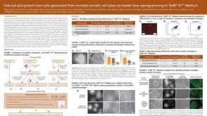 科学海报Induced Pluripotent Stem Cells Generated from Multiple Somatic Cell Types via Feeder Free Reprogramming in TeSR™-E7™ Medium


 沪公网安备31010102008431号