Cipriano AF et al. (APR 2013)
Journal of Materials Science: Materials in Medicine 24 4 989--1003
In vitro degradation of four magnesium-zinc-strontium alloys and their cytocompatibility with human embryonic stem cells
Magnesium alloys have attracted great interest for medical applications due to their unique biodegradable capability and desirable mechanical properties. When designed for medical applications,these alloys must have suitable degradation properties,i.e.,their degradation rate should not exceed the rate at which the degradation products can be excreted from the body. Cellular responses and tissue integration around the Mg-based implants are critical for clinical success. Four magnesium–zinc–strontium (ZSr41) alloys were developed in this study. The degradation properties of the ZSr41 alloys and their cytocompatibility were studied using an in vitro human embryonic stem cell (hESC) model due to the greater sensitivity of hESCs to known toxicants which allows to potentially detect toxicological effects of new biomaterials at an early stage. Four distinct ZSr41 alloys with 4 wt% zinc and a series of strontium compositions (0.15,0.5,1,and 1.5 wt% Sr) were produced through metallurgical processing. Their degradation was characterized by measuring total mass loss of samples and pH change in the cell culture media. The concentration of Mg ions released from ZSr41 alloy into the cell culture media was analyzed using inductively coupled plasma atomic emission spectroscopy. Surface microstructure and composition before and after culturing with hESCs were characterized using field emission scanning electron microscopy and energy dispersive X-ray spectroscopy. Pure Mg was used as a control during cell culture studies. Results indicated that the Mg–Zn–Sr alloy with 0.15 wt% Sr provided slower degradation and improved cytocompatibility as compared with pure Mg control.
View Publication
产品类型:
产品号#:
05850
05857
05870
05875
85850
85857
85870
85875
产品名:
mTeSR™1
mTeSR™1
van der Meer AD et al. (SEP 2013)
Lab on a Chip 13 18 3562--3568
Three-dimensional co-cultures of human endothelial cells and embryonic stem cell-derived pericytes inside a microfluidic device
Organs-on-chips are microengineered in vitro tissue structures that can be used as platforms for physiological and pathological research. They provide tissue-like microenvironments in which different cell types can be co-cultured in a controlled manner to create synthetic organ mimics. Blood vessels are an integral part of all tissues in the human body. Development of vascular structures is therefore an important research topic for advancing the field of organs-on-chips since generated tissues will require a blood or nutrient supply. Here,we have engineered three-dimensional constructs of vascular tissue inside microchannels by injecting a mixture of human umbilical vein endothelial cells,human embryonic stem cell-derived pericytes (the precursors of vascular smooth muscle cells) and rat tail collagen I into a polydimethylsiloxane microfluidic channel with dimensions 500 μm × 120 μm × 1 cm (w × h × l). Over the course of 12 h,the cells organized themselves into a single long tube resembling a blood vessel that followed the contours of the channel. Detailed examination of tube morphology by confocal microscopy revealed a mature endothelial monolayer with complete PECAM-1 staining at cell–cell contacts and pericytes incorporated inside the tubular structures. We also demonstrated that tube formation was disrupted in the presence of a neutralizing antibody against transforming growth factor-beta (TGF-β). The TGF-β signaling pathway is essential for normal vascular development; deletion of any of its components in mouse development results in defective vasculogenesis and angiogenesis and mutations in humans have been linked to multiple vascular genetic diseases. In the engineered microvessels,inhibition of TGF-β signaling resulted in tubes with smaller diameters and higher tortuosity,highly reminiscent of the abnormal vessels observed in patients with one particular vascular disease known as hereditary hemorrhagic telangiectasia (HHT). In summary,we have developed microengineered three-dimensional vascular structures that can be used as a model to test the effects of drugs and study the interaction between different human vascular cell types. In the future,the model may be integrated into larger tissue constructs to advance the development of organs-on-chips.
View Publication
产品类型:
产品号#:
05850
05857
05870
05875
85850
85857
85870
85875
产品名:
mTeSR™1
mTeSR™1
Shah SN et al. (DEC 2016)
PloS one 11 12 e0166657
Evaluation of Stem Cell-Derived Red Blood Cells as a Transfusion Product Using a Novel Animal Model.
Reliance on volunteer blood donors can lead to transfusion product shortages,and current liquid storage of red blood cells (RBCs) is associated with biochemical changes over time,known as 'the storage lesion'. Thus,there is a need for alternative sources of transfusable RBCs to supplement conventional blood donations. Extracorporeal production of stem cell-derived RBCs (stemRBCs) is a potential and yet untapped source of fresh,transfusable RBCs. A number of groups have attempted RBC differentiation from CD34+ cells. However,it is still unclear whether these stemRBCs could eventually be effective substitutes for traditional RBCs due to potential differences in oxygen carrying capacity,viability,deformability,and other critical parameters. We have generated ex vivo stemRBCs from primary human cord blood CD34+ cells and compared them to donor-derived RBCs based on a number of in vitro parameters. In vivo,we assessed stemRBC circulation kinetics in an animal model of transfusion and oxygen delivery in a mouse model of exercise performance. Our novel,chronically anemic,SCID mouse model can evaluate the potential of stemRBCs to deliver oxygen to tissues (muscle) under resting and exercise-induced hypoxic conditions. Based on our data,stem cell-derived RBCs have a similar biochemical profile compared to donor-derived RBCs. While certain key differences remain between donor-derived RBCs and stemRBCs,the ability of stemRBCs to deliver oxygen in a living organism provides support for further development as a transfusion product.
View Publication
产品类型:
产品号#:
70008
70008.1
70008.2
70008.3
70008.4
70008.5
70008.6
200-0000
200-0001
200-0002
产品名:
冻存的人脐带血CD34+细胞
冻存的人脐带血CD34+细胞
冻存的人脐带血CD34+细胞
冻存的人脐带血CD34+细胞
冻存的人脐带血CD34+细胞
冻存的人脐带血CD34+细胞
冻存的人脐带血CD34+细胞
冻存的人脐带血CD34+细胞
冻存的人脐带血CD34+细胞
Zhou S et al. ( 2017)
PloS one 12 1 e0169899
Reprogramming Malignant Cancer Cells toward a Benign Phenotype following Exposure to Human Embryonic Stem Cell Microenvironment.
The embryonic microenvironment is well known to be non-permissive for tumor development because early developmental signals naturally suppress the expression of proto-oncogenes. In an analogous manner,mimicking an early embryonic environment during embryonic stem cell culture has been shown to suppress oncogenic phenotypes of cancer cells. Exosomes derived from human embryonic stem cells harbor substances that mirror the content of the cells of origin and have been reported to reprogram hematopoietic stem/progenitor cells via horizontal transfer of mRNA and proteins. However,the possibility that these embryonic stem cells-derived exosomes might be the main effectors of the anti-tumor effect mediated by the embryonic stem cells has not been explored yet. The present study aims to investigate whether exosomes derived from human embryonic stem cells can reprogram malignant cancer cells to a benign stage and reduce their tumorigenicity. We show that the embryonic stem cell-conditioned medium contains factors that inhibit cancer cell growth and tumorigenicity in vitro and in vivo. Moreover,we demonstrate that exosomes derived from human embryonic stem cells display anti-proliferation and pro-apoptotic effects,and decrease tumor size in a xenograft model. These exosomes are also able to transfer their cargo into target cancer cells,inducing a dose-dependent increase in SOX2,OCT4 and Nanog proteins,leading to a dose-dependent decrease of cancer cell growth and tumorigenicity. This study shows for the first time that human embryonic stem cell-derived exosomes play an important role in the tumor suppressive activity displayed by human embryonic stem cells.
View Publication
Differentiation of human embryonic stem cells to regional specific neural precursors in chemically defined medium conditions.
BACKGROUND: Human embryonic stem cells (hESC) provide a unique model to study early events in human development. The hESC-derived cells can potentially be used to replace or restore different tissues including neuronal that have been damaged by disease or injury.backslashnbackslashnMETHODOLOGY AND PRINCIPAL FINDINGS: The cells of two different hESC lines were converted to neural rosettes using adherent and chemically defined conditions. The progenitor cells were exposed to retinoic acid (RA) or to human recombinant basic fibroblast growth factor (bFGF) in the late phase of the rosette formation. Exposing the progenitor cells to RA suppressed differentiation to rostral forebrain dopamine neural lineage and promoted that of spinal neural tissue including motor neurons. The functional characteristics of these differentiated neuronal precursors under both,rostral (bFGF) and caudalizing (RA) signals were confirmed by patch clamp analysis.backslashnbackslashnCONCLUSIONS/SIGNIFICANCE: These findings suggest that our differentiation protocol has the capacity to generate region-specific and electrophysiologically active neurons under in vitro conditions without embryoid body formation,co-culture with stromal cells and without presence of cells of mesodermal or endodermal lineages.
View Publication
产品类型:
产品号#:
05850
05857
05870
05875
85850
85857
85870
85875
产品名:
mTeSR™1
mTeSR™1
Matsumura-Takeda K et al. (APR 2007)
Stem cells (Dayton,Ohio) 25 4 862--70
CD41+/CD45+ cells without acetylcholinesterase activity are immature and a major megakaryocytic population in murine bone marrow.
Murine megakaryocytes (MKs) are defined by CD41/CD61 expression and acetylcholinesterase (AChE) activity; however,their stages of differentiation in bone marrow (BM) have not been fully elucidated. In murine lineage-negative (Lin(-))/CD45(+) BM cells,we found CD41(+) MKs without AChE activity (AChE(-)) except for CD41(++) MKs with AChE activity (AChE(+)),in which CD61 expression was similar to their CD41 level. Lin(-)/CD41(+)/CD45(+)/AChE(-) MKs could differentiate into AChE(+),with an accompanying increase in CD41/CD61 during in vitro culture. Both proplatelet formation (PPF) and platelet (PLT) production for Lin(-)/CD41(+)/CD45(+)/AChE(-) MKs were observed later than for Lin(-)/CD41(++)/CD45(+)/AChE(+) MKs,whereas MK progenitors were scarcely detected in both subpopulations. GeneChip and semiquantitative polymerase chain reaction analyses revealed that the Lin(-)/CD41(+)/CD45(+)/AChE(-) MKs are assigned at the stage between the progenitor and PPF preparation phases in respect to the many MK/PLT-specific gene expressions,including beta1-tubulin. In normal mice,the number of Lin(-)/CD41(+)/CD45(+)/AChE(-) MKs was 100 times higher than that of AChE(+) MKs in BM. When MK destruction and consequent thrombocytopenia were caused by an antitumor agent,mitomycin-C,Lin(-)/CD41(+)/CD45(+)/AChE(-) MKs led to an increase in AChE(+) MKs and subsequent PLT recovery with interleukin-11 administration. It was concluded that MKs in murine BM at least in part consist of immature Lin(-)/CD41(+)/CD45(+)/AChE(-) MKs and more differentiated Lin(-)/CD41(++)/CD45(+)/AChE(+) MKs. Immature Lin(-)/CD41(+)/CD45(+)/AChE(-) MKs are a major MK population compared with AChE(+) MKs in BM and play an important role in rapid PLT recovery in vivo.
View Publication
产品类型:
产品号#:
03231
04960
04902
04900
04961
04901
04963
04962
04970
04971
产品名:
MethoCult™ M3231
MegaCult™-C胶原和无细胞因子培养基
胶原蛋白溶液
MegaCult™-C无细胞因子培养基
MegaCult™-C胶原和含细胞因子培养基
MegaCult™-C含细胞因子培养基
双室载玻片套件
MegaCult™-C CFU-Mk染色试剂盒
MegaCult™-C无细胞因子全套试剂盒
MegaCult™-C含细胞因子全套试剂盒
Mallon BS et al. (MAR 2014)
Stem Cell Research 12 2 376--386
Comparison of the molecular profiles of human embryonic and induced pluripotent stem cells of isogenic origin
Many studies have compared the genetic and epigenetic profiles of human induced pluripotent stem cells (hiPSCs) to human embryonic stem cells (hESCs) and yet the picture remains unclear. To address this,we derived a population of neural precursor cells (NPCs) from the H1 (WA01) hESC line and generated isogenic iPSC lines by reprogramming. The gene expression and methylation profile of three lines were compared to the parental line and intermediate NPC population. We found no gene probe with expression that differed significantly between hESC and iPSC samples under undifferentiated or differentiated conditions. Analysis of the global methylation pattern also showed no significant difference between the two PSC populations. Both undifferentiated populations were distinctly different from the intermediate NPC population in both gene expression and methylation profiles. One point to note is that H1 is a male line and so extrapolation to female lines should be cautioned. However,these data confirm our previous findings that there are no significant differences between hESCs and hiPSCs at the gene expression or methylation level.
View Publication
产品类型:
产品号#:
05850
05857
05870
05875
85850
85857
85870
85875
产品名:
mTeSR™1
mTeSR™1
Iovino S et al. (DEC 2014)
Diabetes 63 12 4130--4142
Genetic insulin resistance is a potent regulator of gene expression and proliferation in human iPS cells
Insulin resistance is central to diabetes and metabolic syndrome. To define the consequences of genetic insulin resistance distinct from those secondary to cellular differentiation or in vivo regulation,we generated induced pluripotent stem cells (iPSCs) from individuals with insulin receptor mutations and age-appropriate control subjects and studied insulin signaling and gene expression compared with the fibroblasts from which they were derived. iPSCs from patients with genetic insulin resistance exhibited altered insulin signaling,paralleling that seen in the original fibroblasts. Insulin-stimulated expression of immediate early genes and proliferation were also potently reduced in insulin resistant iPSCs. Global gene expression analysis revealed marked differences in both insulin-resistant iPSCs and corresponding fibroblasts compared with control iPSCs and fibroblasts. Patterns of gene expression in patients with genetic insulin resistance were particularly distinct in the two cell types,indicating dependence on not only receptor activity but also the cellular context of the mutant insulin receptor. Thus,iPSCs provide a novel approach to define effects of genetically determined insulin resistance. This study demonstrates that effects of insulin resistance on gene expression are modified by cellular context and differentiation state. Moreover,altered insulin receptor signaling and insulin resistance can modify proliferation and function of pluripotent stem cell populations.
View Publication
产品类型:
产品号#:
05850
05857
05870
05875
85850
85857
85870
85875
产品名:
mTeSR™1
mTeSR™1
Zhao L et al. (SEP 2014)
Stem Cell Research 13 2 342--354
Heterelogous expression of mutated HLA-G decreases immunogenicity of human embryonic stem cells and their epidermal derivatives.
Human embryonic stem cells (hESCs) are capable of extensive self-renewal and expansion and can differentiate into any somatic tissue,making them useful for regenerative medicine applications. Allogeneic transplantation of hESC-derived tissues from results in immunological rejection absent adjunctive immunosuppression. The goal of our study was to generate a universal pluripotent stem cell source by nucleofecting a mutated human leukocyte antigen G (mHLA-G) gene into hESCs using the PiggyBac transposon. We successfully generated stable mHLA-G(EF1$\$)-hESC lines using chEF1$\$ system that stably expressed mHLA-G protein during prolonged undifferentiated proliferation andin differentiated embryoid bodies as well as teratomas. Morphology,karyotype,and telomerase activity of mHLA-G expressing hESC were normal. Immunofluorescence staining and flow cytometry analysis revealed persistent expression of pluripotent markers,OCT-3/4 and SSEA-4,in undifferentiated mHLA-G(EF1$\$)-hESC. Nucleofected hESC formed teratomas and when directed to differentiate into epidermal precursors,expressed high levels of mHLA-G and keratinocyte markers K14 and CD29. Natural killer cell cytotoxicity assays demonstrated a significant decrease in lysis of mHLA-G(EF1a)-hESC targets relative to control cells. Similar results were obtained with mHLA-G(EF1$\$)-hESC-derived epidermal progenitors (hEEP). One way mixed T lymphocyte reactions unveiled that mHLA-G(EF1a)-hESC and -hEEP restrained the proliferative activity of mixed T lymphocytes. We conclude that heterologous expression of mHLA-G decreases immunogenicity of hESCs and their epidermal differentiated derivatives.
View Publication
产品类型:
产品号#:
05850
05857
05870
05875
07913
85850
85857
85870
85875
产品名:
Dispase(5 U/mL)
mTeSR™1
mTeSR™1
Yang L et al. (NOV 2014)
Nature communications 5 5507
Targeted and genome-wide sequencing reveal single nucleotide variations impacting specificity of Cas9 in human stem cells.
CRISPR/Cas9 has demonstrated a high-efficiency in site-specific gene targeting. However,potential off-target effects of the Cas9 nuclease represent a major safety concern for any therapeutic application. Here,we knock out the Tafazzin gene by CRISPR/Cas9 in human-induced pluripotent stem cells with 54% efficiency. We combine whole-genome sequencing and deep-targeted sequencing to characterise the off-target effects of Cas9 editing. Whole-genome sequencing of Cas9-modified hiPSC clones detects neither gross genomic alterations nor elevated mutation rates. Deep sequencing of in silico predicted off-target sites in a population of Cas9-treated cells further confirms high specificity of Cas9. However,we identify a single high-efficiency off-target site that is generated by a common germline single-nucleotide variant (SNV) in our experiment. Based on in silico analysis,we estimate a likelihood of SNVs creating off-target sites in a human genome to be ˜1.5-8.5%,depending on the genome and site-selection method,but also note that mutations might be generated at these sites only at low rates and may not have functional consequences. Our study demonstrates the feasibility of highly specific clonal ex vivo gene editing using CRISPR/Cas9 and highlights the value of whole-genome sequencing before personalised CRISPR design.
View Publication
产品类型:
产品号#:
05850
05857
05870
05875
85850
85857
85870
85875
产品名:
mTeSR™1
mTeSR™1
Jenkins PM et al. (DEC 2015)
Nanoscale research letters 10 1 972
A nerve guidance conduit with topographical and biochemical cues: potential application using human neural stem cells.
Despite major advances in the pathophysiological understanding of peripheral nerve damage,the treatment of nerve injuries still remains an unmet medical need. Nerve guidance conduits present a promising treatment option by providing a growth-permissive environment that 1) promotes neuronal cell survival and axon growth and 2) directs axonal extension. To this end,we designed an electrospun nerve guidance conduit using a blend of polyurea and poly-caprolactone with both biochemical and topographical cues. Biochemical cues were integrated into the conduit by functionalizing the polyurea with RGD to improve cell attachment. Topographical cues that resemble natural nerve tissue were incorporated by introducing intraluminal microchannels aligned with nanofibers. We determined that electrospinning the polymer solution across a two electrode system with dissolvable sucrose fibers produced a polymer conduit with the appropriate biomimetic properties. Human neural stem cells were cultured on the conduit to evaluate its ability to promote neuronal growth and axonal extension. The nerve guidance conduit was shown to enhance cell survival,migration,and guide neurite extension.
View Publication


 EasySep™小鼠TIL(CD45)正选试剂盒





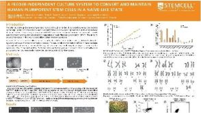 科学海报A Feeder-Independent Culture System to Convert and Maintain Human Pluripotent Stem Cells in a Naïve-Like State
 沪公网安备31010102008431号