Assessing differentiation status of human embryonic stem cells noninvasively using Raman microspectroscopy.
Raman microspectroscopy is an attractive approach for chemical imaging of biological specimens,including live cells,without the need for chemi-selective stains. Using a microspectrometer,near-infrared Raman spectra throughout the range 663 cm(-1) to 1220 cm(-1) were obtained from colonies of CA1 human embryonic stem cells (hESCs) and CA1 cells that had been stimulated to differentiate for 3 weeks by 10% fetal bovine serum on gelatin. Distributions and intensities of spectral bands attributed to proteins varied significantly between undifferentiated and differentiated cells. Importantly,compared to proteins and lipids,the band intensities of nucleic acids were dominant in undifferentiated cells with a dominance-reversal in differentiated cells. Thus,we could identify intensity ratios of particular protein-related bands (e.g.,757 cm(-1) tryptophan) to nucleic acid bands (784 cm(-1) DNA/RNA composite) that were effective in discriminating between spectra of undifferentiated and differentiated cells. We observed no discernible negative effects due to the laser exposure in terms of morphology,proliferation,or pluripotency of the stem cells. We conclude that Raman microscopy and complementary data processing procedures provide a rapid,noninvasive approach that can distinguish hESCs from differentiated cells. This is the first report to identify specific Raman markers for the differentiation status of hESCs.
View Publication
产品类型:
产品号#:
05850
05857
05870
05875
85850
85857
85870
85875
产品名:
mTeSR™1
mTeSR™1
Loewer S et al. (DEC 2010)
Nature genetics 42 12 1113--7
Large intergenic non-coding RNA-RoR modulates reprogramming of human induced pluripotent stem cells.
The conversion of lineage-committed cells to induced pluripotent stem cells (iPSCs) by reprogramming is accompanied by a global remodeling of the epigenome,resulting in altered patterns of gene expression. Here we characterize the transcriptional reorganization of large intergenic non-coding RNAs (lincRNAs) that occurs upon derivation of human iPSCs and identify numerous lincRNAs whose expression is linked to pluripotency. Among these,we defined ten lincRNAs whose expression was elevated in iPSCs compared with embryonic stem cells,suggesting that their activation may promote the emergence of iPSCs. Supporting this,our results indicate that these lincRNAs are direct targets of key pluripotency transcription factors. Using loss-of-function and gain-of-function approaches,we found that one such lincRNA (lincRNA-RoR) modulates reprogramming,thus providing a first demonstration for critical functions of lincRNAs in the derivation of pluripotent stem cells.
View Publication
Widespread Chromatin Accessibility at Repetitive Elements Links Stem Cells with Human Cancer.
Chromatin regulation is critical for differentiation and disease. However,features linking the chromatin environment of stem cells with disease remain largely unknown. We explored chromatin accessibility in embryonic and multipotent stem cells and unexpectedly identified widespread chromatin accessibility at repetitive elements. Integrating genomic and biochemical approaches,we demonstrate that these sites of increased accessibility are associated with well-positioned nucleosomes marked by distinct histone modifications. Differentiation is accompanied by chromatin remodeling at repetitive elements associated with altered expression of genes in relevant developmental pathways. Remarkably,we found that the chromatin environment of Ewing sarcoma,a mesenchymally derived tumor,is shared with primary mesenchymal stem cells (MSCs). Accessibility at repetitive elements in MSCs offers a permissive environment that is exploited by the critical oncogene responsible for this cancer. Our data demonstrate that stem cells harbor a unique chromatin landscape characterized by accessibility at repetitive elements,a feature associated with differentiation and oncogenesis.
View Publication
产品类型:
产品号#:
05850
05857
05870
05875
85850
85857
85870
85875
产品名:
mTeSR™1
mTeSR™1
Wang S et al. (MAR 2015)
Sci Rep 5 9232
Differentiation of human induced pluripotent stem cells to mature functional Purkinje neurons.
It remains a challenge to differentiate human induced pluripotent stem cells (iPSCs) or embryonic stem (ES) cells to Purkinje cells. In this study,we derived iPSCs from human fibroblasts and directed the specification of iPSCs first to Purkinje progenitors,by adding Fgf2 and insulin to the embryoid bodies (EBs) in a time-sensitive manner,which activates the endogenous production of Wnt1 and Fgf8 from EBs that further patterned the cells towards a midbrain-hindbrain-boundary tissue identity. Neph3-positive human Purkinje progenitors were sorted out by using flow cytometry and cultured either alone or with granule cell precursors,in a 2-dimensional or 3-dimensional environment. However,Purkinje progenitors failed to mature further under above conditions. By co-culturing human Purkinje progenitors with rat cerebellar slices,we observed mature Purkinje-like cells with right morphology and marker expression patterns,which yet showed no appropriate membrane properties. Co-culture with human fetal cerebellar slices drove the progenitors to not only morphologically correct but also electrophysiologically functional Purkinje neurons. Neph3-posotive human cells could also survive transplantation into the cerebellum of newborn immunodeficient mice and differentiate to L7- and Calbindin-positive neurons. Obtaining mature human Purkinje cells in vitro has significant implications in studying the mechanisms of spinocerebellar ataxias and other cerebellar diseases.
View Publication
产品类型:
产品号#:
05850
05857
05870
05875
85850
85857
85870
85875
产品名:
mTeSR™1
mTeSR™1
Yea C-H et al. (JAN 2016)
Biomaterials 75 250--259
In situ label-free quantification of human pluripotent stem cells with electrochemical potential
Conventional methods for quantification of undifferentiated pluripotent stem cells such as fluorescence-activated cell sorting and real-time PCR analysis have technical limitations in terms of their sensitivity and recyclability. Herein,we designed a real-time in situ label-free monitoring system on the basis of a specific electrochemical signature of human pluripotent stem cells in vitro. The intensity of the signal of hPSCs highly corresponded to the cell number and remained consistent in a mixed population with differentiated cells. The electrical charge used for monitoring did not markedly affect the proliferation rate or molecular characteristics of differentiated human aortic smooth muscle cells. After YM155 treatment to ablate undifferentiated hPSCs,their specific signal was significantly reduced. This suggests that detection of the specific electrochemical signature of hPSCs would be a valid approach to monitor potential contamination of undifferentiated hPSCs,which can assess the risk of teratoma formation efficiently and economically.
View Publication
产品类型:
产品号#:
05850
05857
05870
05875
37350
37354
85850
85857
85870
85875
产品名:
D-PBS不含钙镁平衡盐溶液
D-PBS不含钙镁平衡盐溶液,10倍浓缩液
mTeSR™1
mTeSR™1
Sigova Aa et al. (FEB 2013)
Proceedings of the National Academy of Sciences of the United States of America 110 8 2876--81
Divergent transcription of long noncoding RNA/mRNA gene pairs in embryonic stem cells.
Many long noncoding RNA (lncRNA) species have been identified in mammalian cells,but the genomic origin and regulation of these molecules in individual cell types is poorly understood. We have generated catalogs of lncRNA species expressed in human and murine embryonic stem cells and mapped their genomic origin. A surprisingly large fraction of these transcripts (textgreater60%) originate from divergent transcription at promoters of active protein-coding genes. The divergently transcribed lncRNA/mRNA gene pairs exhibit coordinated changes in transcription when embryonic stem cells are differentiated into endoderm. Our results reveal that transcription of most lncRNA genes is coordinated with transcription of protein-coding genes.
View Publication
产品类型:
产品号#:
05850
05857
05870
05875
85850
85857
85870
85875
产品名:
mTeSR™1
mTeSR™1
Xue Y et al. (AUG 2013)
PLoS ONE 8 8 e70573
Generating a Non-Integrating Human Induced Pluripotent Stem Cell Bank from Urine-Derived Cells
Induced pluripotent stem cell (iPS cell) holds great potential for applications in regenerative medicine,drug discovery,and disease modeling. We describe here a practical method to generate human iPS cells from urine-derived cells (UCs) under feeder-free,virus-free,serum-free condition and without oncogene c-MYC. We showed that this approach could be applied in a large population with different genetic backgrounds. UCs are easily accessible and exhibit high reprogramming efficiency,offering advantages over other cell types used for the purpose of iPS generation. Using the approach described in this study,we have generated 93 iPS cell lines from 20 donors with diverse genetic backgrounds. The non-viral iPS cell bank with these cell lines provides a valuable resource for iPS cells research,facilitating future applications of human iPS cells.
View Publication
miR-34a contributes to megakaryocytic differentiation of K562 cells independently of p53.
The role of miRNAs in regulating megakaryocyte differentiation was examined using bipotent K562 human leukemia cells. miR-34a is strongly up-regulated during phorbol ester-induced megakaryocyte differentiation,but not during hemin-induced erythrocyte differentiation. Enforced expression of miR-34a in K562 cells inhibits cell proliferation,induces cell-cycle arrest in G(1) phase,and promotes megakaryocyte differentiation as measured by CD41 induction. miR-34a expression is also up-regulated during thrombopoietin-induced differentiation of CD34(+) hematopoietic precursors,and its enforced expression in these cells significantly increases the number of megakaryocyte colonies. miR-34a directly regulates expression of MYB,facilitating megakaryocyte differentiation,and of CDK4 and CDK6,to inhibit the G(1)/S transition. However,these miR-34a target genes are down-regulated rapidly after inducing megakaryocyte differentiation before miR-34a is induced. This suggests that miR-34a is not responsible for the initial down-regulation but may contribute to maintaining their suppression later on. Previous studies have implicated miR-34a as a tumor suppressor gene whose transcription is activated by p53. However,in p53-null K562 cells,phorbol esters induce miR-34a expression independently of p53 by activating an alternative phorbol ester-responsive promoter to produce a longer pri-miR-34a transcript.
View Publication
产品类型:
产品号#:
02696
09850
70008
70008.1
70008.2
70008.3
70008.4
70008.5
70008.6
04971
04902
04901
04963
04962
200-0002
200-0001
200-0000
产品名:
StemSpan™巨核细胞扩增添加物 (100X)
冻存的人脐带血CD34+细胞
冻存的人脐带血CD34+细胞
冻存的人脐带血CD34+细胞
冻存的人脐带血CD34+细胞
冻存的人脐带血CD34+细胞
冻存的人脐带血CD34+细胞
MegaCult™-C含细胞因子全套试剂盒
胶原蛋白溶液
MegaCult™-C含细胞因子培养基
双室载玻片套件
MegaCult™-C CFU-Mk染色试剂盒
冻存的人脐带血CD34+细胞
冻存的人脐带血CD34+细胞
冻存的人脐带血CD34+细胞
Derda R et al. (FEB 2010)
Journal of the American Chemical Society 132 4 1289--1295
High-throughput discovery of synthetic surfaces that support proliferation of pluripotent cells.
Synthetic materials that promote the growth or differentiation of cells have advanced the fields of tissue engineering and regenerative medicine. Most functional biomaterials are based on a handful of peptide sequences derived from protein ligands for cell surface receptors. Because few proteins possess short peptide sequences that alone can engage cell surface receptors,the repertoire of receptors that can be targeted with this approach is limited. Materials that bind diverse classes of receptors,however,may be needed to guide cell growth and differentiation. To provide access to such new materials,we utilized phage display to identify novel peptides that bind to the surface of pluripotent cells. Using human embryonal carcinoma (EC) cells as bait,approximately 3 x 10(4) potential cell-binding phage clones were isolated. The pool was narrowed using an enzyme-linked immunoassay: 370 clones were tested,and seven cell-binding peptides were identified. Of these,six sequences possess EC cell-binding ability. Specifically,when displayed by self-assembled monolayers (SAMs) of alkanethiols on gold,they mediate cell adhesion. The corresponding soluble peptides block this adhesion,indicating that the identified peptide sequences are specific. They also are functional. Synthetic surfaces displaying phage-derived peptides support growth of undifferentiated human embryonic stem (ES) cells. When these cells were cultured on SAMs presenting the sequence TVKHRPDALHPQ or LTTAPKLPKVTR in a chemically defined medium (mTeSR),they expressed markers of pluripotency at levels similar to those of cells cultured on Matrigel. Our results indicate that this screening strategy is a productive avenue for the generation of materials that control the growth and differentiation of cells.
View Publication
产品类型:
产品号#:
05850
05857
05870
05875
85850
85857
85870
85875
产品名:
mTeSR™1
mTeSR™1
Laird DJ et al. (DEC 2005)
Cell 123 7 1351--60
Stem cells are units of natural selection in a colonial ascidian.
Stem cells are highly conserved biological units of development and regeneration. Here we formally demonstrate that stem cell lineages are also legitimate units of natural selection. In a colonial ascidian,Botryllus schlosseri,vascular fusion between genetically distinct individuals results in cellular parasitism of somatic tissues,gametes,or both. We show that genetic hierarchies of somatic and gametic parasitism following fusion can be replicated by transplanting cells between colonies. We prospectively isolate a population of multipotent,self-renewing stem cells that retain their competitive phenotype upon transplantation. Their single-cell contribution to either somatic or germline fates,but not to both,is consistent with separate lineages of somatic and germline stem cells or pluripotent stem cells that differentiate according to the niche in which they land. Since fusion is restricted to individuals that share a fusion/histocompatibility allele,these data suggest that histocompatibility genes in Botryllus evolved to protect the body from parasitic stem cells usurping asexual or sexual inheritance.
View Publication
产品类型:
产品号#:
01700
01705
01701
01702
产品名:
ALDEFLUOR™ 试剂盒
ALDEFLUOR™ DEAB试剂, 1.5 mM, 1 mL
ALDEFLUOR™检测缓冲液
Xing Q et al. (AUG 2014)
Digestive and liver disease : official journal of the Italian Society of Gastroenterology and the Italian Association for the Study of the Liver 46 8 731--737
Hepatectomised patient sera promote hepatocyte differentiation of human-induced pluripotent stem cells.
Background: Human induced pluripotent stem cells,which can be differentiated into hepatocyte-like cells,could provide a source for liver regeneration and bio-artificial liver devices. However,the functionality of hepatocyte-like cells is significantly lower than that of primary hepatocytes. Aims: To investigate whether serum from patients undergoing hepatectomy might promote differentiation from human induced pluripotent stem cells to hepatocyte-like cells. Methods: Serum from patients undergoing hepatectomy (acquired pre-hepatectomy and 3. hours,1 day and 3 days post-hepatectomy) was used to replace foetal bovine serum when differentiating human induced pluripotent stem cells into hepatocyte-like cells. Properties of hepatocyte-like cells were assessed and compared with cells cultured in foetal bovine serum. Results: The differentiation efficiency and functionality of hepatocyte-like cells cultured in human serum 3. hours and 1 day post-hepatectomy were superior to those cultured in foetal bovine serum and human serum pre-hepatectomy. Human serum 3 days post-hepatectomy had an equal effect to that of human serum pre-hepatectomy. Some cytochrome P450 isozyme transcript levels of hepatocyte-like cells cultured in human serum were higher than those cultured in foetal bovine serum. Conclusion: Human serum,particularly that acquired relatively soon after hepatectomy,can enhance the differentiation efficiency and functionality of hepatocyte-like cells derived from human induced pluripotent stem cells. textcopyright 2014 Editrice Gastroenterologica Italiana S.r.l.
View Publication


 EasySep™小鼠TIL(CD45)正选试剂盒





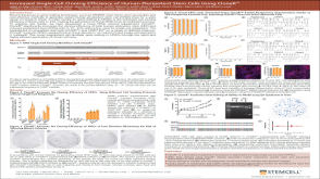 科学海报Increased Single-Cell Cloning Efficiency of Human Pluripotent Stem Cells Using CloneR™
 沪公网安备31010102008431号