Generating human intestinal tissue from pluripotent stem cells in vitro.
Here we describe a protocol for generating 3D human intestinal tissues (called organoids) in vitro from human pluripotent stem cells (hPSCs). To generate intestinal organoids,pluripotent stem cells are first differentiated into FOXA2(+)SOX17(+) endoderm by treating the cells with activin A for 3 d. After endoderm induction,the pluripotent stem cells are patterned into CDX2(+) mid- and hindgut tissue using FGF4 and WNT3a. During this patterning step,3D mid- or hindgut spheroids bud from the monolayer epithelium attached to the tissue culture dish. The 3D spheroids are further cultured in Matrigel along with prointestinal growth factors,and they proliferate and expand over 1-3 months to give rise to intestinal tissue,complete with intestinal mesenchyme and epithelium comprising all of the major intestinal cell types. To date,this is the only method for efficiently directing the differentiation of hPSCs into 3D human intestinal tissue in vitro.
View Publication
产品类型:
产品号#:
05850
05857
05870
05875
85850
85857
85870
85875
产品名:
mTeSR™1
mTeSR™1
Zhou T et al. (DEC 2012)
Nature protocols 7 12 2080--9
Generation of human induced pluripotent stem cells from urine samples.
Human induced pluripotent stem cells (iPSCs) have been generated with varied efficiencies from multiple tissues. Yet,acquiring donor cells is,in most instances,an invasive procedure that requires laborious isolation. Here we present a detailed protocol for generating human iPSCs from exfoliated renal epithelial cells present in urine. This method is advantageous in many circumstances,as the isolation of urinary cells is simple (30 ml of urine are sufficient),cost-effective and universal (can be applied to any age,gender and race). Moreover,the entire procedure is reasonably quick--around 2 weeks for the urinary cell culture and 3-4 weeks for the reprogramming--and the yield of iPSC colonies is generally high--up to 4% using retroviral delivery of exogenous factors. Urinary iPSCs (UiPSCs) also show excellent differentiation potential,and thus represent a good choice for producing pluripotent cells from normal individuals or patients with genetic diseases,including those affecting the kidney.
View Publication
Tan BL et al. (MAR 2003)
The Journal of biological chemistry 278 13 11686--95
Functional and biochemical consequences of abrogating the activation of multiple diverse early signaling pathways in Kit. Role for Src kinase pathway in Kit-induced cooperation with erythropoietin receptor.
Kit receptor tyrosine kinase and erythropoietin receptor (Epo-R) cooperate in regulating blood cell development. Mice that lack the expression of Kit or Epo-R die in utero of severe anemia. Stimulation of Kit by its ligand,stem cell factor activates several distinct early signaling pathways,including phospholipase C gamma,phosphatidylinositol 3-kinase,Src kinase,Grb2,and Grb7. The role of these pathways in Kit-induced growth,proliferation,or cooperation with Epo-R is not known. We demonstrate that inactivation of any one of these early signaling pathways in Kit significantly impairs growth and proliferation. However,inactivation of the Src pathway demonstrated the most profound defect. Combined stimulation with Epo also resulted in impaired cooperation between Src-defective Kit mutant and Epo-R and,to a lesser extent,with Kit mutants defective in the activation of phosphatidylinositol 3-kinase or Grb2. The impaired cooperation between the Src-defective Kit mutant and Epo-R was associated with reduced transphosphorylation of Epo-R and expression of c-Myc. Remarkably,restoration of only the Src pathway in a Kit receptor defective in the activation of all early signaling pathways demonstrated a 50% correction in proliferation in response to Kit stimulation and completely restored the cooperation with Epo-R. These data demonstrate an essential role for Src pathway in regulating growth,proliferation,and cooperation with Epo-R downstream from Kit.
View Publication
产品类型:
产品号#:
03434
03444
产品名:
MethoCult™ GF M3434
MethoCult™ GF M3434
Sareen D et al. (AUG 2014)
Journal of Comparative Neurology 522 12 2707--2728
Human induced pluripotent stem cells are a novel source of neural progenitor cells (iNPCs) that migrate and integrate in the rodent spinal cord
Transplantation of human neural progenitor cells (NPCs) into the brain or spinal cord to replace lost cells,modulate the injury environment,or create a permissive milieu to protect and regenerate host neurons is a promising therapeutic strategy for neurological diseases. Deriving NPCs from human fetal tissue is feasible,although problematic issues include limited sources and ethical concerns. Here we describe a new and abundant source of NPCs derived from human induced pluripotent stem cells (iPSCs). A novel chopping technique was used to transform adherent iPSCs into free-floating spheres that were easy to maintain and were expandable (EZ spheres) (Ebert et al. [2013] Stem Cell Res 10:417–427). These EZ spheres could be differentiated towards NPC spheres with a spinal cord phenotype using a combination of all-trans retinoic acid (RA) and epidermal growth factor (EGF) and fibroblast growth factor-2 (FGF-2) mitogens. Suspension cultures of NPCs derived from human iPSCs or fetal tissue have similar characteristics,although they were not similar when grown as adherent cells. In addition,iPSC-derived NPCs (iNPCs) survived grafting into the spinal cord of athymic nude rats with no signs of overgrowth and with a very similar profile to human fetal-derived NPCs (fNPCs). These results suggest that human iNPCs behave like fNPCs and could thus be a valuable alternative for cellular regenerative therapies of neurological diseases. J. Comp. Neurol. 522:2707–2728,2014. textcopyright 2014 Wiley Periodicals,Inc.
View Publication
产品类型:
产品号#:
05850
05857
05870
05875
85850
85857
85870
85875
产品名:
mTeSR™1
mTeSR™1
Thomas RJ et al. (APR 2009)
Biotechnology and Bioengineering 102 6 1636--1644
Automated, scalable culture of human embryonic stem cells in feeder-free conditions.
Large-scale manufacture of human embryonic stem cells (hESCs) is prerequisite to their widespread use in biomedical applications. However,current hESC culture strategies are labor-intensive and employ highly variable processes,presenting challenges for scaled production and commercial development. Here we demonstrate that passaging of the hESC lines,HUES7,and NOTT1,with trypsin in feeder-free conditions,is compatible with complete automation on the CompacT SelecT,a commercially available and industrially relevant robotic platform. Pluripotency was successfully retained,as evidenced by consistent proliferation during serial passage,expression of stem cell markers (OCT4,NANOG,TRA1-81,and SSEA-4),stable karyotype,and multi-germlayer differentiation in vitro,including to pharmacologically responsive cardiomyocytes. Automation of hESC culture will expedite cell-use in clinical,scientific,and industrial applications.
View Publication
产品类型:
产品号#:
05850
05857
05870
05875
85850
85857
85870
85875
产品名:
mTeSR™1
mTeSR™1
Rodin S et al. (JUN 2010)
Nature biotechnology 28 6 611--5
Long-term self-renewal of human pluripotent stem cells on human recombinant laminin-511.
We describe a system for culturing human embryonic stem (hES) cells and induced pluripotent stem (iPS) cells on a recombinant form of human laminin-511,a component of the natural hES cell niche. The system is devoid of animal products and feeder cells and contains only one undefined component,human albumin. The hES cells self-renewed with normal karyotype for at least 4 months (20 passages),after which the cells could produce teratomas containing cell lineages of all three germ layers. When plated on laminin-511 in small clumps,hES cells spread out in a monolayer,maintaining cellular homogeneity with approximately 97% OCT4-positive cells. Adhesion of hES cells was dependent on alpha6beta1 integrin. The use of homogeneous monolayer hES or iPS cell cultures provides more controllable conditions for the design of differentiation methods. This xeno-free and feeder-free system may be useful for the development of cell lineages for therapeutic purposes.
View Publication
Growth of mesenchymal stem cells on electrospun type I collagen nanofibers.
We reconstituted type I collagen nanofibers prepared by electrospin technology and examined the morphology,growth,adhesion,cell motility,and osteogenic differentiation of human bone marrow-derived mesenchymal stem cells (MSCs) on three nano-sized diameters (50-200,200-500,and 500-1,000 nm). Results from scanning electron microscopy showed that cells on the nanofibers had a more polygonal and flattened cell morphology. MTS (3-[4,5-dimethythiazol-2-yl]-5-[3-carboxy-methoxyphenyl]-2-[4-sul-fophenyl]-2H-tetrazolium compound) assay demonstrated that the MSCs grown on 500-1,000-nm nanofibers had significantly higher cell viability than the tissue culture polystyrene control. A decreased amount of focal adhesion formation was apparent in which quantifiable staining area of the cytoplasmic protein vinculin for the 200-500-nm nanofibers was 39% less compared with control,whereas the area of quantifiable vinculin staining was 45% less for both the 200-500-nm and 500-1,000-nm nanofibers. The distances of cell migration were quantified on green fluorescent protein-nucleofected cells and was 56.7%,37.3%,and 46.3% for 50-200,200-500,and 500-1,000 nm,respectively,compared with those on the control. Alkaline phosphatase activity demonstrated no differences after 12 days of osteogenic differentiation,and reverse transcription-polymerase chain reaction (RT-PCR) analysis showed comparable osteogenic gene expression of osteocalcin,osteonectin,and ostepontin between cells differentiated on polystyrene and nanofiber surfaces. Moreover,single-cell RT-PCR of type I collagen gene expression demonstrated higher expression on cells seeded on the nanofibers. Therefore,type I collagen nanofibers support the growth of MSCs without compromising their osteogenic differentiation capability and can be used as a scaffold for bone tissue engineering to facilitate intramembranous bone formation. Further efforts are necessary to enhance their biomimetic properties.
View Publication
产品类型:
产品号#:
15027
15067
产品名:
RosetteSep™人骨髓祖细胞预富集抗体混合物
RosetteSep™人骨髓祖细胞预富集抗体混合物
Sun Y et al. (SEP 2013)
eLife 2013 2 e00508
Imaging-based chemical screening reveals activity-dependent neural differentiation of pluripotent stem cells
Pluripotent stem cells have the potential to become most of the cell types that make up an organism. However,the signals that trigger these cells to turn into neurons rather than lung cells or muscle cells,for example,are not fully understood. Proteins called growth factors are known to have a role in this process,as are transcription factors,but it is not clear if other factors are also involved. In an attempt to identify additional mechanisms that could contribute to the formation of neurons,Sun et al. screened more than 2,000 small molecules for their ability to transform mouse pluripotent stem cells into neurons in cell culture. Surprisingly,they found that a compound called selamectin,which is used to treat parasitic flatworm infections,also triggered stem cells to turn into neurons. Selamectin works by blocking a particular type of ion channel in flatworms,but this ion channel is not found in vertebrates,which means that selamectin must be promoting the formation of neurons in mice via a different mechanism. Given that a drug related to selamectin is known to act on a subtype of receptors for the neurotransmitter GABA,Sun et al. wondered whether these receptors—known as GABAA receptors—might also underlie the effects of selamectin. Consistent with this idea,drugs that increased GABAA activity stimulated the formation of neurons,whereas drugs that reduced GABAA function blocked the effects of selamectin. In addition,Sun et al. showed that selamectin triggers human embryonic stem cells to become neurons,and that it also promotes the formation of new neurons in developing zebrafish in vivo. As well as revealing an additional mechanism for the formation of neurons from stem cells,the screening technique introduced by Sun et al. could help to identify further pro-neuronal molecules,which could aid the treatment of neurodevelopmental and neurodegenerative disorders. DOI: [http://dx.doi.org/10.7554/eLife.00508.002][1] [1]: /lookup/doi/10.7554/eLife.00508.002
View Publication

 EasySep™小鼠TIL(CD45)正选试剂盒


 57:26
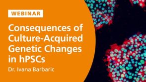
线上讲座Consequences of Culture-Acquired Genetic Changes in Human Pluripotent Stem Cells发布日期: 02/20/2024
57:26
线上讲座Consequences of Culture-Acquired Genetic Changes in Human Pluripotent Stem Cells发布日期: 02/20/2024 实验方案How to Cryopreserve Human Pluripotent Stem Cells in CryoStor® CS10

 沪公网安备31010102008431号