Gomez AM et al. (MAR 2015)
The Journal of Immunology 194 5 2300--8
HIV-1-triggered release of type I IFN by plasmacytoid dendritic cells induces BAFF production in monocytes.
HIV-1 infection leads to numerous B cell abnormalities,including hypergammaglobulinemia,nonspecific B cell activation,nonspecific class switching,increased cell turnover,breakage of tolerance,increased immature/transitional B cells,B cell malignancies,as well as a loss of capacity to generate and maintain memory,all of which contribute to a global impairment of the immune humoral compartment. Several cytokines and soluble factors,which are increased in sera of HIV-1-infected individuals,have been suggested to directly or indirectly contribute to these B cell dysfunctions,and one of these is the B cell-activating factor (BAFF). We report in this study that HIV-1 (X4- and R5-tropic) upregulates BAFF expression and secretion by human monocytes. Moreover,we show that the virus-mediated production of BAFF by monocytes relies on a type I IFN response by a small percentage of plasmacytoid dendritic cells (pDCs) present in the monocyte cultures. HIV-1-induced type I IFN by pDCs triggers BAFF production in both classical and intermediate monocytes,but not in nonclassical monocytes,which nonetheless display a very strong basal BAFF production. We report also that basal BAFF secretion was higher in monocytes obtained from females compared with those from male donors. This study provides a novel mechanistic explanation for the increased BAFF levels observed during HIV-1 infection and highlights the importance of pDC/monocyte crosstalk to drive BAFF secretion.
View Publication
Fogli M et al. (JUL 2008)
PLoS pathogens 4 7 e1000101
Lysis of endogenously infected CD4+ T cell blasts by rIL-2 activated autologous natural killer cells from HIV-infected viremic individuals.
Understanding the cellular mechanisms that ensure an appropriate innate immune response against viral pathogens is an important challenge of biomedical research. In vitro studies have shown that natural killer (NK) cells purified from healthy donors can kill heterologous cell lines or autologous CD4+ T cell blasts exogenously infected with several strains of HIV-1. However,it is not known whether the deleterious effects of high HIV-1 viremia interferes with the NK cell-mediated cytolysis of autologous,endogenously HIV-1-infected CD4+ T cells. Here,we stimulate primary CD4+ T cells,purified ex vivo from HIV-1-infected viremic patients,with PHA and rIL2 (with or without rIL-7). This experimental procedure allows for the significant expansion and isolation of endogenously infected CD4+ T cell blasts detected by intracellular staining of p24 HIV-1 core antigen. We show that,subsequent to the selective down-modulation of MHC class-I (MHC-I) molecules,HIV-1-infected p24(pos) blasts become partially susceptible to lysis by rIL-2-activated NK cells,while uninfected p24(neg) blasts are spared from killing. This NK cell-mediated killing occurs mainly through the NKG2D activation pathway. However,the degree of NK cell cytolytic activity against autologous,endogenously HIV-1-infected CD4+ T cell blasts that down-modulate HLA-A and -B alleles and against heterologous MHC-I(neg) cell lines is particularly low. This phenomenon is associated with the defective surface expression and engagement of natural cytotoxicity receptors (NCRs) and with the high frequency of the anergic CD56(neg)/CD16(pos) subsets of highly dysfunctional NK cells from HIV-1-infected viremic patients. Collectively,our data demonstrate that the chronic viral replication of HIV-1 in infected individuals results in several phenotypic and functional aberrancies that interfere with the NK cell-mediated killing of autologous p24(pos) blasts derived from primary T cells.
View Publication
产品类型:
产品号#:
19052
19052RF
19055
19055RF
产品名:
EasySep™人CD4+ T细胞富集试剂盒
RoboSep™ 人CD4+ T细胞富集试剂盒含滤芯吸头
EasySep™人NK细胞富集试剂盒
RoboSep™ 人NK细胞富集试剂盒含滤芯吸头
Hrecka K et al. (JUL 2016)
Proceedings of the National Academy of Sciences of the United States of America 113 27 E3921--30
HIV-1 and HIV-2 exhibit divergent interactions with HLTF and UNG2 DNA repair proteins.
HIV replication in nondividing host cells occurs in the presence of high concentrations of noncanonical dUTP,apolipoprotein B mRNA-editing,enzyme-catalytic,polypeptide-like 3 (APOBEC3) cytidine deaminases,and SAMHD1 (a cell cycle-regulated dNTP triphosphohydrolase) dNTPase,which maintains low concentrations of canonical dNTPs in these cells. These conditions favor the introduction of marks of DNA damage into viral cDNA,and thereby prime it for processing by DNA repair enzymes. Accessory protein Vpr,found in all primate lentiviruses,and its HIV-2/simian immunodeficiency virus (SIV) SIVsm paralogue Vpx,hijack the CRL4(DCAF1) E3 ubiquitin ligase to alleviate some of these conditions,but the extent of their interactions with DNA repair proteins has not been thoroughly characterized. Here,we identify HLTF,a postreplication DNA repair helicase,as a common target of HIV-1/SIVcpz Vpr proteins. We show that HIV-1 Vpr reprograms CRL4(DCAF1) E3 to direct HLTF for proteasome-dependent degradation independent from previously reported Vpr interactions with base excision repair enzyme uracil DNA glycosylase (UNG2) and crossover junction endonuclease MUS81,which Vpr also directs for degradation via CRL4(DCAF1) E3. Thus,separate functions of HIV-1 Vpr usurp CRL4(DCAF1) E3 to remove key enzymes in three DNA repair pathways. In contrast,we find that HIV-2 Vpr is unable to efficiently program HLTF or UNG2 for degradation. Our findings reveal complex interactions between HIV-1 and the DNA repair machinery,suggesting that DNA repair plays important roles in the HIV-1 life cycle. The divergent interactions of HIV-1 and HIV-2 with DNA repair enzymes and SAMHD1 imply that these viruses use different strategies to guard their genomes and facilitate their replication in the host.
View Publication
产品类型:
产品号#:
19052
19052RF
19051
19051RF
产品名:
EasySep™人CD4+ T细胞富集试剂盒
RoboSep™ 人CD4+ T细胞富集试剂盒含滤芯吸头
EasySep™人T细胞富集试剂盒
RoboSep™ 人T细胞富集试剂盒含滤芯吸头
Kourjian G et al. (MAY 2016)
Journal of Immunology 196 9 3595--607
HIV Protease Inhibitor-Induced Cathepsin Modulation Alters Antigen Processing and Cross-Presentation.
Immune recognition by T cells relies on the presentation of pathogen-derived peptides by infected cells,but the persistence of chronic infections calls for new approaches to modulate immune recognition. Ag cross-presentation,the process by which pathogen Ags are internalized,degraded,and presented by MHC class I,is crucial to prime CD8 T cell responses. The original degradation of Ags is performed by pH-dependent endolysosomal cathepsins. In this article,we show that HIV protease inhibitors (PIs) prescribed to HIV-infected persons variably modulate cathepsin activities in human APCs,dendritic cells and macrophages,and CD4 T cells,three cell subsets infected by HIV. Two HIV PIs acted in two complementary ways on cathepsin hydrolytic activities: directly on cathepsins and indirectly on their regulators by inhibiting Akt kinase activities,reducing NADPH oxidase 2 activation,and lowering phagolysosomal reactive oxygen species production and pH,which led to enhanced cathepsin activities. HIV PIs modified endolysosomal degradation and epitope production of proteins from HIV and other pathogens in a sequence-dependent manner. They altered cross-presentation of Ags by dendritic cells to epitope-specific T cells and T cell-mediated killing. HIV PI-induced modulation of Ag processing partly changed the MHC self-peptidome displayed by primary human cells. This first identification,to our knowledge,of prescription drugs modifying the regulation of cathepsin activities and the MHC-peptidome may provide an alternate therapeutic approach to modulate immune recognition in immune disease beyond HIV.
View Publication


 EasySep™小鼠TIL(CD45)正选试剂盒




 EasySep™非人灵长类T细胞分选试剂盒 通过免疫磁珠负选从包括恒河猴在内的非人灵长类中分离出无磁珠标记的T细胞
 EasySep™非人灵长类CD4+ T细胞分选试剂盒
 EasySep™非人灵长类CD8+ T细胞分选试剂盒
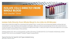 产品手册EasySep™ Direct 仅需20分钟,从全血中分选细胞


 沪公网安备31010102008431号