Kourjian G et al. (MAY 2016)
Journal of Immunology 196 9 3595--607
HIV Protease Inhibitor-Induced Cathepsin Modulation Alters Antigen Processing and Cross-Presentation.
Immune recognition by T cells relies on the presentation of pathogen-derived peptides by infected cells,but the persistence of chronic infections calls for new approaches to modulate immune recognition. Ag cross-presentation,the process by which pathogen Ags are internalized,degraded,and presented by MHC class I,is crucial to prime CD8 T cell responses. The original degradation of Ags is performed by pH-dependent endolysosomal cathepsins. In this article,we show that HIV protease inhibitors (PIs) prescribed to HIV-infected persons variably modulate cathepsin activities in human APCs,dendritic cells and macrophages,and CD4 T cells,three cell subsets infected by HIV. Two HIV PIs acted in two complementary ways on cathepsin hydrolytic activities: directly on cathepsins and indirectly on their regulators by inhibiting Akt kinase activities,reducing NADPH oxidase 2 activation,and lowering phagolysosomal reactive oxygen species production and pH,which led to enhanced cathepsin activities. HIV PIs modified endolysosomal degradation and epitope production of proteins from HIV and other pathogens in a sequence-dependent manner. They altered cross-presentation of Ags by dendritic cells to epitope-specific T cells and T cell-mediated killing. HIV PI-induced modulation of Ag processing partly changed the MHC self-peptidome displayed by primary human cells. This first identification,to our knowledge,of prescription drugs modifying the regulation of cathepsin activities and the MHC-peptidome may provide an alternate therapeutic approach to modulate immune recognition in immune disease beyond HIV.
View Publication
产品类型:
产品号#:
17952
17952RF
100-0696
19654
19654RF
产品名:
EasySep™人CD4+ T细胞分选试剂盒
RoboSep™ 人CD4+ T细胞分选试剂盒
EasySep™人CD4+ T细胞分离试剂盒
EasySep™ Direct 人 PBMC 分选试剂盒
RoboSep™ Direct 人 PBMC 分选试剂盒
Saï et al. (FEB 2016)
PLoS pathogens 12 2 e1005407
HMGB1 Is Involved in IFN-α Production and TRAIL Expression by HIV-1-Exposed Plasmacytoid Dendritic Cells: Impact of the Crosstalk with NK Cells.
Plasmacytoid dendritic cells (pDCs) are innate sensors of viral infections and important mediators of antiviral innate immunity through their ability to produce large amounts of IFN-α. Moreover,Toll-like receptor 7 (TLR7) and 9 (TLR9) ligands,such as HIV and CpG respectively,turn pDCs into TRAIL-expressing killer pDCs able to lyse HIV-infected CD4+ T cells. NK cells can regulate antiviral immunity by modulating pDC functions,and pDC production of IFN-α as well as cell-cell contact is required to promote NK cell functions. Impaired pDC-NK cell crosstalk was reported in the setting of HIV-1 infection,but the impact of HIV-1 on TRAIL expression and innate antiviral immunity during this crosstalk is unknown. Here,we report that low concentrations of CCR5-tropic HIV-1Ba-L promote the release of pro-inflammatory cytokines such as IFN-α,TNF-α,IFN-γ and IL-12,and CCR5-interacting chemokines (MIP-1α and MIP-1β) in NK-pDCs co-cultures. At high HIV-1BaL concentrations,the addition of NK cells did not promote the release of these mediators,suggesting that once efficiently triggered by the virus,pDCs could not integrate new activating signals delivered by NK cells. However,high HIV-1BaL concentrations were required to trigger IFN-α-mediated TRAIL expression at the surface of both pDCs and NK cells during their crosstalk. Interestingly,we identified the alarmin HMGB1,released at pDC-NK cell synapse,as an essential trigger for the secretion of IFN-α and IFN-related soluble mediators during the interplay of HIV-1 exposed pDCs with NK cells. Moreover,HMGB1 was found crucial for mTRAIL translocation to the plasma membrane of both pDCs and NK cells during their crosstalk following pDC exposure to HIV-1. Data from serum analyses of circulating HMGB1,HMGB1-specific antibodies,sTRAIL and IP-10 in a cohort of 67 HIV-1+ patients argue for the in vivo relevance of these observations. Altogether,these findings identify HMGB1 as a trigger for IFN-α-mediated TRAIL expression at the surface of pDCs and NK cells,and they suggest a novel mechanism of innate control of HIV-1 infection.
View Publication
产品类型:
产品号#:
19062
19062RF
19055
19055RF
17977
17977RF
产品名:
EasySep™人浆细胞样DC富集试剂盒
RoboSep™ 人浆细胞样DC富集试剂盒含滤芯吸头
EasySep™人NK细胞富集试剂盒
RoboSep™ 人NK细胞富集试剂盒含滤芯吸头
EasySep™人浆细胞样DC分选试剂盒
RoboSep™ 人浆细胞样DC分选试剂盒
Xu X et al. ( 2014)
The Journal of Immunology 193 8 4125--4136
IFN-Stimulated Gene LY6E in Monocytes Regulates the CD14/TLR4 Pathway but Inadequately Restrains the Hyperactivation of Monocytes during Chronic HIV-1 Infection
Owing to ongoing recognition of pathogen-associated molecular patterns,immune activation and upregulation of IFN-stimulated genes (ISGs) are sustained in the chronically infected host. Albeit most ISGs are important effectors for containing viral replication,some might exert compensatory immune suppression to limit pathological dysfunctions,although the mechanisms are not fully understood. In this study,we report that the ISG lymphocyte Ag 6 complex,locus E (LY6E) is a negative immune regulator of monocytes. LY6E in monocytes negatively modulated CD14 expression and subsequently dampened the responsiveness to LPS stimulation in vitro. In the setting of chronic HIV infection,the upregulation of LY6E was correlated with reduced CD14 level on monocytes; however,the immunosuppressive effect of LY6E was not adequate to remedy the hyperresponsiveness of activated monocytes. Taken together,the regulatory LY6E pathway in monocytes represents one of negative feedback mechanisms that counterbalance monocyte activation,which might be caused by LPS translocation through the compromised gastrointestinal tract during persistent HIV-1 infection and may serve as a potential target for immune intervention.
View Publication
Lin S et al. (SEP 2010)
Journal of virology 84 18 9487--96
HIV infection upregulates caveolin 1 expression to restrict virus production.
Caveolin 1 (Cav-1) is a major protein of a specific membrane lipid raft known as caveolae. Cav-1 interacts with the gp41 of the human immunodeficiency virus (HIV) envelope,but the role of Cav-1 in HIV replication and pathogenesis is not known. In this report,we demonstrate that HIV infection in primary human monocyte-derived macrophages (MDMs),THP-1 macrophages,and U87-CD4 cells results in a dramatic upregulation of Cav-1 expression mediated by HIV Tat. The activity of p53 is essential for Tat-induced Cav-1 expression,as our findings show enhanced phosphorylation of serine residues at amino acid positions 15 and 46 in the presence of Tat with a resulting Cav-1 upregulation. Furthermore,inhibition of p38 mitogen-activated protein kinase (MAPK) blocked phosphorylation of p53 in the presence of Tat. Infection studies of Cav-1-overexpressing cells reveal a significant reduction of HIV production. Taken together,these results suggest that HIV infection enhances the expression of Cav-1,which subsequently causes virus reduction,suggesting that Cav-1 may contribute to persistent infection in macrophages.
View Publication
产品类型:
产品号#:
19058
19058RF
100-1525
19059
19059RF
产品名:
EasySep™人单核细胞富集试剂盒(不去除CD16)
RoboSep™ 人单核细胞富集试剂盒(不去除CD16)含滤芯吸头
EasySep™人单核细胞富集试剂盒(不去除CD16)
EasySep™人单核细胞富集试剂盒
RoboSep™ 人单核细胞富集试剂盒含滤芯吸头
Sá et al. (JUN 2010)
Nature protocols 5 6 1033--41
Ex vivo T cell-based HIV suppression assay to evaluate HIV-specific CD8+ T-cell responses.
To advance T cell-based HIV vaccine development,it is necessary to evaluate the immune correlates of a protective CD8(+) T-cell response. We have developed an assay that assesses the capacity ex vivo of HIV-specific CD8(+) T cells to suppress HIV-1 infection of autologous CD4(+) T cells. This assay directly reflects the ultimate effector function of CD8(+) T cells,the elimination of infected cells,and accurately differentiates the effective CD8(+) T-cell response in spontaneous HIV controllers from ineffective responses in other patients. In this article,we describe all the steps from cell purification to assessment of viral replication by HIV-p24 ELISA and analysis,along with conditions for cell culturing,and how to choose the viral infectious dose that gives the most reliable results. We also depict the conditions of a rapid assay on the basis of flow cytometry analysis of intracellular HIV-Gag products. These procedures take 14-17 d when the p24 ELISA assay is used,or 6 d with the intracellular Gag assay.
View Publication
产品类型:
产品号#:
21000
20119
20155
19053
19053RF
20104
20124
产品名:
RoboSep™- S
RoboSep™ 吸头组件抛光剂
RoboSep™分选管套装(9个塑料管)
EasySep™人CD8+ T细胞富集试剂盒
RoboSep™ 人CD8+ T细胞富集试剂盒含滤芯吸头
RoboSep™ 缓冲液
RoboSep™ 缓冲液 (5X浓缩液)
Fogli M et al. (JUL 2008)
PLoS pathogens 4 7 e1000101
Lysis of endogenously infected CD4+ T cell blasts by rIL-2 activated autologous natural killer cells from HIV-infected viremic individuals.
Understanding the cellular mechanisms that ensure an appropriate innate immune response against viral pathogens is an important challenge of biomedical research. In vitro studies have shown that natural killer (NK) cells purified from healthy donors can kill heterologous cell lines or autologous CD4+ T cell blasts exogenously infected with several strains of HIV-1. However,it is not known whether the deleterious effects of high HIV-1 viremia interferes with the NK cell-mediated cytolysis of autologous,endogenously HIV-1-infected CD4+ T cells. Here,we stimulate primary CD4+ T cells,purified ex vivo from HIV-1-infected viremic patients,with PHA and rIL2 (with or without rIL-7). This experimental procedure allows for the significant expansion and isolation of endogenously infected CD4+ T cell blasts detected by intracellular staining of p24 HIV-1 core antigen. We show that,subsequent to the selective down-modulation of MHC class-I (MHC-I) molecules,HIV-1-infected p24(pos) blasts become partially susceptible to lysis by rIL-2-activated NK cells,while uninfected p24(neg) blasts are spared from killing. This NK cell-mediated killing occurs mainly through the NKG2D activation pathway. However,the degree of NK cell cytolytic activity against autologous,endogenously HIV-1-infected CD4+ T cell blasts that down-modulate HLA-A and -B alleles and against heterologous MHC-I(neg) cell lines is particularly low. This phenomenon is associated with the defective surface expression and engagement of natural cytotoxicity receptors (NCRs) and with the high frequency of the anergic CD56(neg)/CD16(pos) subsets of highly dysfunctional NK cells from HIV-1-infected viremic patients. Collectively,our data demonstrate that the chronic viral replication of HIV-1 in infected individuals results in several phenotypic and functional aberrancies that interfere with the NK cell-mediated killing of autologous p24(pos) blasts derived from primary T cells.
View Publication
Gomez AM et al. (MAR 2015)
The Journal of Immunology 194 5 2300--8
HIV-1-triggered release of type I IFN by plasmacytoid dendritic cells induces BAFF production in monocytes.
HIV-1 infection leads to numerous B cell abnormalities,including hypergammaglobulinemia,nonspecific B cell activation,nonspecific class switching,increased cell turnover,breakage of tolerance,increased immature/transitional B cells,B cell malignancies,as well as a loss of capacity to generate and maintain memory,all of which contribute to a global impairment of the immune humoral compartment. Several cytokines and soluble factors,which are increased in sera of HIV-1-infected individuals,have been suggested to directly or indirectly contribute to these B cell dysfunctions,and one of these is the B cell-activating factor (BAFF). We report in this study that HIV-1 (X4- and R5-tropic) upregulates BAFF expression and secretion by human monocytes. Moreover,we show that the virus-mediated production of BAFF by monocytes relies on a type I IFN response by a small percentage of plasmacytoid dendritic cells (pDCs) present in the monocyte cultures. HIV-1-induced type I IFN by pDCs triggers BAFF production in both classical and intermediate monocytes,but not in nonclassical monocytes,which nonetheless display a very strong basal BAFF production. We report also that basal BAFF secretion was higher in monocytes obtained from females compared with those from male donors. This study provides a novel mechanistic explanation for the increased BAFF levels observed during HIV-1 infection and highlights the importance of pDC/monocyte crosstalk to drive BAFF secretion.
View Publication
产品类型:
产品号#:
19062
19062RF
19058
19058RF
100-1525
产品名:
EasySep™人浆细胞样DC富集试剂盒
RoboSep™ 人浆细胞样DC富集试剂盒含滤芯吸头
EasySep™人单核细胞富集试剂盒(不去除CD16)
RoboSep™ 人单核细胞富集试剂盒(不去除CD16)含滤芯吸头
EasySep™人单核细胞富集试剂盒(不去除CD16)
Apps R et al. (MAY 2016)
Cell Host & Microbe 19 5 686--95
HIV-1 Vpu Mediates HLA-C Downregulation.
Many pathogens evade cytotoxic T lymphocytes (CTLs) by downregulating HLA molecules on infected cells,but the loss of HLA can trigger NK cell-mediated lysis. HIV-1 is thought to subvert CTLs while preserving NK cell inhibition by Nef-mediated downregulation of HLA-A and -B but not HLA-C molecules. We find that HLA-C is downregulated by most primary HIV-1 clones,including transmitted founder viruses,in contrast to the laboratory-adapted NL4-3 virus. HLA-C reduction is mediated by viral Vpu and reduces the ability of HLA-C restricted CTLs to suppress viral replication in CD4+ cells in vitro. HLA-A/B are unaffected by Vpu,and primary HIV-1 clones vary in their ability to downregulate HLA-C,possibly in response to whether CTLs or NK cells dominate immune pressure through HLA-C. HIV-2 also suppresses HLA-C expression through distinct mechanisms,underscoring the immune pressure HLA-C exerts on HIV. This viral immune evasion casts new light on the roles of CTLs and NK cells in immune responses against HIV.
View Publication

 EasySep™小鼠TIL(CD45)正选试剂盒


 产品手册细胞分选相关工具
 产品手册使用RoboSep™ 进行全自动细胞分选
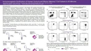 科学海报Immunomagnetic Purification of Human Central and Effector Memory T Cell Subsets in 45 Minutes
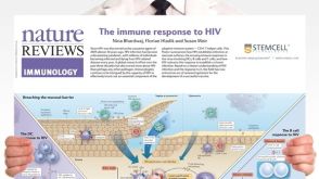 挂图The Immune Response to HIV Poster Summary of how HIV subverts the immune response to establish a chronic infection
 沪公网安备31010102008431号