
 EasySep™小鼠TIL(CD45)正选试剂盒


技术资料
-
-
-
-
 研究综述Mesenchymal Stromal Cells: Markers, Isolation and Culture, Differentiation, and Therapeutic Potential
细胞类型:
间充质干祖细胞
发布日期: 06/01/2015 -
-
-
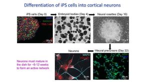
 研究综述Neural Stem Cells: Identification, Function, Culture, and Isolation
细胞类型:
神经干祖细胞
发布日期: 04/01/2015


 沪公网安备31010102008431号