Quintarelli C et al. (MAR 2011)
Blood 117 12 3353--62
High-avidity cytotoxic T lymphocytes specific for a new PRAME-derived peptide can target leukemic and leukemic-precursor cells.
The cancer testis antigen (CTA) preferentially expressed antigen of melanoma (PRAME) is overexpressed by many hematologic malignancies,but is absent on normal tissues,including hematopoietic progenitor cells,and may therefore be an appropriate candidate for T cell-mediated immunotherapy. Because it is likely that an effective antitumor response will require high-avidity,PRAME-specific cytotoxic T lymphocytes (CTLs),we attempted to generate such CTLs using professional and artificial antigen-presenting cells loaded with a peptide library spanning the entire PRAME protein and consisting of 125 synthetic pentadecapeptides overlapping by 11 amino acids. We successfully generated polyclonal,PRAME-specific CTL lines and elicited high-avidity CTLs,with a high proportion of cells recognizing a previously uninvestigated HLA-A*02-restricted epitope,P435-9mer (NLTHVLYPV). These PRAME-CTLs could be generated both from normal donors and from subjects with PRAME(+) hematologic malignancies. The cytotoxic activity of our PRAME-specific CTLs was directed not only against leukemic blasts,but also against leukemic progenitor cells as assessed by colony-forming-inhibition assays,which have been implicated in leukemia relapse. These PRAME-directed CTLs did not affect normal hematopoietic progenitors,indicating that this approach may be of value for immunotherapy of PRAME(+) hematologic malignancies.
View Publication
产品号#:
产品名:
Le Dieu R et al. (AUG 2009)
Journal of immunological methods 348 1-2 95--100
Negative immunomagnetic selection of T cells from peripheral blood of presentation AML specimens.
To date,studies on T cells in acute myeloid leukemia (AML) have been limited to flow cytometric analysis of whole peripheral blood mononuclear cell (PBMC) specimens or functional work looking at the impact of AML myeloblasts on normal or remission T cells. This lack of information on T cells at the time of presentation with disease is due in part to the difficulty in isolating sufficiently pure T cells from these specimens for further study. Negative immunomagnetic selection has been the method of choice for isolating immune cells for functional studies due to concerns that binding antibodies to the cell surface may induce cellular activation,block ligand-receptor interactions or result in immune clearance. In order specifically to study T cells in presentation AML specimens,we set out to develop a method of isolating highly pure CD4 and CD8 T cells by negative selection from the peripheral blood (PB) of newly diagnosed AML patients. This technique,unlike T cell selection from PB from normal individuals or from patients with chronic lymphocytic leukaemia,was extremely problematic due to properties of the leukaemic myeloblasts. A successful method was eventually optimized requiring the use of a custom antibody cocktail consisting of CD33,CD34,CD123,CD11c and CD36,to deplete myeloblasts.
View Publication
产品号#:
产品名:
Staton PJ et al. (APR 2006)
Journal of immunology (Baltimore,Md. : 1950) 176 7 3978--86
IL-7 is a critical factor in modulating lesion development in Skn-directed autoimmunity.
In a murine model of autoimmunity targeted against the epidermal cell Ags,Skn,adoptive transfer of Skn-immune T cells to immunosuppressed recipients elicits skin lesions in areas of mild epidermal trauma. In this study,we examined peripheral regulation of Skn-induced autoreactivity disrupted by rendering the mice immunoincompetent. We found that regulation of Skn-directed autoimmunity was restored by cotransfer of normal syngeneic spleen cells at twice the concentration of Skn-immune cells and was evidenced by significantly reduced lesion severity by days 5-7 post-cotransfer compared with animals given injections of Skn-immune cells alone. Enrichment and depletion of normal CD4(+) or CD8(+) spleen cells and RT-PCR analysis of selected cytokines identified CD4(+) cells as the regulatory cells in the cotransfer inoculum; however,significant reduction in lesion severity was observed only when there was a concomitant increase in levels of IL-7. The role of IL-7 was further supported in that mice cotransferred with Skn-immune cells plus normal spleen cells,but also treated with anti-IL-7 Ab,no longer exhibited reduced lesion severity. To determine whether IL-7 expression without normal spleen cell cotransfer could modulate lesion development,an IL-7-encoding plasmid (pCMV-Tag1-IL-7) was topically delivered to sites flanking the stressed skin site in Skn-induced autoimmune mice. Daily application of 15 mug of pCMV-Tag1-IL-7 significantly suppressed lesion severity. Our results support a mechanism for CD4(+) T cells and IL-7 in contributing to the control of autoreactivity.
View Publication

 EasySep™小鼠TIL(CD45)正选试剂盒


 挂图Frequencies of Immune Cells in Rat Tissue Lists the estimated frequencies of more than 15 immune cell types in Sprague Dawley rats
 专家访谈Cori Fain Overcoming Life Circumstances to Pursue Neuroimmunology Research
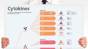 挂图Human Immune Cytokines Infographic of key cytokines for expansion, differentiation and characterization of major immune cell types


 沪公网安备31010102008431号