Simons MP et al. (MAR 2008)
Journal of leukocyte biology 83 3 621--9
TNF-related apoptosis-inducing ligand (TRAIL) is expressed throughout myeloid development, resulting in a broad distribution among neutrophil granules.
TRAIL induces apoptosis in a variety of tumor cells. Our laboratory found that human neutrophils contain an intracellular reservoir of prefabricated TRAIL that is released after stimulation with Mycobacterium bovis bacillus Calmette-Guérin. In this study,we examined the subcellular distribution of TRAIL in freshly isolated neutrophils. Neutrophil granules,secretory vesicles (SV),and plasma membrane vesicles were isolated by subcellular fractionation,followed by free-flow electrophoresis,and examined by ELISA and immunoblot. TRAIL was found in all membrane-bound fractions with the highest amounts in the fractions enriched in azurophilic granule (AG) and SV. Immunofluorescence confocal microscopy showed that TRAIL colocalized independently with myeloperoxidase (MPO),lactoferrin (LF),and albumin,respective markers of AG,specific granules,and SV. Furthermore,immunotransmission electron microscopy demonstrated that TRAIL colocalized intracellularly with MPO and albumin. We examined TRAIL expression in PLB-985 cells induced with dimethylformamide and in CD34-positive stem cells treated with G-CSF. Quantitative RT-PCR analysis showed that TRAIL was expressed in each stage of development,whereas MPO and LF were only expressed at distinct times during differentiation. Collectively,these findings suggest that TRAIL is expressed throughout neutrophil development,resulting in a broad distribution among different granule subtypes.
View Publication
产品号#:
09600
09650
09850
产品名:
StemSpan™ SFEM
StemSpan™ SFEM
Shah SN et al. (DEC 2016)
PloS one 11 12 e0166657
Evaluation of Stem Cell-Derived Red Blood Cells as a Transfusion Product Using a Novel Animal Model.
Reliance on volunteer blood donors can lead to transfusion product shortages,and current liquid storage of red blood cells (RBCs) is associated with biochemical changes over time,known as 'the storage lesion'. Thus,there is a need for alternative sources of transfusable RBCs to supplement conventional blood donations. Extracorporeal production of stem cell-derived RBCs (stemRBCs) is a potential and yet untapped source of fresh,transfusable RBCs. A number of groups have attempted RBC differentiation from CD34+ cells. However,it is still unclear whether these stemRBCs could eventually be effective substitutes for traditional RBCs due to potential differences in oxygen carrying capacity,viability,deformability,and other critical parameters. We have generated ex vivo stemRBCs from primary human cord blood CD34+ cells and compared them to donor-derived RBCs based on a number of in vitro parameters. In vivo,we assessed stemRBC circulation kinetics in an animal model of transfusion and oxygen delivery in a mouse model of exercise performance. Our novel,chronically anemic,SCID mouse model can evaluate the potential of stemRBCs to deliver oxygen to tissues (muscle) under resting and exercise-induced hypoxic conditions. Based on our data,stem cell-derived RBCs have a similar biochemical profile compared to donor-derived RBCs. While certain key differences remain between donor-derived RBCs and stemRBCs,the ability of stemRBCs to deliver oxygen in a living organism provides support for further development as a transfusion product.
View Publication
产品号#:
70008
70008.1
70008.2
70008.3
70008.4
70008.5
70008.6
200-0000
200-0001
200-0002
产品名:
冻存的人脐带血CD34+细胞
冻存的人脐带血CD34+细胞
冻存的人脐带血CD34+细胞
冻存的人脐带血CD34+细胞
冻存的人脐带血CD34+细胞
冻存的人脐带血CD34+细胞
冻存的人脐带血CD34+细胞
冻存的人脐带血CD34+细胞
冻存的人脐带血CD34+细胞
Keller G et al. (JAN 1993)
Molecular and cellular biology 13 1 473--86
Hematopoietic commitment during embryonic stem cell differentiation in culture.
We report that embryonic stem cells efficiently undergo differentiation in vitro to mesoderm and hematopoietic cells and that this in vitro system recapitulates days 6.5 to 7.5 of mouse hematopoietic development. Embryonic stem cells differentiated as embryoid bodies (EBs) develop erythroid precursors by day 4 of differentiation,and by day 6,more than 85% of EBs contain such cells. A comparative reverse transcriptase-mediated polymerase chain reaction profile of marker genes for primitive endoderm (collagen alpha IV) and mesoderm (Brachyury) indicates that both cell types are present in the developing EBs as well in normal embryos prior to the onset of hematopoiesis. GATA-1,GATA-3,and vav are expressed in both the EBs and embryos just prior to and/or during the early onset of hematopoiesis,indicating that they could play a role in the early stages of hematopoietic development both in vivo and in vitro. The initial stages of hematopoietic development within the EBs occur in the absence of added growth factors and are not significantly influenced by the addition of a broad spectrum of factors,including interleukin-3 (IL-3),IL-1,IL-6,IL-11,erythropoietin,and Kit ligand. At days 10 and 14 of differentiation,EB hematopoiesis is significantly enhanced by the addition of both Kit ligand and IL-11 to the cultures. Kinetic analysis indicates that hematopoietic precursors develop within the EBs in an ordered pattern. Precursors of the primitive erythroid lineage appear first,approximately 24 h before precursors of the macrophage and definitive erythroid lineages. Bipotential neutrophil/macrophage and multilineage precursors appear next,and precursors of the mast cell lineage develop last. The kinetics of precursor development,as well as the growth factor responsiveness of these early cells,is similar to that found in the yolk sac and early fetal liver,indicating that the onset of hematopoiesis within the EBs parallels that found in the embryo.
View Publication
产品号#:
06902
06952
00321
00322
00323
00324
00325
产品名:
Miyoshi H et al. (JAN 1999)
Science (New York,N.Y.) 283 5402 682--6
Transduction of human CD34+ cells that mediate long-term engraftment of NOD/SCID mice by HIV vectors.
Efficient gene transfer into human hematopoietic stem cells (HSCs) is an important goal in the study of the hematopoietic system as well as for gene therapy of hematopoietic disorders. A lentiviral vector based on the human immunodeficiency virus (HIV) was able to transduce human CD34+ cells capable of stable,long-term reconstitution of nonobese diabetic/severe combined immunodeficient (NOD/SCID) mice. High-efficiency transduction occurred in the absence of cytokine stimulation and resulted in transgene expression in multiple lineages of human hematopoietic cells for up to 22 weeks after transplantation.
View Publication
Diekmann F et al. (FEB 2012)
Nephrology,dialysis,transplantation : official publication of the European Dialysis and Transplant Association - European Renal Association 27 2 537--41
mTOR inhibition and erythropoiesis: microcytosis or anaemia?
BACKGROUND: Anaemia and microcytosis are common post kidney transplantation. The aim of this study was to evaluate the potential role of mammalian target of rapamycin (mTOR) inhibition in the development of anaemia and microcytosis in healthy animals and in human erythroid cultures in vitro. METHODS: Rats with normal kidney function were treated with sirolimus (n = 7) or vehicle (n = 8) for 15 weeks. Hemograms were determined thereafter. In the sirolimus withdrawal part of the study,rats received sirolimus (SRL) for 67 days (n = 4) 1 mg/kg three times per week or for 30 days (n = 4) and were observed until Day 120. Hemograms were performed regularly. Peripheral blood mononuclear cells from healthy controls (HC; n = 8),kidney transplant patients with sirolimus treatment with (SRL + MC; n = 8) or without microcytosis (SRL - MC; n = 8) were isolated and cultured in the absence or presence of SRL (5 ng/mL). RESULTS: SRL-treated animals had a reduced mean corpuscular volume (MCV) and elevated erythrocyte count compared with control animals after 15 weeks of treatment. This effect was evident as early as 4 weeks (MCV: 61.5 ± 1.8 versus 57 ± 1.7 fL; P = 0.0156; Red blood count 7.4 ± 0.3 × 10(9)/L versus 8.6 ± 0.5 × 10(9)/L; P = 0.0156) and was reversible 90 days after SRL withdrawal. SRL in the culture medium of erythroid cultures led to fewer colonies in cultures from HC as well as from kidney transplant patients (without SRL: 34.2 ± 11.4 versus with SRL: 27.5 ± 9.9 BFU-E-derived colonies P = 0.03),regardless if the cultures were derived from recipients with normocytic or with microcytic erythrocytes. The presence of tacrolimus in the culture medium had no influence on the number and size of colonies. CONCLUSION: mTOR inhibition induces microcytosis and polyglobulia,but not anaemia in healthy rats. This might be caused by growth inhibition of erythroid precursor cells.
View Publication
产品号#:
04531
产品名:
MethoCult™ H4531
Gasparetto M et al. (OCT 2012)
Experimental hematology 40 10 857--66.e5
Varying levels of aldehyde dehydrogenase activity in adult murine marrow hematopoietic stem cells are associated with engraftment and cell cycle status.
Aldehyde dehydrogenase (ALDH) activity is a widely used marker for human hematopoietic stem cells (HSCs),yet its relevance and role in murine HSCs remain unclear. We found that murine marrow cells with a high level of ALDH activity as measured by Aldefluor staining (ALDH(br) cells) do not contain known HSCs or progenitors. In contrast,highly enriched murine HSCs defined by the CD48(-)EPCR(+) and other phenotypes contain two subpopulations,one that stains dimly with Aldefluor (ALDH(dim)) and one that stains at intermediate levels (ALDH(int)). The CD48(-)EPCR(+)ALDH(dim) cells are virtually all in G(0) and yield high levels of engraftment via both intravenous and intrabone routes. In contrast the CD48(-)EPCR(+)ALDH(int) cells are virtually all in G(1),have little intravenous engraftment potential,and yet can engraft long-term after intrabone transplantation. These data demonstrate that Aldefluor staining of unfractionated murine marrow does not identify known HSCs or progenitors. However,varying levels of Aldefluor staining when combined with CD48 and EPCR detection can identify novel populations in murine marrow including a highly enriched population of resting HSCs and a previously unknown HSC population in G(1) with an intravenous engraftment defect.
View Publication
产品号#:
01700
01705
01702
产品名:
ALDEFLUOR™ 试剂盒
ALDEFLUOR™ DEAB试剂, 1.5 mM, 1 mL
ALDEFLUOR™检测缓冲液
Mahbub AA et al. (DEC 2013)
Anti-cancer agents in medicinal chemistry 13 10 1601--13
Differential effects of polyphenols on proliferation and apoptosis in human myeloid and lymphoid leukemia cell lines.
BACKGROUND Mortality rates for leukemia are high despite considerable improvements in treatment. Since polyphenols exert pro-apoptotic effects in solid tumors,our study investigated the effects of polyphenols in haematological malignancies. The effect of eight polyphenols (quercetin,chrysin,apigenin,emodin,aloe-emodin,rhein,cis-stilbene and trans-stilbene) were studied on cell proliferation,cell cycle and apoptosis in four lymphoid and four myeloid leukemic cells lines,together with normal haematopoietic control cells. METHODS Cellular proliferation was measured by CellTiter-Glo(®) luminescent assay; and cell cycle arrest was assessed using flow cytometry of propidium iodide stained cells. Apoptosis was investigated by caspase-3 activity assay using flow cytometry and apoptotic morphology was confirmed by Hoescht 33342 staining. RESULTS Emodin,quercetin,and cis-stilbene were the most effective polyphenols at decreasing cell viability (IC50 values of 5-22 μM,8-33 μM,and 25-85 μM respectively) and inducing apoptosis (AP50 values (the concentration which 50% of cells undergo apoptosis) of 2-27 μM,19-50 μM,and 8-50 μM respectively). Generally,lymphoid cell lines were more sensitive to polyphenol treatment compared to myeloid cell lines,however the most resistant myeloid (KG-1a and K562) cell lines were still found to respond to emodin and quercetin treatment at low micromolar levels. Non-tumor cells were less sensitive to all polyphenols compared to the leukemia cells. CONCLUSIONS These findings suggest that polyphenols have anti-tumor activity against leukemia cells with differential effects. Importantly,the differential sensitivity of emodin,quercetin,and cis-stilbene between leukemia and normal cells suggests that polyphenols are potential therapeutic agents for leukemia.
View Publication
产品号#:
70008
70008.1
70008.2
70008.3
70008.4
70008.5
70008.6
200-0002
200-0001
200-0000
产品名:
冻存的人脐带血CD34+细胞
冻存的人脐带血CD34+细胞
冻存的人脐带血CD34+细胞
冻存的人脐带血CD34+细胞
冻存的人脐带血CD34+细胞
冻存的人脐带血CD34+细胞
冻存的人脐带血CD34+细胞
冻存的人脐带血CD34+细胞
冻存的人脐带血CD34+细胞
Li L et al. (AUG 2011)
Blood 118 6 1504--15
A critical role for SHP2 in STAT5 activation and growth factor-mediated proliferation, survival, and differentiation of human CD34+ cells.
SHP2,a cytoplasmic protein-tyrosine phosphatase encoded by the PTPN11 gene,plays a critical role in developmental hematopoiesis in the mouse,and gain-of-function mutations of SHP2 are associated with hematopoietic malignancies. However,the role of SHP2 in adult hematopoiesis has not been addressed in previous studies. In addition,the role of SHP2 in human hematopoiesis has not been described. These questions are of considerable importance given the interest in development of SHP2 inhibitors for cancer treatment. We used shRNA-mediated inhibition of SHP2 expression to investigate the function of SHP2 in growth factor (GF) signaling in normal human CD34(+) cells. SHP2 knockdown resulted in markedly reduced proliferation and survival of cells cultured with GF,and reduced colony-forming cell growth. Cells expressing gain-of-function SHP2 mutations demonstrated increased dependency on SHP2 expression for survival compared with cells expressing wild-type SHP2. SHP2 knockdown was associated with significantly reduced myeloid and erythroid differentiation with retention of CD34(+) progenitors with enhanced proliferative capacity. Inhibition of SHP2 expression initially enhanced and later inhibited STAT5 phosphorylation and reduced expression of the antiapoptotic genes MCL1 and BCLXL. These results indicate an important role for SHP2 in STAT5 activation and GF-mediated proliferation,survival,and differentiation of human progenitor cells.
View Publication

 EasySep™小鼠TIL(CD45)正选试剂盒


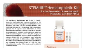
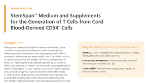 技术公告StemSpan™ Medium and Supplements for the Generation of T Cells from Cord Blood-Derived CD34+ Cells

 沪公网安备31010102008431号