Bruserud &O et al. (APR 2004)
Haematologica 89 4 391--402
Osteoblasts increase proliferation and release of pro-angiogenic interleukin 8 by native human acute myelogenous leukemia blasts.
BACKGROUND AND OBJECTIVES: Interactions between acute myelogenous leukemia (AML) blasts and non-leukemic cells in the bone marrow seem to be important for both disease development and susceptibility to chemotherapy. Recent studies have focused on the endothelial cells,but other non-leukemic cells may also be involved. In the present study we investigated how osteoblasts affect native human AML blasts. DESIGN AND METHODS: AML cells were derived from a large group of consecutive patients. The AML blasts and osteoblastic sarcoma cell lines (Cal72,SJSA-1) were incubated together in different chambers separated by a semipermeable membrane. We investigated effects of co-culture on proliferation,apoptosis and cytokine release. RESULTS: The cross-talk between these two cell populations,achieved via release of soluble mediators,resulted in increased AML blast proliferation,including increased proliferation of clonogenic progenitors,but did not affect spontaneous in vitro apoptosis. Both interleukin (IL) 1-b and granulocyte-macrophage colony-stimulating factor were involved in this growth-enhancing cross-talk,and normal osteoblasts could also increase the AML blast proliferation. Furthermore,co-culture of AML blasts with osteoblastic sarcoma cells as well as normal osteoblasts increased the levels of the pro-angiogenic mediator IL8. INTERPRETATION AND CONCLUSIONS: Our in vitro results suggest that the release of soluble mediators by osteoblasts supports leukemic hematopoiesis through two major mechanisms: (i) direct enhancement of AML blast proliferation; and (ii) enhanced angiogenesis caused by increased IL8 levels.
View Publication
Pesce M et al. (SEP 2003)
Circulation research 93 5 e51--62
Myoendothelial differentiation of human umbilical cord blood-derived stem cells in ischemic limb tissues.
Human umbilical cord blood (UCB) contains high numbers of endothelial progenitors cells (EPCs) characterized by coexpression of CD34 and CD133 markers. Prior studies have shown that CD34+/CD133+ EPCs from the cord or peripheral blood (PB) can give rise to endothelial cells and induce angiogenesis in ischemic tissues. In the present study,it is shown that freshly isolated human cord blood CD34+ cells injected into ischemic adductor muscles gave rise to endothelial and,unexpectedly,to skeletal muscle cells in mice. In fact,the treated limbs exhibited enhanced arteriole length density and regenerating muscle fiber density. Under similar experimental conditions,CD34- cells did not enhance the formation of new arterioles and regenerating muscle fibers. In nonischemic limbs CD34+ cells increased arteriole length density but did not promote formation of new muscle fibers. Endothelial and myogenic differentiation ability was maintained in CD34+ cells after ex vivo expansion. Myogenic conversion of human cord blood CD34+ cells was also observed in vitro by coculture onto mouse myoblasts. These results show that human cord blood CD34+ cells differentiate into endothelial and skeletal muscle cells,thus providing an indication of human EPCs plasticity. The full text of this article is available online at http://www.circresaha.org.
View Publication
A role for thrombopoietin in hemangioblast development.
Vascular endothelial growth factor (VEGF) and stem cell factor (SCF) act as growth factors for the hemangioblast,an embryonic progenitor of the hematopoietic and endothelial lineages. Because thrombopoietin (TPO) and its receptor,c-Mpl,regulate primitive hematopoietic populations,including bone marrow hematopoietic stem cells,we investigated whether TPO acts on the hemangioblasts that derive from differentiation of embryonic stem cells in vitro. Reverse transcriptase polymerase chain reaction analysis detected expression of c-Mpl beginning on day 3 of embryoid body differentiation when the hemangioblast first arises. In assays of the hemangioblast colony-forming cell (BL-CFC),TPO alone supported BL-CFC formation and nearly doubled the number of BL-CFC when added together with VEGF and SCF. When replated under the appropriate conditions,TPO-stimulated BL-CFC gave rise to secondary hematopoietic colonies,as well as endothelial cells,confirming their nature as hemangioblasts. Addition of a neutralizing anti-VEGF antibody did not block TPO enhancement of BL-CFC formation,suggesting that TPO acts independently of VEGF. These results establish that Mpl signaling plays a role in the earliest stages of hematopoietic development and that TPO represents a third growth factor influencing hemangioblast formation.
View Publication
Tamaki T et al. (MAY 2002)
The Journal of cell biology 157 4 571--7
Identification of myogenic-endothelial progenitor cells in the interstitial spaces of skeletal muscle.
Putative myogenic and endothelial (myo-endothelial) cell progenitors were identified in the interstitial spaces of murine skeletal muscle by immunohistochemistry and immunoelectron microscopy using CD34 antigen. Enzymatically isolated cells were characterized by fluorescence-activated cell sorting on the basis of cell surface antigen expression,and were sorted as a CD34+ and CD45- fraction. Cells in this fraction were approximately 94% positive for Sca-1,and mostly negative (textless3% positive) for CD14,31,49,144,c-kit,and FLK-1. The CD34+/45- cells formed colonies in clonal cell cultures and colony-forming units displayed the potential to differentiate into adipocytes,endothelial,and myogenic cells. The CD34+/45- cells fully differentiated into vascular endothelial cells and skeletal muscle fibers in vivo after transplantation. Immediately after sorting,CD34+/45- cells expressed only c-met mRNA,and did not express any other myogenic cell-related markers such as MyoD,myf-5,myf-6,myogenin,M-cadherin,Pax-3,and Pax-7. However,after 3 d of culture,these cells expressed mRNA for all myogenic markers. CD34+/45- cells were distinct from satellite cells,as they expressed Bcrp1/ABCG2 gene mRNA (Zhou et al.,2001). These findings suggest that myo-endothelial progenitors reside in the interstitial spaces of mammalian skeletal muscles,and that they can potentially contribute to postnatal skeletal muscle growth.
View Publication
Sata M et al. (APR 2002)
Nature medicine 8 4 403--9
Hematopoietic stem cells differentiate into vascular cells that participate in the pathogenesis of atherosclerosis.
Excessive accumulation of smooth-muscle cells (SMCs) has a key role in the pathogenesis of vascular diseases. It has been assumed that SMCs derived from the outer medial layer migrate,proliferate and synthesize extracellular matrix components on the luminal side of the vessel. Although much effort has been devoted to targeting migration and proliferation of medial SMCs,there is no effective therapy that prevents occlusive vascular remodeling. We show here that in models of post-angioplasty restenosis,graft vasculopathy and hyperlipidemia-induced atherosclerosis,bone-marrow cells give rise to most of the SMCs that contribute to arterial remodeling. Notably,purified hematopoietic stem cells differentiate into SMCs in vitro and in vivo. Our findings indicate that somatic stem cells contribute to pathological remodeling of remote organs,and may provide the basis for the development of new therapeutic strategies for vascular diseases through targeting mobilization,homing,differentiation and proliferation of bone marrow-derived vascular progenitor cells.
View Publication

 EasySep™小鼠TIL(CD45)正选试剂盒


 挂图Small Molecules, Big Impact in Pluripotent Stem Cell Research Overview of signaling pathways and small molecules in pluripotent stem cell research
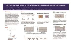 科学海报The Effect of Age and Gender on the Frequency of Peripheral Blood Endothelial Precursor Cells

 沪公网安备31010102008431号