CCAAT/Enhancer binding proteins repress the leukemic phenotype of acute myeloid leukemia.
CCAAT/enhancer binding proteins (C/EBPs) are a family of factors that regulate cell growth and differentiation. These factors,particularly C/EBPalpha and C/EBPepsilon,have important roles in normal myelopoiesis. In addition,loss of C/EBP activity appears to have a role in the pathogenesis of myeloid disorders including acute myeloid leukemia (AML). Acute promyelocytic leukemia (APL) is a subtype of AML in which a role for C/EBPs has been postulated. In almost all cases of APL,a promyelocytic leukemia-retinoic acid receptor alpha (PML-RARalpha) fusion protein is expressed as a result of a t(15;17)(q22;q12) chromosomal translocation. PML-RARalpha inhibits expression of C/EBPepsilon,whereas all-trans retinoic acid (tRA),a differentiating agent to which APL is particularly susceptible,induces C/EBPepsilon expression. PML-RARalpha may also inhibit C/EBPalpha activity. Thus,the effects of PML-RARalpha on C/EBPs may contribute to both the development of leukemia and the unique sensitivity of APL to tRA. We tested the hypothesis that increasing the activity of C/EBPs would revert the leukemic phenotype. C/EBPalpha and C/EBPepsilon were introduced into the FDC-P1 myeloid cell line and into leukemic cells from PML-RARA transgenic mice. C/EBP factors suppressed growth and induced partial differentiation in vitro. In vivo,enhanced expression of C/EBPs prolonged survival. By using a tamoxifen-responsive version of C/EBPepsilon,we observed that C/EBPepsilon could mimic the effect of tRA,driving neutrophilic differentiation in leukemic animals. Our results support the hypothesis that induction of C/EBP activity is a critical effect of tRA in APL. Furthermore,our findings suggest that targeted modulation of C/EBP activities could provide a new approach to therapy of AML.
View Publication
产品号#:
05350
产品名:
Liu E et al. (APR 2003)
Blood 101 8 3294--301
Discrimination of polycythemias and thrombocytoses by novel, simple, accurate clonality assays and comparison with PRV-1 expression and BFU-E response to erythropoietin.
Essential thrombocythemia (ET) and polycythemia vera (PV) are clonal myeloproliferative disorders that are often difficult to distinguish from other causes of elevated blood cell counts. Assays that could reliably detect clonal hematopoiesis would therefore be extremely valuable for diagnosis. We previously reported 3 X-chromosome transcription-based clonality assays (TCAs) involving the G6PD,IDS,and MPP1 genes,which together were informative in about 65% of female subjects. To increase our ability to detect clonality,we developed simple TCA for detecting the transcripts of 2 additional X-chromosome genes: Bruton tyrosine kinase (BTK) and 4-and-a-half LIM domain 1 (FHL1). The combination of TCA established the presence or absence of clonal hematopoiesis in about 90% of female subjects. We show that both genes are subject to X-chromosome inactivation and are polymorphic in all major US ethnic groups. The 5 TCAs were used to examine clonality in 46 female patients along with assays for erythropoietin-independent erythroid colonies (EECs) and granulocyte PRV-1 mRNA levels to discriminate polycythemias and thrombocytoses. Of these,all 19 patients with familial polycythemia or thrombocytosis had polyclonal hematopoiesis,whereas 22 of 26 patients with clinical evidence of myeloproliferative disorder and 1 patient with clinically obscure polycythemia were clonal. Interestingly,interferon alpha therapy in 2 patients with PV was associated with reversion of clonal to polyclonal hematopoiesis. EECs were observed in 14 of 14 patients with PV and 4 of 12 with ET,and increased granulocyte PRV-1 mRNA levels were found in 9 of 13 patients with PV and 2 of 12 with ET. Thus,these novel clonality assays are useful in the diagnosis and follow-up of polycythemic conditions and disorders with increased platelet levels.
View Publication
产品号#:
04531
15021
15061
产品名:
MethoCult™ H4531
RosetteSep™人T细胞富集抗体混合物
RosetteSep™人T细胞富集抗体混合物
Kumagai T et al. (JUN 2003)
Journal of the National Cancer Institute 95 12 896--905
Vitamin D2 analog 19-nor-1,25-dihydroxyvitamin D2: antitumor activity against leukemia, myeloma, and colon cancer cells.
BACKGROUND: 1,25-Dihydroxyvitamin D(3) inhibits growth of several types of human cancer cells in vitro,but its therapeutic use is hampered because it causes hypercalcemia. 19-nor-1,25-Dihydroxyvitamin D(2) (paricalcitol) is a noncalcemic vitamin D analog that is approved by the Food and Drug Administration for the treatment of secondary hyperparathyroidism. We investigated the antitumor activity and mechanism of action of paricalcitol in vitro and in vivo. METHODS: Effects of paricalcitol on proliferation,the cell cycle,differentiation,and apoptosis were examined in cancer cell lines. Effects on tumor growth were examined with colon cancer cell xenografts in nude mice (five in the experimental group and five in the control group). The interaction of paricalcitol with the vitamin D receptor (VDR) in mononuclear spleen cells and myeloid stem cells from wild-type and VDR knockout mice was examined. All statistical tests were two-sided. RESULTS: Paricalcitol inhibited the proliferation of myeloid leukemia cell lines HL-60,NB-4,and THP-1 cells at an effective dose that inhibited growth 50% (ED(50)) of 2.4-5.8 x 10(-9) M by inducing cell cycle arrest and differentiation. Paricalcitol inhibited the proliferation of NCI-H929 myeloma cells at an ED(50) of 2.0 x 10(-10) M by inducing cell cycle arrest and apoptosis. Paricalcitol also inhibited the proliferation of colon cancer cell lines HT-29 (ED(50) = 1.7 x 10(-8) M) and SW837 (ED(50) = 3.2 x 10(-8) M). HT-29 colon cancer xenografts in paricalcitol-treated nude mice were smaller (1044 mm(3) and 1752 mm(3),difference = 708 mm(3),95% confidence interval = 311 to 1104 mm(3); P =.03) and weighed less (1487 mg and 4162 mg,difference = 2675 mg,95% confidence interval = 2103 to 3248 mg; Ptextless.001) than those in vehicle-treated mice. Paricalcitol induced committed myeloid hematopoietic stem cells from wild-type but not from VDR knockout mice to differentiate as macrophages. CONCLUSION: Paricalcitol has anticancer activity against myeloid leukemia,myeloma,and colon cancer cells that may be mediated through the VDR. Because it has been approved by the Food and Drug Administration,clinical trials of this agent in certain cancers are reasonable.
View Publication
产品号#:
03234
产品名:
MethoCult™ M3234
Deonarain R et al. (NOV 2003)
Proceedings of the National Academy of Sciences of the United States of America 100 23 13453--8
Critical roles for IFN-beta in lymphoid development, myelopoiesis, and tumor development: links to tumor necrosis factor alpha.
We have generated mice null for IFN-beta and report the diverse consequences of IFN-beta for both the innate and adaptive arms of immunity. Despite no abnormalities in the proportional balance of CD4 and CD8 T cell populations in the peripheral blood,thymus,and spleen of IFN-beta-/- mice,activated lymph node and splenic T lymphocytes exhibit enhanced T cell proliferation and decreased tumor necrosis factor alpha production,relative to IFN-beta+/+ mice. Notably,constitutive and induced expression of tumor necrosis factor alpha is reduced in the spleen and bone marrow (BM) macrophages,respectively,of IFN-beta-/- mice. We also observe an altered splenic architecture in IFN-beta-/- mice and a reduction in resident macrophages. We identify a potential defect in B cell maturation in IFN-beta-/- mice,associated with a decrease in B220+ve/high/CD43-ve BM-derived cells and a reduction in BP-1,IgM,and CD23 expression. Circulating IgM-,Mac-1-,and Gr-1-positive cells are also substantially decreased in IFN-beta-/- mice. The decrease in the numbers of circulating macrophages and granulocytes likely reflects defective maturation of primitive BM hematopoiesis in mice,shown by the reduction of colony-forming units,granulocyte-macrophage. We proceeded to evaluate the in vivo growth of malignant cells in the IFN-beta-/- background and give evidence that Lewis lung carcinoma-specific tumor growth is more aggressive in IFN-beta-/- mice. Taken altogether,our data suggest that,in addition to the direct growth-inhibitory effects on tumor cells,IFN-beta is required during different stages of maturation in the development of the immune system.
View Publication
产品号#:
03434
03444
产品名:
MethoCult™ GF M3434
MethoCult™ GF M3434
Thanopoulou E et al. (JUN 2004)
Blood 103 11 4285--93
Engraftment of NOD/SCID-beta2 microglobulin null mice with multilineage neoplastic cells from patients with myelodysplastic syndrome.
The development of immunodeficient mouse xenograft models has greatly facilitated the investigation of some human hematopoietic malignancies,but application of this approach to the myelodysplastic syndromes (MDSs) has proven difficult. We now show that cells from most MDS patients (including all subtypes) repopulate nonobese diabetic-severe combined immunodeficient (scid)/scid-beta2 microglobulin null (NOD/SCID-beta2m(-/-)) mice at least transiently and produce abnormal differentiation patterns in this model. Normal marrow transplants initially produce predominantly erythroid cells and later predominantly B-lymphoid cells in these mice,whereas most MDS samples produced predominantly granulopoietic cells. In 4 of 4 MDS cases,the regenerated cells showed the same clonal markers (trisomy 8,n = 3; and 5q-,n = 1) as the original sample and,in one instance,regenerated trisomy 8(+) B-lymphoid as well as myeloid cells were identified. Interestingly,the enhanced growth of normal marrow obtained in NOD/SCID-beta2m(-/-) mice engineered to produce human interleukin-3,granulocyte-macrophage colony-stimulating factor,and Steel factor was seen only with 1 of 7 MDS samples. These findings support the concept that human MDS originates in a transplantable multilineage hematopoietic stem cell whose genetic alteration may affect patterns of differentiation and responsiveness to hematopoietic growth factors. They also demonstrate the potential of this new murine xenotransplant model for future investigations of MDS.
View Publication
产品号#:
04100
产品名:
MethoCult™ H4100
Hess DA et al. (SEP 2004)
Blood 104 6 1648--55
Functional characterization of highly purified human hematopoietic repopulating cells isolated according to aldehyde dehydrogenase activity.
Human hematopoietic stem cells (HSCs) are commonly purified by the expression of cell surface markers such as CD34. Because cell phenotype can be altered by cell cycle progression or ex vivo culture,purification on the basis of conserved stem cell function may represent a more reliable way to isolate various stem cell populations. We have purified primitive HSCs from human umbilical cord blood (UCB) by lineage depletion (Lin(-)) followed by selection of cells with high aldehyde dehydrogenase (ALDH) activity. ALDH(hi)Lin(-) cells contained 22.6% +/- 3.0% of the Lin(-) population and highly coexpressed primitive HSC phenotypes (CD34(+) CD38(-) and CD34(+)CD133(+)). In vitro hematopoietic progenitor function was enriched in the ALDH(hi)Lin(-) population,compared with ALDH(lo)Lin(-) cells. Multilineage human hematopoietic repopulation was observed exclusively after transplantation of ALDH(hi)Lin(-) cells. Direct comparison of repopulation with use of the nonobese diabetic/severe combined immunodeficient (NOD/SCID) and NOD/SCID beta2 microglobulin (beta2M) null models demonstrated that 10-fold greater numbers of ALDH(hi)-Lin(-) cells were needed to engraft the NOD/SCID mouse as compared with the more permissive NOD/SCID beta2M null mouse,suggesting that the ALDH(hi)Lin(-) population contained committed progenitors as well as primitive repopulating cells. Cell fractionation according to lineage depletion and ALDH activity provides a viable and prospective purification of HSCs on the basis of cell function rather than cell surface phenotype.
View Publication
产品号#:
01700
01705
04434
04444
01702
产品名:
ALDEFLUOR™ 试剂盒
ALDEFLUOR™ DEAB试剂, 1.5 mM, 1 mL
MethoCult™ H4434 Classic
MethoCult™ H4434 Classic
ALDEFLUOR™检测缓冲液
Fang B et al. (APR 2005)
Blood 105 7 2733--40
Identification of human chronic myelogenous leukemia progenitor cells with hemangioblastic characteristics.
Overwhelming evidence from leukemia research has shown that the clonal population of neoplastic cells exhibits marked heterogeneity with respect to proliferation and differentiation. There are rare stem cells within the leukemic population that possess extensive proliferation and self-renewal capacity not found in the majority of the leukemic cells. These leukemic stem cells are necessary and sufficient to maintain the leukemia. Interestingly,the BCR/ABL fusion gene,which is present in chronic myelogenous leukemia (CML),was also detected in the endothelial cells of patients with CML,suggesting that CML might originate from hemangioblastic progenitor cells that can give rise to both blood cells and endothelial cells. Here we isolated fetal liver kinase-1-positive (Flk1+) cells carrying the BCR/ABL fusion gene from the bone marrow of 17 Philadelphia chromosome-positive (Ph+) patients with CML and found that these cells could differentiate into malignant blood cells and phenotypically defined endothelial cells at the single-cell level. These findings provide direct evidence for the first time that rearrangement of the BCR/ABL gene might happen at or even before the level of hemangioblastic progenitor cells,thus resulting in detection of the BCR/ABL fusion gene in both blood and endothelial cells.
View Publication

 EasySep™小鼠TIL(CD45)正选试剂盒


 挂图Small Molecules, Big Impact in Pluripotent Stem Cell Research Overview of signaling pathways and small molecules in pluripotent stem cell research
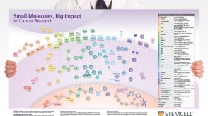 挂图Small Molecules, Big Impact in Cancer Research Overview of signaling pathways and small molecules in cancer research
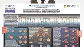 挂图Natural Killer Cells Overview of NK cell receptors, subsets, activation and function
 沪公网安备31010102008431号