
 EasySep™小鼠TIL(CD45)正选试剂盒


技术资料
-

-

-
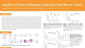 科学海报Isolation of Tumor-Infiltrating Leukocytes from Mouse Tumors
Conference:
AAI 2020
发布日期: 10/22/2020 -
-
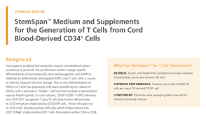 技术公告StemSpan™ Medium and Supplements for the Generation of T Cells from Cord Blood-Derived CD34+ Cells
细胞类型:
CD4+ T细胞,CD8+ T细胞,T细胞,造血干祖细胞
发布日期: 08/24/2020


 沪公网安备31010102008431号