
 EasySep™小鼠TIL(CD45)正选试剂盒


技术资料
-
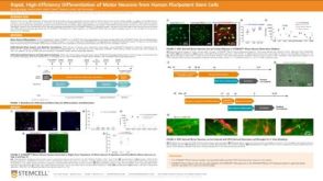 科学海报Rapid, High-Efficiency Differentiation of Motor Neurons from Human Pluripotent Stem Cells
Conference:
FENS 2022
过滤器
筛选结果
研究领域
- HIV 70 项目
- HLA 52 项目
- 上皮细胞生物学 269 项目
- 免疫 1012 项目
- 内皮细胞研究 1 项目
- 呼吸系统研究 48 项目
- 嵌合体 25 项目
- 干细胞生物学 2827 项目
- 感染性疾病(传染病) 7 项目
- 抗体制备 7 项目
- 新陈代谢 7 项目
- 杂交瘤制备 2 项目
- 疾病建模 248 项目
- 癌症 6 项目
- 神经科学 650 项目
- 移植研究 100 项目
- 类器官 178 项目
- 细胞外囊泡研究 10 项目
- 细胞治疗开发 18 项目
- 细胞疗法开发 113 项目
- 细胞系制备 191 项目
- 脐带血库 64 项目
- 血管生成细胞研究 1 项目
- 传染病 64 项目
- 内皮细胞生物学 7 项目
- 杂交瘤生成 14 项目
- 癌症研究 724 项目
- 血管生成细胞研究 51 项目
Show More
Show Less
产品系列
- BrainPhys 1 项目
- MyoCult 1 项目
- STEMdiff 1 项目
Show More
Show Less
细胞类型
- B 细胞 229 项目
- CD4+ T细胞 46 项目
- CD8+ T细胞 29 项目
- CHO细胞 15 项目
- HEK-293细胞(人胚肾293细胞) 2 项目
- NK 细胞 162 项目
- PSC衍生 37 项目
- T 细胞 440 项目
- 上皮细胞 143 项目
- 中胚层 5 项目
- 乳腺细胞 95 项目
- 先天性淋巴细胞 32 项目
- 全血 10 项目
- 其他子集 1 项目
- 其他细胞系 10 项目
- 内皮细胞 11 项目
- 内胚层 4 项目
- 前列腺细胞 18 项目
- 单个核细胞 93 项目
- 单核细胞 178 项目
- 多能干细胞 1986 项目
- 小胶质细胞 13 项目
- 巨噬细胞 42 项目
- 巨核细胞 10 项目
- 心肌细胞 21 项目
- 成骨细胞 10 项目
- 星形胶质细胞 14 项目
- 杂交瘤细胞 92 项目
- 树突状细胞(DCs) 118 项目
- 气道细胞 4 项目
- 浆细胞 3 项目
- 淋巴细胞 73 项目
- 癌细胞及细胞系 149 项目
- 癌细胞和细胞系 1 项目
- 白细胞 24 项目
- 白细胞单采样本 13 项目
- 白血病/淋巴瘤细胞 14 项目
- 监管 1 项目
- 真皮细胞 3 项目
- 神经元 1 项目
- 神经干/祖细胞 465 项目
- 神经细胞 12 项目
- 粒细胞及其亚群 96 项目
- 红系细胞 12 项目
- 红细胞 13 项目
- 肌源干/祖细胞 11 项目
- 肝细胞 40 项目
- 肠道细胞 103 项目
- 肾细胞 4 项目
- 肿瘤细胞 27 项目
- 胰腺细胞 17 项目
- 脂肪细胞 6 项目
- 脑肿瘤干细胞 103 项目
- 血小板 4 项目
- 血管生成细胞 1 项目
- 角质形成细胞 1 项目
- 调节性T细胞 10 项目
- 软骨细胞 9 项目
- 造血干/祖细胞 968 项目
- 造血干祖细胞 6 项目
- 造血细胞 4 项目
- 间充质基质细胞 25 项目
- 间充质干/祖细胞 188 项目
- 间充质干祖细胞 1 项目
- 间充质细胞 3 项目
- 骨髓基质细胞 1 项目
- 髓系细胞 135 项目
- 肾脏细胞 8 项目
- PSC衍生上皮细胞 39 项目
- PSC衍生中胚层 25 项目
- PSC衍生内皮细胞 20 项目
- PSC衍生内胚层 28 项目
- PSC衍生心肌细胞 26 项目
- PSC衍生神经细胞 130 项目
- PSC衍生肝细胞 18 项目
- PSC衍生造血干细胞 39 项目
- PSC衍生间充质细胞 27 项目
- 其他T细胞亚型 31 项目
- 呼吸道细胞 96 项目
- 多巴胺能神经元 6 项目
- 小鼠胚胎成纤维细胞 1 项目
- 神经元 201 项目
Show More
Show Less
资源类别
- 科学海报 1 项目
Show More
Show Less

 沪公网安备31010102008431号