Hou Y et al. (MAY 2014)
Neurobiology of Aging 35 5 975--989
Permeability transition pore-mediated mitochondrial superoxide flashes mediate an early inhibitory effect of amyloid beta1 42 on neural progenitor cell proliferation
Cellular damage by reactive oxygen species and altered neurogenesis are implicated in the etiology of AD and the pathogenic actions of amyloid β-peptide (Aβ); the underlying mechanisms and the early oxidative intracellular events triggered by Aβ are not established. In the present study,we found that mouse embryonic cortical neural progenitor cells exhibit intermittent spontaneous mitochondrial superoxide (SO) flashes that require transient opening of mitochondrial permeability transition pores (mPTPs). The incidence of mitochondria SO flash activity in neural progenitor cells (NPCs) increased during the first 6-24 hours of exposure to aggregating amyloid β-peptide (Aβ1-42),indicating an increase in transient mPTP opening. Subsequently,the SO flash frequency progressively decreased and ceased between 48 and 72 hours of exposure to Aβ1-42,during which time global cellular reactive oxygen species increased,mitochondrial membrane potential decreased,cytochrome C was released from mitochondria and the cells degenerated. Inhibition of mPTPs and selective reduction in mitochondrial SO flashes significantly ameliorated the negative effects of Aβ1-42 on NPC proliferation and survival. Our findings suggest that mPTP-mediated bursts of mitochondrial SO production is a relatively early and pivotal event in the adverse effects of Aβ1-42 on NPCs. If Aβ inhibits NPC proliferation in the brains of AD patients by a similar mechanism,then interventions that inhibit mPTP-mediated superoxide flashes would be expected to protect NPCs against the adverse effects of Aβ.
View Publication
产品号#:
05707
产品名:
NeuroCult™化学解离试剂盒(小鼠)
Hotta R et al. (APR 2016)
Neurogastroenterology and motility : the official journal of the European Gastrointestinal Motility Society 28 4 498--512
Isogenic enteric neural progenitor cells can replace missing neurons and glia in mice with Hirschsprung disease.
BACKGROUND Transplanting autologous patient-derived enteric neuronal stem/progenitor cells (ENSCs) is an innovative approach to replacing missing enteric neurons in patients with Hirschsprung disease (HSCR). Using autologous cells eliminates immunologic and ethical concerns raised by other cell sources. However,whether postnatal aganglionic bowel is permissive for transplanted ENSCs and whether ENSCs from HSCR patients can be successfully isolated,cultured,and transplanted in vivo remains unknown. METHODS ENSCs isolated from the ganglionic intestine of Ednrb(-/-) mice (HSCR-ENSCs) were characterized immunohistochemically and evaluated for their capacity to proliferate and differentiate in vitro. Fluorescently labeled ENSCs were co-cultured ex vivo with aganglionic Ednrb(-/-) colon. For in vivo transplantation,HSCR-ENSCs were labeled with lentivirus expressing green fluorescent protein (GFP) and implanted into aganglionic embryonic chick gut in ovo and postnatal aganglionic Ednrb(-/-) rectum in vivo. KEY RESULTS HSCR-ENSCs maintain normal capacity self-renewal and neuronal differentiation. Moreover,the Ednrb(-/-) aganglionic environment is permissive to engraftment by wild-type ENSCs ex vivo and supports migratrion and neuroglial differentiation of these cells following transplantation in vivo. Lentiviral GFP-labeled HSCR-ENSCs populated embryonic chick hindgut and postnatal colon of Ednrb(-/-) HSCR,with cells populating the intermuscular layer and forming enteric neurons and glia. CONCLUSIONS & INFERENCES ENSCs can be isolated and cultured from mice with HSCR,and transplanted into the aganglionic bowel of HSCR littermates to generate enteric neuronal networks. These results in an isogenic model establish the potential of using autologous-derived stem cells to treat HSCR and other intestinal neuropathies.
View Publication
产品号#:
05700
产品名:
NeuroCult™ 基础培养基(小鼠&大鼠)
Hotta R et al. (MAY 2016)
Biomaterials 88 1--11
Delivery of enteric neural progenitors with 5-HT4 agonist-loaded nanoparticles and thermosensitive hydrogel enhances cell proliferation and differentiation following transplantation in vivo.
Cell therapy offers an innovative approach for treating enteric neuropathies. Postnatal gut-derived enteric neural stem/progenitor cells (ENSCs) represent a potential autologous source,but have a limited capacity for proliferation and neuronal differentiation. Since serotonin (5-HT) promotes enteric neuronal growth during embryonic development,we hypothesized that serotonin receptor agonism would augment growth of neurons from transplanted ENSCs. Postnatal ENSCs were isolated from 2 to 4 week-old mouse colon and cultured with 5-HT4 receptor agonist (RS67506)-loaded liposomal nanoparticles. ENSCs were co-cultured with mouse colon explants in the presence of RS67506-loaded (n = 3) or empty nanoparticles (n = 3). ENSCs were also transplanted into mouse rectum in vivo with RS67506-loaded (n = 8) or blank nanoparticles (n = 4) confined in a thermosensitive hydrogel,Pluronic F-127. Neuronal density and proliferation were analyzed immunohistochemically. Cultured ENSCs gave rise to significantly more neurons in the presence of RS67506-loaded nanoparticles. Similarly,colon explants had significantly increased neuronal density when RS67506-loaded nanoparticles were present. Finally,following in vivo cell delivery,co-transplantation of ENSCs with 5-HT4 receptor agonist-loaded nanoparticles led to significantly increased neuronal density and proliferation. We conclude that optimization of postnatal ENSCs can support their use in cell-based therapies for neurointestinal diseases.
View Publication
产品号#:
05700
05701
05702
05704
产品名:
NeuroCult™ 基础培养基(小鼠&大鼠)
NeuroCult™ 扩增添加物 (小鼠&大鼠)
NeuroCult™ 扩增试剂盒 (小鼠&大鼠)
NeuroCult™ 分化试剂盒 (小鼠&大鼠)
Hothi P et al. (OCT 2012)
Oncotarget 3 10 1124--36
High-Throughput Chemical Screens Identify Disulfiram as an Inhibitor of Human Glioblastoma Stem Cells
Glioblastoma Multiforme (GBM) continues to have a poor patient prognosis despite optimal standard of care. Glioma stem cells (GSCs) have been implicated as the presumed cause of tumor recurrence and resistance to therapy. With this in mind,we screened a diverse chemical library of 2,000 compounds to identify therapeutic agents that inhibit GSC proliferation and therefore have the potential to extend patient survival. High-throughput screens (HTS) identified 78 compounds that repeatedly inhibited cellular proliferation,of which 47 are clinically approved for other indications and 31 are experimental drugs. Several compounds (such as digitoxin,deguelin,patulin and phenethyl caffeate) exhibited high cytotoxicity,with half maximal inhibitory concentrations (IC50) in the low nanomolar range. In particular,the FDA approved drug for the treatment of alcoholism,disulfiram (DSF),was significantly potent across multiple patient samples (IC50 of 31.1 nM). The activity of DSF was potentiated by copper (Cu),which markedly increased GSC death. DSF-Cu inhibited the chymotrypsin-like proteasomal activity in cultured GSCs,consistent with inactivation of the ubiquitin-proteasome pathway and the subsequent induction of tumor cell death. Given that DSF is a relatively non-toxic drug that can penetrate the blood-brain barrier,we suggest that DSF should be tested (as either a monotherapy or as an adjuvant) in pre-clinical models of human GBM. Data also support targeting of the ubiquitin-proteasome pathway as a therapeutic approach in the treatment of GBM.
View Publication
产品号#:
05750
05751
产品名:
NeuroCult™ NS-A 基础培养基(人)
NeuroCult™ NS-A 扩增试剂盒(人)
Holmberg Olausson K et al. ( 2014)
PloS one 9 9 e106694
Prominin-1 (CD133) defines both stem and non-stem cell populations in CNS development and gliomas.
Prominin-1 (CD133) is a commonly used cancer stem cell marker in central nervous system (CNS) tumors including glioblastoma (GBM). Expression of Prom1 in cancer is thought to parallel expression and function in normal stem cells. Using RNA in situ hybridization and antibody tools capable of detecting multiple isoforms of Prom1,we find evidence for two distinct Prom1 cell populations in mouse brain. Prom1 RNA is first expressed in stem/progenitor cells of the ventricular zone in embryonic brain. Conversely,in adult mouse brain Prom1 RNA is low in SVZ/SGZ stem cell zones but high in a rare but widely distributed cell population (Prom1(hi)). Lineage marker analysis reveals Prom1(hi) cells are Olig2+Sox2+ glia but Olig1/2 knockout mice lacking oligodendroglia retain Prom1(hi) cells. Bromodeoxyuridine labeling identifies Prom1(hi) as slow-dividing distributed progenitors distinct from NG2+Olig2+ oligodendrocyte progenitors. In adult human brain,PROM1 cells are rarely positive for OLIG2,but express astroglial markers GFAP and SOX2. Variability of PROM1 expression levels in human GBM and patient-derived xenografts (PDX) - from no expression to strong,uniform expression--highlights that PROM1 may not always be associated with or restricted to cancer stem cells. TCGA and PDX data show that high expression of PROM1 correlates with poor overall survival. Within proneural subclass tumors,high PROM1 expression correlates inversely with IDH1 (R132H) mutation. These findings support PROM1 as a tumor cell-intrinsic marker related to GBM survival,independent of its stem cell properties,and highlight potentially divergent roles for this protein in normal mouse and human glia.
View Publication
产品号#:
05750
05751
产品名:
NeuroCult™ NS-A 基础培养基(人)
NeuroCult™ NS-A 扩增试剂盒(人)
Hjelm BE et al. (SEP 2013)
Human Molecular Genetics 22 17 3534--3546
In vitro-differentiated neural cell cultures progress towards donor-identical brain tissue
Multiple research groups have observed neuropathological phenotypes and molecular symptoms in vitro using induced pluripotent stem cell (iPSC)-derived neural cell cultures (i.e. patient-specific neurons and glia). However,the global differences/similarities that may exist between in vitro neural cells and their tissue-derived counterparts remain largely unknown. In this study,we compared temporal series of iPSC-derived in vitro neural cell cultures to endogenous brain tissue from the same autopsy donor. Specifically,we utilized RNA sequencing (RNA-Seq) to evaluate the transcriptional progression of in vitro-differentiated neural cells (over a timecourse of 0,35,70,105 and 140 days),and compared this with donor-identical temporal lobe tissue. We observed in vitro progression towards the reference brain tissue,and the following three results support this conclusion: (i) there was a significant increasing monotonic correlation between the days of our timecourse and the number of actively transcribed protein-coding genes and long intergenic non-coding RNAs (lincRNAs) (P < 0.05),consistent with the transcriptional complexity of the brain; (ii) there was an increase in CpG methylation after neural differentiation that resembled the epigenomic signature of the endogenous tissue; and (iii) there was a significant decreasing monotonic correlation between the days of our timecourse and the percent of in vitro to brain-tissue differences (P < 0.05) for tissue-specific protein-coding genes and all putative lincRNAs. Taken together,these results are consistent with in vitro neural development and physiological progression occurring predominantly by transcriptional activation of downregulated genes rather than deactivation of upregulated genes.
View Publication
产品号#:
05750
05751
05752
产品名:
NeuroCult™ NS-A 基础培养基(人)
NeuroCult™ NS-A 扩增试剂盒(人)
NeuroCult™ NS-A 分化试剂盒 (人)
Harlow DE et al. (JAN 2014)
Journal of Neuroscience 34 4 1333--1343
Expression of Proteolipid Protein Gene in Spinal Cord Stem Cells and Early Oligodendrocyte Progenitor Cells Is Dispensable for Normal Cell Migration and Myelination
Plp1 gene expression occurs very early in development,well before the onset of myelination,creating a conundrum with regard to the function of myelin proteolipid protein (PLP),one of the major proteins in compact myelin. Using PLP-EGFP mice to investigate Plp1 promoter activity,we found that,at very early time points,PLP-EGFP was expressed in Sox2+ undifferentiated precursors in the spinal cord ventricular zone (VZ),as well as in the progenitors of both neuronal and glial lineages. As development progressed,most PLP-EGFP-expressing cells gave rise to oligodendrocyte progenitor cells (OPCs). The expression of PLP-EGFP in the spinal cord was quite dynamic during development. PLP-EGFP was highly expressed as cells delaminated from the VZ. Expression was downregulated as cells moved laterally through the cord,and then robustly upregulated as OPCs differentiated into mature myelinating oligodendrocytes. The presence of PLP-EGFP expression in OPCs raises the question of its role in this migratory population. We crossed PLP-EGFP reporter mice into a Plp1-null background to investigate the role of PLP in early OPC development. In the absence of PLP,normal numbers of OPCs were generated and their distribution throughout the spinal cord was unaffected. However,the orientation and length of OPC processes during migration was abnormal in Plp1-null mice,suggesting that PLP plays a role either in the structural integrity of OPC processes or in their response to extracellular cues that orient process outgrowth.
View Publication

 EasySep™小鼠TIL(CD45)正选试剂盒


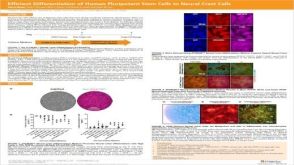 科学海报Efficient Generation of Human Pluripotent Stem Cells to Neural Crest Cells


 沪公网安备31010102008431号