Lee S-HH et al. (JUN 2000)
Nature biotechnology 18 6 675--9
Efficient generation of midbrain and hindbrain neurons from mouse embryonic stem cells.
Embryonic stem (ES) cells are clonal cell lines derived from the inner cell mass of the developing blastocyst that can proliferate extensively in vitro and are capable of adopting all the cell fates in a developing embryo. Clinical interest in the use of ES cells has been stimulated by studies showing that isolated human cells with ES properties from the inner cell mass or developing germ cells can provide a source of somatic precursors. Previous studies have defined in vitro conditions for promoting the development of specific somatic fates,specifically,hematopoietic,mesodermal,and neurectodermal. In this study,we present a method for obtaining dopaminergic (DA) and serotonergic neurons in high yield from mouse ES cells in vitro. Furthermore,we demonstrate that the ES cells can be obtained in unlimited numbers and that these neuron types are generated efficiently. We generated CNS progenitor populations from ES cells,expanded these cells and promoted their differentiation into dopaminergic and serotonergic neurons in the presence of mitogen and specific signaling molecules. The differentiation and maturation of neuronal cells was completed after mitogen withdrawal from the growth medium. This experimental system provides a powerful tool for analyzing the molecular mechanisms controlling the functions of these neurons in vitro and in vivo,and potentially for understanding and treating neurodegenerative and psychiatric diseases.
View Publication
产品号#:
06902
06952
07152
07157
00321
00322
00323
00324
00325
产品名:
N2 添加物-A
Stanurova J et al. (AUG 2016)
Scientific reports 6 August 30792
Angelman syndrome-derived neurons display late onset of paternal UBE3A silencing.
Genomic imprinting is an epigenetic phenomenon resulting in parent-of-origin-specific gene expression that is regulated by a differentially methylated region. Gene mutations or failures in the imprinting process lead to the development of imprinting disorders,such as Angelman syndrome. The symptoms of Angelman syndrome are caused by the absence of functional UBE3A protein in neurons of the brain. To create a human neuronal model for Angelman syndrome,we reprogrammed dermal fibroblasts of a patient carrying a defined three-base pair deletion in UBE3A into induced pluripotent stem cells (iPSCs). In these iPSCs,both parental alleles are present,distinguishable by the mutation,and express UBE3A. Detailed characterization of these iPSCs demonstrated their pluripotency and exceptional stability of the differentially methylated region regulating imprinted UBE3A expression. We observed strong induction of SNHG14 and silencing of paternal UBE3A expression only late during neuronal differentiation,in vitro. This new Angelman syndrome iPSC line allows to study imprinted gene regulation on both parental alleles and to dissect molecular pathways affected by the absence of UBE3A protein.
View Publication
产品号#:
05850
05857
05870
05875
07174
85850
85857
85870
85875
100-0485
100-1077
产品名:
mTeSR™1
mTeSR™1
温和细胞解离试剂
ReLeSR™
Brohawn DG et al. (AUG 2016)
PloS one 11 8 e0160520
RNAseq Analyses Identify Tumor Necrosis Factor-Mediated Inflammation as a Major Abnormality in ALS Spinal Cord.
ALS is a rapidly progressive,devastating neurodegenerative illness of adults that produces disabling weakness and spasticity arising from death of lower and upper motor neurons. No meaningful therapies exist to slow ALS progression,and molecular insights into pathogenesis and progression are sorely needed. In that context,we used high-depth,next generation RNA sequencing (RNAseq,Illumina) to define gene network abnormalities in RNA samples depleted of rRNA and isolated from cervical spinal cord sections of 7 ALS and 8 CTL samples. We aligned textgreater50 million 2X150 bp paired-end sequences/sample to the hg19 human genome and applied three different algorithms (Cuffdiff2,DEseq2,EdgeR) for identification of differentially expressed genes (DEG's). Ingenuity Pathways Analysis (IPA) and Weighted Gene Co-expression Network Analysis (WGCNA) identified inflammatory processes as significantly elevated in our ALS samples,with tumor necrosis factor (TNF) found to be a major pathway regulator (IPA) and TNF$$-induced protein 2 (TNFAIP2) as a major network hub" gene (WGCNA). Using the oPOSSUM algorithm�
View Publication
产品号#:
05850
05857
05870
05875
85850
85857
85870
85875
产品名:
mTeSR™1
mTeSR™1
Orellana DI et al. (OCT 2016)
EMBO molecular medicine 8 10 1197--1211
Coenzyme A corrects pathological defects in human neurons of PANK2-associated neurodegeneration.
Pantothenate kinase-associated neurodegeneration (PKAN) is an early onset and severely disabling neurodegenerative disease for which no therapy is available. PKAN is caused by mutations in PANK2,which encodes for the mitochondrial enzyme pantothenate kinase 2. Its function is to catalyze the first limiting step of Coenzyme A (CoA) biosynthesis. We generated induced pluripotent stem cells from PKAN patients and showed that their derived neurons exhibited premature death,increased ROS production,mitochondrial dysfunctions-including impairment of mitochondrial iron-dependent biosynthesis-and major membrane excitability defects. CoA supplementation prevented neuronal death and ROS formation by restoring mitochondrial and neuronal functionality. Our findings provide direct evidence that PANK2 malfunctioning is responsible for abnormal phenotypes in human neuronal cells and indicate CoA treatment as a possible therapeutic intervention.
View Publication
产品号#:
05850
05857
05870
05875
05872
05873
85850
85857
85870
85875
100-0483
100-0484
产品名:
mTeSR™1
mTeSR™1
Hausser Scientificᵀᴹ 明线血球计数板
ReLeSR™
Kandasamy M et al. (MAR 2017)
Cell and Tissue Research 368 3 531--549
Glycoconjugates reveal diversity of human neural stem cells (hNSCs) derived from human induced pluripotent stem cells (hiPSCs)
Neural stem cells (NSCs) have the ability to self-renew and to differentiate into various cell types of the central nervous system. This potential can be recapitulated by human induced pluripotent stem cells (hiPSCs) in vitro. The differentiation capacity of hiPSCs is characterized by several stages with distinct morphologies and the expression of various marker molecules. We used the monoclonal antibodies (mAbs) 487(LeX),5750(LeX) and 473HD to analyze the expression pattern of particular carbohydrate motifs as potential markers at six differentiation stages of hiPSCs. Mouse ESCs were used as a comparison. At the pluripotent stage,487(LeX)-,5750(LeX)- and 473HD-related glycans were differently expressed. Later,cells of the three germ layers in embryoid bodies (hEBs) and,even after neuralization of hEBs,subpopulations of cells were labeled with these surface antibodies. At the human rosette-stage of NSCs (hR-NSC),LeX- and 473HD-related epitopes showed antibody-specific expression patterns. We also found evidence that these surface antibodies could be used to distinguish the hR-NSCs from the hSR-NSCs stages. Characterization of hNSCs(FGF-2/EGF) derived from hSR-NSCs revealed that both LeX antibodies and the 473HD antibody labeled subpopulations of hNSCs(FGF-2/EGF). Finally,we identified potential LeX carrier molecules that were spatiotemporally regulated in early and late stages of differentiation. Our study provides new insights into the regulation of glycoconjugates during early human stem cell development. The mAbs 487(LeX),5750(LeX) and 473HD are promising tools for identifying distinct stages during neural differentiation.
View Publication
产品号#:
05832
05850
05857
05870
05875
85850
85857
85870
85875
产品名:
STEMdiff™ 神经花环选择试剂
mTeSR™1
mTeSR™1
Bain G et al. (APR 1995)
Developmental biology 168 2 342--57
Embryonic stem cells express neuronal properties in vitro.
Mouse embryonic stem (ES) cells cultured as aggregates and exposed to retinoic acid are induced to express multiple phenotypes normally associated with neurons. A large percentage of treated aggregates produce a rich neuritic outgrowth. Dissociating the induced aggregates with trypsin and plating the cells as a monolayer results in cultures in which a sizable percentage of the cells have a neuronal appearance. These neuron-like cells express class III beta-tubulin and the neurofilament M subunit. Induced cultures express transcripts for neural-associated genes including the neurofilament L subunit,glutamate receptor subunits,the transcription factor Brn-3,and GFAP. Levels of neurofilament L and GAD67 and GAD65 transcripts rise dramatically upon induction. Physiological studies show that the neuron-like cells generate action potentials and express TTX-sensitive sodium channels,as well as voltage-gated potassium channels and calcium channels. We conclude that a complex system of neuronal gene expression can be activated in cultured ES cells. This system should be favorable for investigating some of the mechanisms that regulate neuronal differentiation.
View Publication
产品号#:
06902
06952
00321
00322
00323
00324
00325
产品名:
Nakamura H et al. (OCT 2013)
Herpesviridae 4 1 2
Human cytomegalovirus induces apoptosis in neural stem/progenitor cells derived from induced pluripotent stem cells by generating mitochondrial dysfunction and endoplasmic reticulum stress
BACKGROUND Congenital human cytomegalovirus (HCMV) infection,a leading cause of birth defects,is most often manifested as neurological disorders. The pathogenesis of HCMV-induced neurological disorders is,however,largely unresolved,primarily because of limited availability of model systems to analyze the effects of HCMV infection on neural cells. METHODS An induced pluripotent stem cell (iPSC) line was established from the human fibroblast line MRC5 by introducing the Yamanaka's four factors and then induced to differentiate into neural stem/progenitor cells (NSPCs) by dual inhibition of the SMAD signaling pathway using Noggin and SB-431542. RESULTS iPSC-derived NSPCs (NSPC/iPSCs) were susceptible to HCMV infection and allowed the expression of both early and late viral gene products. HCMV-infected NSPC/iPSCs underwent apoptosis with the activation of caspase-3 and -9 as well as positive staining by the terminal deoxynucleotidyl transferase-mediated dUTP nick-end labeling (TUNEL). Cytochrome c release from mitochondria to cytosol was observed in these cells,indicating the involvement of mitochondrial dysfunction in their apoptosis. In addition,phosphorylation of proteins involved in the unfolded protein response (UPR),such as PKR-like eukaryotic initiation factor 2a kinase (PERK),c-Jun NH2-terminal kinase (JNK),inositol-requiring enzyme 1 (IRE1),and the alpha subunit of eukaryotic initiation factor 2 (eIF2$$) was observed in HCMV-infected NSPC/iPSCs. These results,coupled with the finding of increased expression of mRNA encoding the C/EBP-homologous protein (CHOP) and the detection of a spliced form of X-box binding protein 1 (XBP1) mRNA,suggest that endoplasmic reticulum (ER) stress is also involved in HCMV-induced apoptosis of these cells. CONCLUSIONS iPSC-derived NSPCs are thought to be a useful model to study HCMV neuropathogenesis and to analyze the mechanisms of HCMV-induced apoptosis in neural cells.
View Publication


 EasySep™小鼠TIL(CD45)正选试剂盒




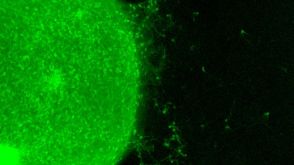 实验方案How to Generate AssemBloids™ from hPSC-Derived Dorsal and Ventral Forebrain Organoid Co-Cultures

 沪公网安备31010102008431号