
 EasySep™小鼠TIL(CD45)正选试剂盒


技术资料
-
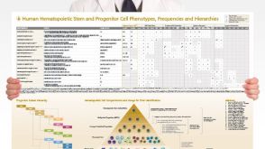 挂图Human Hematopoietic Stem and Progenitor Cell Phenotyping Overview of subset surface markers, frequencies and assays for analysis
 -
-
-

-
-
-
-

-
-

-


 沪公网安备31010102008431号