Induced Pluripotency of Human Prostatic Epithelial Cells
Induced pluripotent stem (iPS) cells are a valuable resource for discovery of epigenetic changes critical to cell type-specific differentiation. Although iPS cells have been generated from other terminally differentiated cells,the reprogramming of normal adult human basal prostatic epithelial (E-PZ) cells to a pluripotent state has not been reported. Here,we attempted to reprogram E-PZ cells by forced expression of Oct4,Sox2,c-Myc,and Klf4 using lentiviral vectors and obtained embryonic stem cell (ESC)-like colonies at a frequency of 0.01%. These E-PZ-iPS-like cells with normal karyotype gained expression of pluripotent genes typical of iPS cells (Tra-1-81,SSEA-3,Nanog,Sox2,and Oct4) and lost gene expression characteristic of basal prostatic epithelial cells (CK5,CK14,and p63). E-PZ-iPS-like cells demonstrated pluripotency by differentiating into ectodermal,mesodermal,and endodermal cells in vitro,although lack of teratoma formation in vivo and incomplete demethylation of pluripotency genes suggested only partial reprogramming. Importantly,E-PZ-iPS-like cells re-expressed basal epithelial cell markers (CD44,p63,MAO-A) in response to prostate-specific medium in spheroid culture. Androgen induced expression of androgen receptor (AR),and co-culture with rat urogenital sinus further induced expression of prostate-specific antigen (PSA),a hallmark of secretory cells,suggesting that E-PZ-iPS-like cells have the capacity to differentiate into prostatic basal and secretory epithelial cells. Finally,when injected into mice,E-PZ-iPS-like cells expressed basal epithelial cell markers including CD44 and p63. When co-injected with rat urogenital mesenchyme,E-PZ-iPS-like cells expressed AR and expression of p63 and CD44 was repressed. DNA methylation profiling identified epigenetic changes in key pathways and genes involved in prostatic differentiation as E-PZ-iPS-like cells converted to differentiated AR- and PSA-expressing cells. Our results suggest that iPS-like cells derived from prostatic epithelial cells are pluripotent and capable of prostatic differentiation; therefore,provide a novel model for investigating epigenetic changes involved in prostate cell lineage specification.
View Publication
产品号#:
05850
05857
05870
05875
85850
85857
85870
85875
产品名:
mTeSR™1
mTeSR™1
Panova AV et al. (APR 2013)
Acta Naturae 5 17 54--61
Late Replication of the Inactive X Chromosome Is Independent of the Compactness of Chromosome Territory in Human Pluripotent Stem Cells
Dosage compensation of the X chromosomes in mammals is performed via the formation of facultative heterochromatin on extra X chromosomes in female somatic cells. Facultative heterochromatin of the inactivated X (Xi),as well as constitutive heterochromatin,replicates late during the S-phase. It is generally accepted that Xi is always more compact in the interphase nucleus. The dense chromosomal folding has been proposed to define the late replication of Xi. In contrast to mouse pluripotent stem cells (PSCs),the status of X chromosome inactivation in human PSCs may vary significantly. Fluorescence in situ hybridization with a whole X-chromosome- specific DNA probe revealed that late-replicating Xi may occupy either compact or dispersed territory in human PSCs. Thus,the late replication of the Xi does not depend on the compactness of chromosome territory in human PSCs. However,the Xi reactivation and the synchronization in the replication timing of X chromosomes upon reprogramming are necessarily accompanied by the expansion of X chromosome territory.
View Publication
产品号#:
05850
05857
05870
05875
85850
85857
85870
85875
产品名:
mTeSR™1
mTeSR™1
Diniz B et al. (JUL 2013)
Investigative Ophthalmology and Visual Science 54 7 5087--5096
Subretinal Implantation of Retinal Pigment Epithelial Cells Derived From Human Embryonic Stem Cells: Improved Survival When Implanted as a Monolayer
PURPOSE: To evaluate cell survival and tumorigenicity of human embryonic stem cell-derived retinal pigment epithelium (hESC-RPE) transplantation in immunocompromised nude rats. Cells were transplanted as a cell suspension (CS) or as a polarized monolayer plated on a parylene membrane (PM).backslashnbackslashnMETHODS: Sixty-nine rats (38 male,31 female) were surgically implanted with CS (n = 33) or PM (n = 36). Cohort subsets were killed at 1,6,and 12 months after surgery. Both ocular tissues and systemic organs (brain,liver,kidneys,spleen,heart,and lungs) were fixed in 4% paraformaldehyde,embedded in paraffin,and sectioned. Every fifth section was stained with hematoxylin and eosin and analyzed histologically. Adjacent sections were processed for immunohistochemical analysis (as needed) using the following antibodies: anti-RPE65 (RPE-specific marker),anti-TRA-1-85 (human cell marker),anti-Ki67 (proliferation marker),anti-CD68 (macrophage),and anti-cytokeratin (epithelial marker).backslashnbackslashnRESULTS: The implanted cells were immunopositive for the RPE65 and TRA-1-85. Cell survival (P = 0.006) and the presence of a monolayer (P textless 0.001) of hESC-RPE were significantly higher in eyes that received the PM. Gross morphological and histological analysis of the eye and the systemic organs after the surgery revealed no evidence of tumor or ectopic tissue formation in either group.backslashnbackslashnCONCLUSIONS: hESC-RPE can survive for at least 12 months in an immunocompromised animal model. Polarized monolayers of hESC-RPE show improved survival compared to cell suspensions. The lack of teratoma or any ectopic tissue formation in the implanted rats bodes well for similar results with respect to safety in human subjects.
View Publication
产品号#:
05850
05857
05870
05875
85850
85857
85870
85875
产品名:
mTeSR™1
mTeSR™1
Almeida S et al. (SEP 2013)
Acta Neuropathologica 126 3 385--399
Modeling key pathological features of frontotemporal dementia with C9ORF72 repeat expansion in iPSC-derived human neurons
The recently identified GGGGCC repeat expansion in the noncoding region of C9ORF72 is the most common pathogenic mutation in patients with frontotemporal dementia (FTD) or amyotrophic lateral sclerosis (ALS). We generated a human neuronal model and investigated the pathological phenotypes of human neurons containing GGGGCC repeat expansions. Skin biopsies were obtained from two subjects who had textgreater1,000 GGGGCC repeats in C9ORF72 and their respective fibroblasts were used to generate multiple induced pluripotent stem cell (iPSC) lines. After extensive characterization,two iPSC lines from each subject were selected,differentiated into postmitotic neurons,and compared with control neurons to identify disease-relevant phenotypes. Expanded GGGGCC repeats exhibit instability during reprogramming and neuronal differentiation of iPSCs. RNA foci containing GGGGCC repeats were present in some iPSCs,iPSC-derived human neurons and primary fibroblasts. The percentage of cells with foci and the number of foci per cell appeared to be determined not simply by repeat length but also by other factors. These RNA foci do not seem to sequester several major RNA-binding proteins. Moreover,repeat-associated non-ATG (RAN) translation products were detected in human neurons with GGGGCC repeat expansions and these neurons showed significantly elevated p62 levels and increased sensitivity to cellular stress induced by autophagy inhibitors. Our findings demonstrate that key neuropathological features of FTD/ALS with GGGGCC repeat expansions can be recapitulated in iPSC-derived human neurons and also suggest that compromised autophagy function may represent a novel underlying pathogenic mechanism.
View Publication
DNA targeting specificity of RNA-guided Cas9 nucleases.
The Streptococcus pyogenes Cas9 (SpCas9) nuclease can be efficiently targeted to genomic loci by means of single-guide RNAs (sgRNAs) to enable genome editing. Here,we characterize SpCas9 targeting specificity in human cells to inform the selection of target sites and avoid off-target effects. Our study evaluates textgreater700 guide RNA variants and SpCas9-induced indel mutation levels at textgreater100 predicted genomic off-target loci in 293T and 293FT cells. We find that SpCas9 tolerates mismatches between guide RNA and target DNA at different positions in a sequence-dependent manner,sensitive to the number,position and distribution of mismatches. We also show that SpCas9-mediated cleavage is unaffected by DNA methylation and that the dosage of SpCas9 and sgRNA can be titrated to minimize off-target modification. To facilitate mammalian genome engineering applications,we provide a web-based software tool to guide the selection and validation of target sequences as well as off-target analyses.
View Publication
产品号#:
05850
05857
05870
05875
85850
85857
85870
85875
产品名:
mTeSR™1
mTeSR™1
Yang L et al. (OCT 2013)
Nucleic Acids Research 41 19 9049--9061
Optimization of scarless human stem cell genome editing
Efficient strategies for precise genome editing in human-induced pluripotent cells (hiPSCs) will enable sophisticated genome engineering for research and clinical purposes. The development of programmable sequence-specific nucleases such as Transcription Activator-Like Effectors Nucleases (TALENs) and Cas9-gRNA allows genetic modifications to be made more efficiently at targeted sites of interest. However,many opportunities remain to optimize these tools and to enlarge their spheres of application. We present several improvements: First,we developed functional re-coded TALEs (reTALEs),which not only enable simple one-pot TALE synthesis but also allow TALE-based applications to be performed using lentiviral vectors. We then compared genome-editing efficiencies in hiPSCs mediated by 15 pairs of reTALENs and Cas9-gRNA targeting CCR5 and optimized ssODN design in conjunction with both methods for introducing specific mutations. We found Cas9-gRNA achieved 7-8× higher non-homologous end joining efficiencies (3%) than reTALENs (0.4%) and moderately superior homology-directed repair efficiencies (1.0 versus 0.6%) when combined with ssODN donors in hiPSCs. Using the optimal design,we demonstrated a streamlined process to generated seamlessly genome corrected hiPSCs within 3 weeks.
View Publication
产品号#:
05850
05857
05870
05875
85850
85857
85870
85875
产品名:
mTeSR™1
mTeSR™1
Akizu N et al. (AUG 2013)
Cell 154 3 505--517
AMPD2 Regulates GTP Synthesis and Is Mutated in a Potentially Treatable Neurodegenerative Brainstem Disorder
Purine biosynthesis and metabolism,conserved in all living organisms,is essential for cellular energy homeostasis and nucleic acid synthesis. The de novo synthesis of purine precursors is under tight negative feedback regulation mediated by adenosine and guanine nucleotides. We describe a distinct early-onset neurodegenerative condition resulting from mutations in the adenosine monophosphate deaminase 2 gene (AMPD2). Patients have characteristic brain imaging features of pontocerebellar hypoplasia (PCH) due to loss of brainstem and cerebellar parenchyma. We found that AMPD2 plays an evolutionary conserved role in the maintenance of cellular guanine nucleotide pools by regulating the feedback inhibition of adenosine derivatives on de novo purine synthesis. AMPD2 deficiency results in defective GTP-dependent initiation of protein translation,which can be rescued by administration of purine precursors. These data suggest AMPD2-related PCH as a potentially treatable early-onset neurodegenerative disease. ?? 2013 Elsevier Inc.
View Publication
产品号#:
05850
05857
05870
05875
85850
85857
85870
85875
产品名:
mTeSR™1
mTeSR™1
Xue Y et al. (AUG 2013)
PLoS ONE 8 8 e70573
Generating a Non-Integrating Human Induced Pluripotent Stem Cell Bank from Urine-Derived Cells
Induced pluripotent stem cell (iPS cell) holds great potential for applications in regenerative medicine,drug discovery,and disease modeling. We describe here a practical method to generate human iPS cells from urine-derived cells (UCs) under feeder-free,virus-free,serum-free condition and without oncogene c-MYC. We showed that this approach could be applied in a large population with different genetic backgrounds. UCs are easily accessible and exhibit high reprogramming efficiency,offering advantages over other cell types used for the purpose of iPS generation. Using the approach described in this study,we have generated 93 iPS cell lines from 20 donors with diverse genetic backgrounds. The non-viral iPS cell bank with these cell lines provides a valuable resource for iPS cells research,facilitating future applications of human iPS cells.
View Publication
产品号#:
05850
05857
05870
05875
85850
85857
85870
85875
产品名:
mTeSR™1
mTeSR™1
Zhu H et al. (OCT 2013)
Nucleic Acids Research 41 19 e180
Baculoviral transduction facilitates TALEN-mediated targeted transgene integration and Cre/LoxP cassette exchange in human-induced pluripotent stem cells
Safety and reliability of transgene integration in human genome continue to pose challenges for stem cell-based gene therapy. Here,we report a baculovirus-transcription activator-like effector nuclease system for AAVS1 locus-directed homologous recombination in human induced pluripotent stem cells (iPSCs). This viral system,when optimized in human U87 cells,provided a targeted integration efficiency of 95.21% in incorporating a Neo-eGFP cassette and was able to mediate integration of DNA insert up to 13.5 kb. In iPSCs,targeted integration with persistent transgene expression was achieved without compromising genomic stability. The modified iPSCs continued to express stem cell pluripotency markers and maintained the ability to differentiate into three germ lineages in derived embryoid bodies. Using a baculovirus-Cre/LoxP system in the iPSCs,the Neo-eGFP cassette at the AAVS1 locus could be replaced by a Hygro-mCherry cassette,demonstrating the feasibility of cassette exchange. Moreover,as assessed by measuring γ-H2AX expression levels,genome toxicity associated with chromosomal double-strand breaks was not detectable after transduction with moderate doses of baculoviral vectors expressing transcription activator-like effector nucleases. Given high targeted integration efficiency,flexibility in transgene exchange and low genome toxicity,our baculoviral transduction-based approach offers great potential and attractive option for precise genetic manipulation in human pluripotent stem cells.
View Publication

 EasySep™小鼠TIL(CD45)正选试剂盒


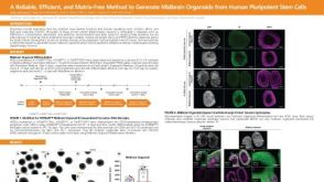 科学海报A Reliable, Efficient, and Matrix-Free Method to Generate Midbrain Organoids from Human Pluripotent Stem Cells


 沪公网安备31010102008431号