Dobo I et al. (JAN 2001)
The hematology journal : the official journal of the European Haematology Association / EHA 2 6 396--403
Comparison of four serum-free, cytokine-free media for analysis of endogenous erythroid colony growth in polycythemia vera and essential thrombocythemia.
INTRODUCTION: The assay of endogenous erythroid colony formation (EEC),a characteristic of polycythemia vera and essential thrombocythemia,is not standardized. In this multicentric study,we tested four semisolid,serum-free,cytokine-free media based on either methylcellulose (M1,M2) or collagen (C1,C2) commercialized for the EEC assay. MATERIALS AND METHODS: Bone marrow mononuclear cells (BMMC) from 73 individuals (62 patients with either polycythemia vera (26),essential thrombocythemia (19),secondary polyglobuly (17) or chronic myeloid leukemia (2) and 11 healthy donors) were grown in parallel in the four media without,or with 0.01 U/ml erythropoietin (EPo). RESULTS: In all four media EEC formation was specific,as it was not observed in cultures of patients with secondary polyglobuly or chronic myeloid leukemia,nor of healthy donors. Analysis of fresh or MGG-stained collagen gel cultures allowed detection of EEC formation significantly more frequently than methylcellulose-based media; addition of 0.01 U/ml of EPo had little or no effect on EEC formation. Collagen-based medium C1 gave better results than the other media tested: the 'C1' EEC assay was positive for 68.2% of polycythemia vera cultures with significantly higher median EEC numbers (6.5/10(5) BMMC for patients with one major criteria of polycythemia vera and 19 and 21/10(5) BMMC for patients with two or three major criteria,respectively). Medium C1 was also better for essential thrombocythemia cultures with 47.4% of positive results but with a low median EEC number (6.7/10(5) BMMC). When associated with the ELISA dosage of serum EPo,the 'C1' EEC assay allowed confirmation or elimination of the diagnosis of polycythemia vera for 91% (20/22) of polyglobulic patients. CONCLUSION: We propose that serum-free collagen-based culture systems be considered to standardize the EEC assay,now part of the new criteria of polycythemia vera.
View Publication
产品号#:
04961
04965
04962
04915
04807
04809
04906
04913
04803
04804
04905
04850
04974
04902
04960
04900
04901
04963
04970
04971
产品名:
MegaCult™-C胶原和含细胞因子培养基
MegaCult™-C CFU-Mk染色试剂盒
MegaCult-C 10% BSA, 6mL
MegaCult-C Human Serum, 6mL
Alkaline Phosphatase Substrate Tabs, pk
Biotin/Conjugate Goat Anti-Mu lgG, 125uL
MegaCult-C Evans Blue Stain, 5mL
Primary Ab, Anti-HuAnti-GPIIb/IIIa 360uL
MegaCult-C Control Antibody, 100 µL
Avidin-Alk Phosphatase Conjugate, 200 uL
MegaCult™-C含脂质培养基
MegaCult™-C胶原和含脂质培养基
胶原蛋白溶液
MegaCult™-C胶原和无细胞因子培养基
MegaCult™-C无细胞因子培养基
MegaCult™-C含细胞因子培养基
双室载玻片套件
MegaCult™-C无细胞因子全套试剂盒
MegaCult™-C含细胞因子全套试剂盒
Malerba I et al. (OCT 2002)
Toxicological sciences : an official journal of the Society of Toxicology 69 2 433--8
In vitro myelotoxicity of propanil and 3,4-dichloroaniline on murine and human CFU-E/BFU-E progenitors.
Because of the wide use of pesticides for domestic and industrial purposes,the evaluation of their potential effects is of major concern for public health. The myelotoxicity of the herbicide propanil (3,4-dichloroproprioanilide) and its metabolite 3,4-dichloroaniline (DCA) is well documented in mice,but evidence that pesticides may severely compromise hematopoiesis in humans is lacking. In this study,an interspecies comparison of in vitro toxicity of these two compounds on murine and human burst- and colony-forming unit-erythrocyte (BFU-E,CFU-E) and colony-forming unit-granulocyte/macrophage (CFU-GM) progenitors,has been carried out. Murine bone marrow progenitors and human cord blood cells were exposed to propanil or DCA in doses ranging from 10 micro M to 1000 micro M,and the toxic effect was detected by a clonogenic assay with continuous exposure to the compounds. The results on murine cells indicate that the erythrocytic lineage is the most sensitive target for propanil and DCA. On the other hand,human progenitors seem to be less sensitive to the toxic effects of both compounds than murine progenitors at the same concentrations (IC(50) values are 305.2 +/- 22.6 micro M [total erythroid colonies] and textgreater500 micro M [CFU-GM] for propanil). Propanil was significantly more toxic to human erythroid progenitors than to human CFU-GM progenitors,as was found for the murine cells,emphasizing the role of the heme pathway as the target for propanil. These data confirm the evidence that the compounds investigated interfere with erythroid colony formation at different stages of the differentiation pathway and have different effects according to the dose.
View Publication
产品号#:
04564
04534
04544
产品名:
MethoCult™ H4534 Classic 无 EPO 入门试剂盒
MethoCult™ H4534 Classic(不含 EPO)
MethoCult™ H4534 Classic(不含 EPO)
Truong B-TH et al. (FEB 2003)
Blood 101 3 1141--8
CCAAT/Enhancer binding proteins repress the leukemic phenotype of acute myeloid leukemia.
CCAAT/enhancer binding proteins (C/EBPs) are a family of factors that regulate cell growth and differentiation. These factors,particularly C/EBPalpha and C/EBPepsilon,have important roles in normal myelopoiesis. In addition,loss of C/EBP activity appears to have a role in the pathogenesis of myeloid disorders including acute myeloid leukemia (AML). Acute promyelocytic leukemia (APL) is a subtype of AML in which a role for C/EBPs has been postulated. In almost all cases of APL,a promyelocytic leukemia-retinoic acid receptor alpha (PML-RARalpha) fusion protein is expressed as a result of a t(15;17)(q22;q12) chromosomal translocation. PML-RARalpha inhibits expression of C/EBPepsilon,whereas all-trans retinoic acid (tRA),a differentiating agent to which APL is particularly susceptible,induces C/EBPepsilon expression. PML-RARalpha may also inhibit C/EBPalpha activity. Thus,the effects of PML-RARalpha on C/EBPs may contribute to both the development of leukemia and the unique sensitivity of APL to tRA. We tested the hypothesis that increasing the activity of C/EBPs would revert the leukemic phenotype. C/EBPalpha and C/EBPepsilon were introduced into the FDC-P1 myeloid cell line and into leukemic cells from PML-RARA transgenic mice. C/EBP factors suppressed growth and induced partial differentiation in vitro. In vivo,enhanced expression of C/EBPs prolonged survival. By using a tamoxifen-responsive version of C/EBPepsilon,we observed that C/EBPepsilon could mimic the effect of tRA,driving neutrophilic differentiation in leukemic animals. Our results support the hypothesis that induction of C/EBP activity is a critical effect of tRA in APL. Furthermore,our findings suggest that targeted modulation of C/EBP activities could provide a new approach to therapy of AML.
View Publication
产品号#:
05350
产品名:
Portis T and Longnecker R (JAN 2003)
Journal of virology 77 1 105--14
Epstein-Barr virus LMP2A interferes with global transcription factor regulation when expressed during B-lymphocyte development.
Epstein-Barr virus (EBV) is associated with the development of malignant lymphomas and lymphoproliferative disorders in immunocompromised individuals. The LMP2A protein of EBV is thought to play a central role in this process by allowing the virus to persist in latently infected B lymphocytes. We have demonstrated that LMP2A,when expressed in B cells of transgenic mice,allows normal B-cell developmental checkpoints to be bypassed. To identify cellular genes targeted by LMP2A that are involved in this process,we have utilized DNA microarrays to compare gene transcription in B cells from wild-type versus LMP2A transgenic mice. In B cells from LMP2A transgenic mice,we observed decreased expression of many genes associated with normal B-cell development as well as reduced levels of the transcription factors that regulate their expression. In particular,expression of the transcription factor E2A was down-regulated in bone marrow and splenic B cells. Furthermore,E2A activity was inhibited in these cells as determined by decreased DNA binding and reduced expression of its target genes,including the transcription factors early B-cell factor and Pax-5. Expression of two E2A inhibitors,Id2 and SCL,was up-regulated in splenic B cells expressing LMP2A,suggesting a possible mechanism for E2A inhibition. These results indicate that LMP2A deregulates transcription factor expression and activity in developing B cells,and this likely allows for a bypass of normal signaling events required for proper B-cell development. The ability of LMP2A to interfere with B-cell transcription factor regulation has important implications regarding its role in EBV latency.
View Publication
产品号#:
03630
产品名:
MethoCult™ M3630
Tan BL et al. (MAR 2003)
The Journal of biological chemistry 278 13 11686--95
Functional and biochemical consequences of abrogating the activation of multiple diverse early signaling pathways in Kit. Role for Src kinase pathway in Kit-induced cooperation with erythropoietin receptor.
Kit receptor tyrosine kinase and erythropoietin receptor (Epo-R) cooperate in regulating blood cell development. Mice that lack the expression of Kit or Epo-R die in utero of severe anemia. Stimulation of Kit by its ligand,stem cell factor activates several distinct early signaling pathways,including phospholipase C gamma,phosphatidylinositol 3-kinase,Src kinase,Grb2,and Grb7. The role of these pathways in Kit-induced growth,proliferation,or cooperation with Epo-R is not known. We demonstrate that inactivation of any one of these early signaling pathways in Kit significantly impairs growth and proliferation. However,inactivation of the Src pathway demonstrated the most profound defect. Combined stimulation with Epo also resulted in impaired cooperation between Src-defective Kit mutant and Epo-R and,to a lesser extent,with Kit mutants defective in the activation of phosphatidylinositol 3-kinase or Grb2. The impaired cooperation between the Src-defective Kit mutant and Epo-R was associated with reduced transphosphorylation of Epo-R and expression of c-Myc. Remarkably,restoration of only the Src pathway in a Kit receptor defective in the activation of all early signaling pathways demonstrated a 50% correction in proliferation in response to Kit stimulation and completely restored the cooperation with Epo-R. These data demonstrate an essential role for Src pathway in regulating growth,proliferation,and cooperation with Epo-R downstream from Kit.
View Publication
产品号#:
03434
03444
产品名:
MethoCult™ GF M3434
MethoCult™ GF M3434
Liu E et al. (APR 2003)
Blood 101 8 3294--301
Discrimination of polycythemias and thrombocytoses by novel, simple, accurate clonality assays and comparison with PRV-1 expression and BFU-E response to erythropoietin.
Essential thrombocythemia (ET) and polycythemia vera (PV) are clonal myeloproliferative disorders that are often difficult to distinguish from other causes of elevated blood cell counts. Assays that could reliably detect clonal hematopoiesis would therefore be extremely valuable for diagnosis. We previously reported 3 X-chromosome transcription-based clonality assays (TCAs) involving the G6PD,IDS,and MPP1 genes,which together were informative in about 65% of female subjects. To increase our ability to detect clonality,we developed simple TCA for detecting the transcripts of 2 additional X-chromosome genes: Bruton tyrosine kinase (BTK) and 4-and-a-half LIM domain 1 (FHL1). The combination of TCA established the presence or absence of clonal hematopoiesis in about 90% of female subjects. We show that both genes are subject to X-chromosome inactivation and are polymorphic in all major US ethnic groups. The 5 TCAs were used to examine clonality in 46 female patients along with assays for erythropoietin-independent erythroid colonies (EECs) and granulocyte PRV-1 mRNA levels to discriminate polycythemias and thrombocytoses. Of these,all 19 patients with familial polycythemia or thrombocytosis had polyclonal hematopoiesis,whereas 22 of 26 patients with clinical evidence of myeloproliferative disorder and 1 patient with clinically obscure polycythemia were clonal. Interestingly,interferon alpha therapy in 2 patients with PV was associated with reversion of clonal to polyclonal hematopoiesis. EECs were observed in 14 of 14 patients with PV and 4 of 12 with ET,and increased granulocyte PRV-1 mRNA levels were found in 9 of 13 patients with PV and 2 of 12 with ET. Thus,these novel clonality assays are useful in the diagnosis and follow-up of polycythemic conditions and disorders with increased platelet levels.
View Publication

 EasySep™小鼠TIL(CD45)正选试剂盒


 挂图Small Molecules, Big Impact in Pluripotent Stem Cell Research Overview of signaling pathways and small molecules in pluripotent stem cell research
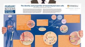 挂图The Identity and Properties of Mesenchymal Stem Cells Overview of MSC expansion, differentiation, immunoregulatory properties and therapeutic potential
 挂图Stem Cell States: Naive to Primed Pluripotency Properties of naive (ground) and primed pluripotent stem cells


 沪公网安备31010102008431号