Trevisan M et al. (JAN 2017)
International journal of molecular sciences 18 1
Reprogramming Methods Do Not Affect Gene Expression Profile of Human Induced Pluripotent Stem Cells.
Induced pluripotent stem cells (iPSCs) are pluripotent cells derived from adult somatic cells. After the pioneering work by Yamanaka,who first generated iPSCs by retroviral transduction of four reprogramming factors,several alternative methods to obtain iPSCs have been developed in order to increase the yield and safety of the process. However,the question remains open on whether the different reprogramming methods can influence the pluripotency features of the derived lines. In this study,three different strategies,based on retroviral vectors,episomal vectors,and Sendai virus vectors,were applied to derive iPSCs from human fibroblasts. The reprogramming efficiency of the methods based on episomal and Sendai virus vectors was higher than that of the retroviral vector-based approach. All human iPSC clones derived with the different methods showed the typical features of pluripotent stem cells,including the expression of alkaline phosphatase and stemness maker genes,and could give rise to the three germ layer derivatives upon embryoid bodies assay. Microarray analysis confirmed the presence of typical stem cell gene expression profiles in all iPSC clones and did not identify any significant difference among reprogramming methods. In conclusion,the use of different reprogramming methods is equivalent and does not affect gene expression profile of the derived human iPSCs.
View Publication
产品号#:
85850
85857
85870
85875
产品名:
mTeSR™1
mTeSR™1
Dai D-F et al. ( 2017)
Stem cells international 2017 5153625
Mitochondrial Maturation in Human Pluripotent Stem Cell Derived Cardiomyocytes.
Human pluripotent stem cells derived cardiomyocytes (PSC-CMs) have been widely used for disease modeling,drug safety screening,and preclinical cell therapy to regenerate myocardium. Most studies have utilized PSC-CM grown in vitro for a relatively short period after differentiation. These PSC-CMs demonstrated structural,electrophysiological,and mechanical features of primitive cardiomyocytes. A few studies have extended in vitro PSC-CM culture time and reported improved maturation of structural and electromechanical properties. The degree of mitochondrial maturation,however,remains unclear. This study characterized the development of mitochondria during prolonged in vitro culture. PSC-CM demonstrated an improved mitochondrial maturation with prolonged culture,in terms of increased mitochondrial relative abundance,enhanced membrane potential,and increased activity of several mitochondrial respiratory complexes. These are in parallel with the maturation of other cellular components. However,the maturation of mitochondria in PSC-CMs grown for extended in vitro culture exhibits suboptimal maturation when compared with the maturation of mitochondria observed in the human fetal heart during similar time interval.
View Publication
产品号#:
85850
85857
85870
85875
产品名:
mTeSR™1
mTeSR™1
Ghezzi S et al. (APR 2017)
Antiviral research 140 13--17
Heparin prevents Zika virus induced-cytopathic effects in human neural progenitor cells.
The recent Zika virus (ZIKV) outbreak,which mainly affected Brazil and neighbouring states,demonstrated the paucity of information concerning the epidemiology of several flaviruses,but also highlighted the lack of available agents with which to treat such emerging diseases. Here,we show that heparin,a widely used anticoagulant,while exerting a modest inhibitory effect on Zika Virus replication,fully prevents virus-induced cell death of human neural progenitor cells (NPCs).
View Publication
R. G. Walton et al. (dec 2019)
Aging cell 18 6 e13039
Metformin blunts muscle hypertrophy in response to progressive resistance exercise training in older adults: A randomized, double-blind, placebo-controlled, multicenter trial: The MASTERS trial.
Progressive resistance exercise training (PRT) is the most effective known intervention for combating aging skeletal muscle atrophy. However,the hypertrophic response to PRT is variable,and this may be due to muscle inflammation susceptibility. Metformin reduces inflammation,so we hypothesized that metformin would augment the muscle response to PRT in healthy women and men aged 65 and older. In a randomized,double-blind trial,participants received 1,700 mg/day metformin (N = 46) or placebo (N = 48) throughout the study,and all subjects performed 14 weeks of supervised PRT. Although responses to PRT varied,placebo gained more lean body mass (p = .003) and thigh muscle mass (p {\textless} .001) than metformin. CT scan showed that increases in thigh muscle area (p = .005) and density (p = .020) were greater in placebo versus metformin. There was a trend for blunted strength gains in metformin that did not reach statistical significance. Analyses of vastus lateralis muscle biopsies showed that metformin did not affect fiber hypertrophy,or increases in satellite cell or macrophage abundance with PRT. However,placebo had decreased type I fiber percentage while metformin did not (p = .007). Metformin led to an increase in AMPK signaling,and a trend for blunted increases in mTORC1 signaling in response to PRT. These results underscore the benefits of PRT in older adults,but metformin negatively impacts the hypertrophic response to resistance training in healthy older individuals. ClinicalTrials.gov Identifier: NCT02308228.
View Publication

 EasySep™小鼠TIL(CD45)正选试剂盒


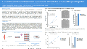 科学海报A Serum-Free Workflow for the Isolation, Expansion and Differentiation of Human Myogenic Progenitors

 沪公网安备31010102008431号