
 EasySep™小鼠TIL(CD45)正选试剂盒


技术资料
-
-
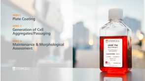
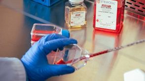 实验方案Transitioning from Feeder-Free Media to mTeSR™ Plus for Human Pluripotent Stem Cell Culture
研究方向:
干细胞生物学
发布日期: 02/10/2020 -
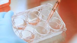
 实验方案Enzyme-Free Passaging of Human Pluripotent Stem Cells Using ReLeSR™
研究方向:
干细胞生物学
发布日期: 02/10/2020 -
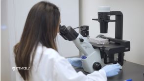
 实验方案How to Coat Cultureware with Vitronectin XF™ for Pluripotent Stem Cell Culture
研究方向:
干细胞生物学
发布日期: 02/10/2020


 沪公网安备31010102008431号