
 EasySep™小鼠TIL(CD45)正选试剂盒


技术资料
-
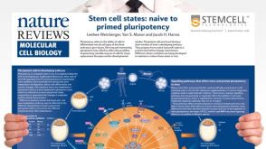 挂图Stem Cell States: Naive to Primed Pluripotency Properties of naive (ground) and primed pluripotent stem cells
 -
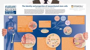 挂图The Identity and Properties of Mesenchymal Stem Cells Overview of MSC expansion, differentiation, immunoregulatory properties and therapeutic potential
 -
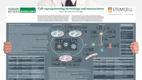 挂图Cell-Reprogramming Technology and Neuroscience Details on human iPSC-derived models of neuropsychiatric and neurodegenerative disorders
 -
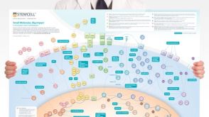 挂图Small Molecules, Big Impact in Pluripotent Stem Cell Research Overview of signaling pathways and small molecules in pluripotent stem cell research
 -
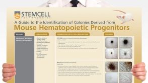 挂图Identification of Colonies Derived from Mouse Hematopoietic Progenitors Representative colony images and tips for identifying progenitor subtypes in CFU assays
 -
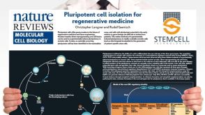 挂图Pluripotent Stem Cell Biology Nuclear transfer, reprogramming and cell fusion to induce pluripotency in somatic cells
 -
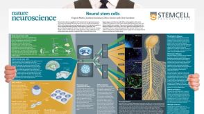 挂图Neural Stem Cells Overview of the types of NSCs and their potential use as therapeutic agents for disease
 -
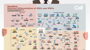 挂图Directed Differentiation of Pluripotent Stem Cells Strategies for differentiating ES and iPS cells into ectoderm, endoderm or mesoderm
 -
-


 沪公网安备31010102008431号