
 EasySep™小鼠TIL(CD45)正选试剂盒


产品号 #05008_C
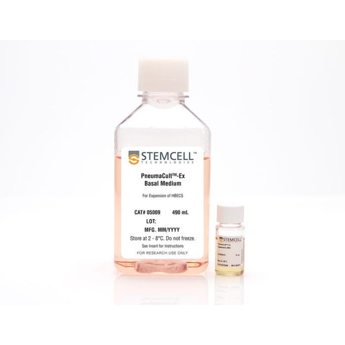
用于扩增原代人呼吸道上皮细胞的无血清和无BPE培养基
若您需要咨询产品或有任何技术问题,请通过官方电话 400 885 9050 或邮箱 info.cn@stemcell.com 与我们联系。
用于扩增原代人呼吸道上皮细胞的无血清和无BPE培养基
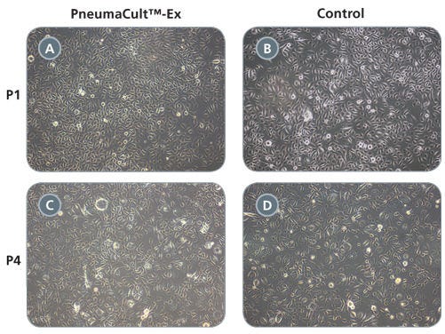
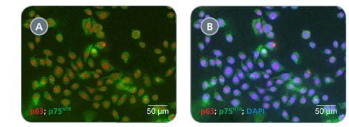
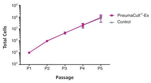
PneumaCult™- Ex是一款成份明确的,不含血清和BPE的细胞培养基,支持人呼吸道上皮细胞的快速扩增。使用PneumaCult™-Ex培养的原代呼吸道上皮细胞在至少3代内迅速扩增,同时保持鹅卵石状形态和基底细胞标记物p63和p75NTR的均匀表达。此外,这些细胞在 PneumaCult™-ALI 的气-液界面条件下可进一步分化,形成具黏液纤毛特征的假复层上皮结构。
PneumaCult™-ALI Medium和PneumaCult™-Ex Plus Medium(产品号:#05040)构成了一个完整、无BPE的人源呼吸道上皮体外建模系统,也与原代人鼻上皮细胞兼容。这一稳定、定义明确的培养系统是开展基础呼吸研究、毒理学研究和药物开发的宝贵工具。
欢迎观看我们的肺部研究课程,了解如何在 ALI 条件下培养人呼吸道上皮细胞,或浏览我们的常见问题解答(FAQs)关于使用PneumaCult™的ALI培养工作流程。
分类
专用培养基
细胞类型
气道细胞
种属
人
应用
细胞培养,扩增,培养
品牌
PneumaCult
研究领域
上皮细胞研究
制剂类别
无血清
请在《产品说明书》中查找相关支持信息和使用说明,或浏览下方更多实验方案。
本产品专为以下研究领域设计,适用于工作流程中的高亮阶段。探索这些工作流程,了解更多我们为各研究领域提供的其他配套产品。
| 物种 | 人 |
|---|---|
| 配方 | 无血清 |
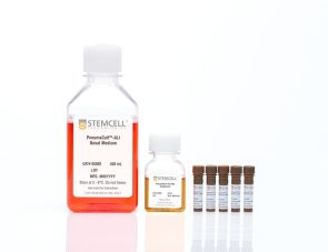
用于在气液界面培养的人呼吸道上皮细胞的无血清和无BPE培养基
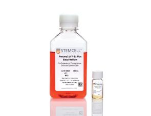
用于扩增人原代呼吸道上皮细胞的无血清和无BPE培养基

哺乳动物活细胞计数试剂
在线联系


 沪公网安备31010102008431号