Kim J et al. (NOV 2013)
Stem Cell Research 11 3 978--989
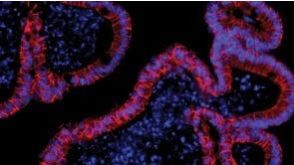
Alginate microcapsule as a 3D platform for the efficient differentiation of human embryonic stem cells to dopamine neurons
Human embryonic stem cells (hESCs) are emerging as an attractive alternative source for cell replacement therapy since the cells can be expanded in culture indefinitely and differentiated into any cell types in the body. In order to optimize cell-to-cell interaction,cell proliferation and differentiation into specific lineages as well as tissue organization,it is important to provide a microenvironment for the hESCs which mimics the stem cell niche. One approach is to provide a three-dimensional (3D) environment such as encapsulation. We present an approach to culture and differentiate hESCs into midbrain dopamine (mdDA) neurons in a 3D microenvironment using alginate microcapsules for the first time. A detailed gene and protein expression analysis during neuronal differentiation showed an increased gene and protein expression of various specific DA neuronal markers,particularly tyrosine hydroxylase (TH) by textgreater100 folds after 2weeks and at least 50% higher expression after 4weeks respectively,compared to cells differentiated under conventional two-dimensional (2D) platform. The encapsulated TH+ cells co-expressed mdDA neuronal markers,forkhead box protein A-2 (FOXA2) and pituitary homeobox-3 (PITX3) after 4weeks and secreted approximately 60pg/ml/106 cells higher DA level when induced. We propose that the 3D platform facilitated an early onset of DA neuronal generation compared to that with conventional 2D system which also secretes more DA under potassium-induction. It is a very useful model to study the proliferation and directed differentiation of hESCs to various lineages,particularly to mdDA neurons. This 3D system also allows the separation of feeder cells from hESCs during the process of differentiation and also has potential for immune-isolation during transplantation studies. ?? 2013 Elsevier B.V.
View Publication
产品类型:
产品号#:
05850
05857
05870
05875
07923
85850
85857
85870
85875
产品名:
Dispase (1 U/mL)
mTeSR™1
mTeSR™1
Hannum C et al. (APR 1994)
Nature 368 6472 643--8
Ligand for FLT3/FLK2 receptor tyrosine kinase regulates growth of haematopoietic stem cells and is encoded by variant RNAs.
The FLT3/FLK2 receptor tyrosine kinase is closely related to two receptors,c-Kit and c-Fms,which function with their respective ligands,Kit ligand and macrophage colony-stimulating factor to control differentiation of haematopoietic and non-haematopoietic cells. FLT3/FLK2 is thought to be present on haematopoietic stem cells and found in brain,placenta and testis. We have purified to homogeneity and partially sequenced a soluble form of the FLT3/FLK2 ligand produced by mouse thymic stromal cells. We isolated several mouse and human complementary DNAs that encode polypeptides with identical N termini and different C termini. Some variants contain hydrophobic transmembrane segments,suggesting that processing may be required to release soluble ligand. The purified ligand enhances the response of mouse stem cells and a primitive human progenitor cell population to other growth factors such as interleukins IL-3 and IL-6 and to granulocyte-macrophage colony-stimulating factor,and also stimulates fetal thymocytes.
View Publication
产品类型:
产品号#:
02640
02840
产品名:
E. Yamashita et al. (Sep 2025)
The FASEB Journal 39 17
Red Blood Cell‐Mediated Enhancement of Hematopoietic Stem Cell Functions via a Hes1‐Dependent Pathway
In bone marrow,cell numbers are balanced between production and loss. After chemotherapy,blood cell counts decrease initially but later recover as hematopoietic progenitor cells expand,although the mechanisms underlying this recovery are still unclear. We investigated the influence of red blood cells (RBCs) on hematopoietic stem cell (HSC) function during bone marrow recovery. Following chemotherapy,RBC concentrations in bone marrow peaked on day 5 posttreatment,coinciding with the recovery of hematopoiesis. Coculture of HSCs with RBCs resulted in a significant increase in hematopoiesis. Direct contact between RBCs and HSCs was essential for enhancement of hematopoiesis,and HSCs precultured with RBCs resulted in greater numbers of donor‐derived mature hematopoietic cells after transplantation. RNA‐sequencing analysis showed that Hes1 was the most significantly upregulated transcription factor in RBC coculture,and the response to RBC‐induced hematopoiesis of Hes1‐deficient HSCs was reduced. These findings imply a role of RBCs and Hes1 in the enhancement of hematopoietic recovery following bone marrow stress.
View Publication
产品类型:
产品号#:
03436
产品名:
MethoCult™ SF M3436
Pirson L et al. (JUL 2006)
Stem cells (Dayton,Ohio) 24 7 1814--21
Despite inhibition of hematopoietic progenitor cell growth in vitro, the tyrosine kinase inhibitor imatinib does not impair engraftment of human CD133+ cells into NOD/SCIDbeta2mNull mice.
There is potential interest for combining allogeneic hematopoietic cell transplantation (HCT),and particularly allogeneic HCT with a nonmyeloablative regimen,to the tyrosine kinase inhibitor imatinib (Glivec; Novartis,Basel,Switzerland,http://www.novartis.com) in order to maximize anti-leukemic activity against Philadelphia chromosome-positive leukemias. However,because imatinib inhibits c-kit,the stem cell factor receptor,it could interfere with bone marrow engraftment. In this study,we examined the impact of imatinib on normal progenitor cell function. Imatinib decreased the colony-forming capacity of mobilized peripheral blood human CD133(+) cells but not that of long-term culture-initiating cells. Imatinib also decreased the proliferation of cytokine-stimulated CD133(+) cells but did not induce apoptosis of these cells. Expression of very late antigen (VLA)-4,VLA-5,and CXCR4 of CD133(+) cells was not modified by imatinib,but imatinib decreased the ability of CD133(+) cells to migrate. Finally,imatinib did not decrease engraftment of CD133(+) cells into irradiated nonobese diabetic/severe combined immunodeficient/beta2m(null) mice conditioned with 3 or 1 Gy total body irradiation. In summary,our results suggest that,despite inhibition of hematopoietic progenitor cell growth in vitro,imatinib does not interfere with hematopoietic stem cell engraftment.
View Publication
G. Schiroli et al. (apr 2019)
Cell stem cell 24 4 551--565.e8
Precise Gene Editing Preserves Hematopoietic Stem Cell Function following Transient p53-Mediated DNA Damage Response.
Precise gene editing in hematopoietic stem and progenitor cells (HSPCs) holds promise for treating genetic diseases. However,responses triggered by programmable nucleases in HSPCs are poorly characterized and may negatively impact HSPC engraftment and long-term repopulation capacity. Here,we induced either one or several DNA double-stranded breaks (DSBs) with optimized zinc-finger and CRISPR/Cas9 nucleases and monitored DNA damage response (DDR) foci induction,cell-cycle progression,and transcriptional responses in HSPC subpopulations,with up to single-cell resolution. p53-mediated DDR pathway activation was the predominant response to even single-nuclease-induced DSBs across all HSPC subtypes analyzed. Excess DSB load and/or adeno-associated virus (AAV)-mediated delivery of DNA repair templates induced cumulative p53 pathway activation,constraining proliferation,yield,and engraftment of edited HSPCs. However,functional impairment was reversible when DDR burden was low and could be overcome by transient p53 inhibition. These findings provide molecular and functional evidence for feasible and seamless gene editing in HSPCs.
View Publication
产品类型:
产品号#:
04434
04444
72912
72914
产品名:
MethoCult™ H4434 Classic
MethoCult™ H4434 Classic
Jones C et al. (MAY 2004)
Cancer research 64 9 3037--45
Expression profiling of purified normal human luminal and myoepithelial breast cells: identification of novel prognostic markers for breast cancer.
The normal duct-lobular system of the breast is lined by two epithelial cell types,inner luminal secretory cells and outer contractile myoepithelial cells. We have generated comprehensive expression profiles of the two normal cell types,using immunomagnetic cell separation and gene expression microarray analysis. The cell-type specificity was confirmed at the protein level by immunohistochemistry in normal breast tissue. New prognostic markers for survival were identified when the luminal- and myoepithelial-specific molecules were evaluated on breast tumor tissue microarrays. Nuclear expression of luminal epithelial marker galectin 3 correlated with a shorter overall survival in these patients,and the expression of SPARC (osteonectin),a myoepithelial marker,was an independent marker of poor prognosis in breast cancers as a whole. These data provide a framework for the interpretation of breast cancer molecular profiling experiments,the identification of potential new diagnostic markers,and development of novel indicators of prognosis.
View Publication
产品类型:
产品号#:
01434
产品名:
H. Cai et al. (Jan 2026)
International Journal of Molecular Sciences 27 1
NGR1 Pretreatment Enhances the Therapeutic Efficacy of Transplanting Cardiomyocytes Derived from Human Induced Pluripotent Stem Cells for Myocardial Infarction
Human induced pluripotent stem cells (hiPSCs) offer significant potential for differentiation and research applications in cardiovascular diseases. When induced differentiated hiPSC-derived cardiomyocytes (hiPSC-CMs) are transplanted into the infarcted myocardial region,they exhibit extremely low survival rates and unsatisfactory therapeutic effects due to ischemia,hypoxia,and immune inflammation in the surrounding environment. To address this issue,we used Panax notoginseng saponin R1 (NGR1),which has demonstrated significant protective effects in prior research,to pretreat hiPSC-CMs before transplantation. Utilizing an in vitro H2O2 oxidative stress model and a nude mouse myocardial infarction (MI) model,we investigated the mechanism through which NGR1 pretreatment enhances the therapeutic efficacy of hiPSC-CM transplantation. The results revealed that the hiPSC-CMs expressed cTnT. NGR1 did not promote the proliferation of hiPSC-CMs but instead induced elevated levels of p-Akt protein in these cells. Compared to hiPSC-CM transplantation alone,transplantation of hiPSC-CMs pretreated with NGR1 exhibited higher ejection fraction (EF) and fractional shortening (FS) values,along with reduced infarct size and collagen deposition. Additionally,there were more HNA-positive cardiomyocytes in the cardiac tissue,fewer TUNEL-positive signals,and increased VWF-positive and Lyve1-positive signals. Furthermore,the gene expression levels of VEGFC,IGF-1,and SDF-1 were higher. Therefore,NGR1 pretreatment improves the survival of transplanted hiPSC-CMs in tissues,reduces myocardial apoptosis,enhances cardiac function,decreases infarct size and collagen deposition,promotes angiogenesis and lymphangiogenesis,and stimulates paracrine secretion.
View Publication
产品类型:
产品号#:
85850
85857
产品名:
mTeSR™1
mTeSR™1
(Nov 2024)
International Journal of Molecular Sciences 25 22
The Generation of Genetically Engineered Human Induced Pluripotent Stem Cells Overexpressing IFN-? for Future Experimental and Clinically Oriented Studies
Induced pluripotent stem cells (iPSCs) can be generated from various adult cells,genetically modified and differentiated into diverse cell populations. Type I interferons (IFN-Is) have multiple immunotherapeutic applications; however,their systemic administration can lead to severe adverse outcomes. One way of overcoming the limitation is to introduce cells able to enter the site of pathology and to produce IFN-Is locally. As a first step towards the generation of such cells,here,we aimed to generate human iPSCs overexpressing interferon-beta (IFNB,IFNB-iPSCs). IFNB-iPSCs were obtained by CRISPR/Cas9 editing of the previously generated iPSC line K7-4Lf. IFNB-iPSCs overexpressed IFNB RNA and produced a functionally active IFN-?. The cells displayed typical iPSC morphology and expressed pluripotency markers. Following spontaneous differentiation,IFNB-iPSCs formed embryoid bodies and upregulated endoderm,mesoderm,and some ectoderm markers. However,an upregulation of key neuroectoderm markers,PAX6 and LHX2,was compromised. A negative effect of IFN-? on iPSC neuroectoderm differentiation was confirmed in parental iPSCs differentiated in the presence of a recombinant IFN-?. The study describes new IFN-?-producing iPSC lines suitable for the generation of various types of IFN-?-producing cells for future experimental and clinical applications,and it unravels an inhibitory effect of IFN-? on stem cell neuroectoderm differentiation.
View Publication
产品类型:
产品号#:
85850
85857
产品名:
mTeSR™1
mTeSR™1
J. L. D. Andrés et al. (Sep 2024)
Materials Today Bio 29 6
A bioengineered tumor matrix-based scaffold for the evaluation of melatonin efficacy on head and neck squamous cancer stem cells
Head and neck squamous cell carcinoma (HNSCC) presents a significant challenge worldwide due to its aggressiveness and high recurrence rates post-treatment,often linked to cancer stem cells (CSCs). Melatonin shows promise as a potent tumor suppressor; however,the effects of melatonin on CSCs remain unclear,and the development of models that closely resemble tumor heterogeneity could help to better understand the effects of this molecule. This study developed a tumor scaffold based on patient fibroblast-derived decellularized extracellular matrix that mimics the HNSCC microenvironment. Our study investigates the antitumoral effects of melatonin within this context. We validated its strong antiproliferative effect on HNSCC CSCs and the reduction of tumor invasion and migration markers,even in a strongly chemoprotective environment,as it is required to increase the minimum doses necessary to impact tumor viability compared to the non-scaffolded tumorspheres culture. Moreover,melatonin exhibited no cytotoxic effects on healthy cells co-cultured in the tumor hydrogel. This scaffold-based platform allows an in vitro study closer to HNSCC tumor reality,including CSCs,stromal component,and a biomimetic matrix,providing a new valuable research tool in precision oncology.
View Publication
产品类型:
产品号#:
01700
产品名:
ALDEFLUOR™ 试剂盒
Jin S et al. (JUN 2016)
Stem Cells
A Novel Role for miR-1305 in Regulation of Pluripotency-Differentiation Balance, Cell Cycle, and Apoptosis in Human Pluripotent Stem Cells
Human embryonic stem cells (hESCs) and human induced pluripotent stem cells (hiPSCs) are defined as pluripotent in view of their self-renewal ability and potential to differentiate to cells of all three germ layers. Recent studies have indicated that microRNAs (miRNAs) play an important role in the maintenance of pluripotency and cell cycle regulation. We used a microarray based approach to identify miRNAs that were enriched in hESCs when compared to differentiated cells and at the same time showed significant expression changes between different phases of cell cycle. We identified 34 candidate miRNAs and performed functional studies on one of these,miR-1305,which showed the highest expression change during cell cycle transition. Overexpression of miR-1305 induced differentiation of pluripotent stem cells,increased cell apoptosis and sped up G1/S transition,while its downregulation facilitated the maintenance of pluripotency and increased cell survival. Using target prediction software and luciferase based reporter assays we identified POLR3G as a downstream target by which miR-1305 regulates the fine balance between maintenance of pluripotency and onset of differentiation. Overexpression of POLR3G rescued pluripotent stem cell differentiation induced by miR-1305 overexpression. In contrast,knock-down of POLR3G expression abolished the miR-1305-knockdown mediated enhancement of pluripotency,thus validating its role as miR-1305 target in human pluripotent stem cells. Together our data point to an important role for miR-1305 as a novel regulator of pluripotency,cell survival and cell cycle and uncovers new mechanisms and networks by which these processes are intertwined in human pluripotent stem cells. This article is protected by copyright. All rights reserved.
View Publication
产品类型:
产品号#:
05850
05857
05870
05875
85850
85857
85870
85875
产品名:
mTeSR™1
mTeSR™1
Sebaa M et al. (JAN 2015)
Journal of Biomedical Materials Research - Part A 103 1 25--37
The effects of poly(3,4-ethylenedioxythiophene) coating on magnesium degradation and cytocompatibility with human embryonic stem cells for potential neural applications
Magnesium (Mg) is a promising conductive metallic biomaterial due to its desirable mechanical properties for load bearing and biodegradability in human body. Controlling the rapid degradation of Mg in physiological environment continues to be the key challenge toward clinical translation. In this study,we investigated the effects of conductive poly(3,4-ethylenedioxythiophene) (PEDOT) coating on the degradation behavior of Mg substrates and their cytocompatibility. Human embryonic stem cells (hESCs) were used as the in vitro model system to study cellular responses to Mg degradation because they are sensitive and can potentially differentiate into many cell types of interest (e.g.,neurons) for regenerative medicine. The PEDOT was deposited on Mg substrates using electrochemical deposition. The greater number of cyclic voltammetry (CV) cycles yielded thicker PEDOT coatings on Mg substrates. Specifically,the coatings produced by 2,5,and 10 CV cycles (denoted as 2×-PEDOT-Mg,5×-PEDOT-Mg,and 10×-PEDOT-Mg) had an average thickness of 31,63,and 78 µm,respectively. Compared with non-coated Mg samples,all PEDOT coated Mg samples showed slower degradation rates,as indicated by Tafel test results and Mg ion concentrations in the post-culture media. The 5×-PEDOT-Mg showed the best coating adhesion and slowest Mg degradation among the tested samples. Moreover,hESCs survived for the longest period when cultured with the 5×-PEDOT-Mg samples compared with the non-coated Mg and 2×-PEDOT-Mg. Overall,the results of this study showed promise in using PEDOT coating on biodegradable Mg-based implants for potential neural recording,stimulation and tissue engineering applications,thus encouraging further research.
View Publication

 EasySep™小鼠TIL(CD45)正选试剂盒


 沪公网安备31010102008431号